Online Gather.town Pitches
Diffusion & Susceptibility I
Joint Annual Meeting ISMRM-ESMRMB & ISMRT 31st Annual Meeting • 07-12 May 2022 • London, UK

Online Gather.town Pitches
Diffusion & Susceptibility I
| Booth # | ||||
|---|---|---|---|---|
3299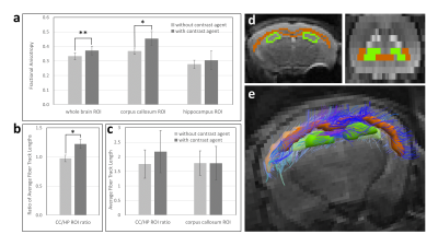 |
1 | The effect of primed intravenous infusion of magnetic resonance contrast agent on diffusion tensor imaging in vivo
Norman Arthur Lapin1, Xiaodong Wen1, and Janaka Wansapura1
1Advanced Imaging Research Center, UT Southwestern Medical Center, Dallas, TX, United States
The challenge of conducting diffusion tensor imaging in the presence of systemic contrast agent has been noted clinically. Here, we investigate primed infusion as a means of stabilizing contrast enhancement in the mouse brain during image acquisition. Our results show that with infusion-sustained contrast enhancement, fractional anisotropy (FA) was significantly elevated in whole brain and corpus callosum ROIs, but not in hippocampus ROI. Contrast enhancement significantly increased tractography metric of relative mean fiber-track length in corpus callosum versus hippocampus. Thus, contrast enhancement may have a preferential effect on anisotropic brain regions and could potentially be applied to amplify FA selectively.
|
||
3300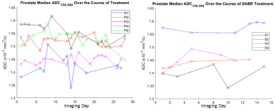 |
2 | Daily Measurement of Prostate ADC in Patients Undergoing SABR and Conventional Treatment for Prostate Cancer on an MR-Linac
Chris Moore1, Joe Stickley1, Abigael Clough2, Claire Nelder2, Robert Chuter1, Ananya Choudhury3, Damien McHugh1, and Michael Dubec1,4
1CMPE, The Christie NHS Foundation Trust, Manchester, United Kingdom, 2Radiotherapy Services, The Christie NHS Foundation Trust, Manchester, United Kingdom, 3Research and Development, The Christie NHS Foundation Trust, Manchester, United Kingdom, 4University of Manchester, Manchester, United Kingdom
MR-Linacs enable daily functional imaging to be performed in patients being treated with radiotherapy. In this work, the diffusion sequence developed by the MRL Consortium biomarkers working group underwent technical validation, showing accurate and repeatable phantom ADC measurements. Daily, whole-prostate ADC mapping was also performed in patients undergoing treatment for prostate cancer on an MRL. No significant differences were observed in whole-prostate ADC during the course of treatment for conventional and stereotactic ablative body radiotherapy (SABR) treatments. Absence of significant change in ADC could be related to the fact that patients were treated with neo-adjuvant hormone therapy prior to radiotherapy.
|
||
3301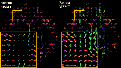 |
3 | Mr CSD – Motion-robust Constrained Spherical Deconvolution for multi-shell multi-tissue brain modelling of infants and other restless patients
Viljami Sairanen1
1BABA Center, Pediatric Research Center, Department of Clinical Neurophysiology, Children’s Hospital, Helsinki University Hospital and University of Helsinki, University of Helsinki, Helsinki, Finland
Investigation of brain structure of infants or other uncooperative patients using diffusion-weighted MRI is challenging due to subject motion artefacts. This work proposes a novel robust augmentation to current state-of-the-art multi-shell multi-tissue (MSMT) constrained spherical deconvolution (CSD) pipeline that accounts for these artefacts in both response function estimation and in deconvolution. A proof-of-concept is shown using multi-shell infant dataset and it is compared against the normal MSMT-CSD. The results indicate that motion artefacts can result in incorrect tissue type segmentations and fiber orientation distribution (FOD) estimates which could affect negatively any following analysis such as tractography or microstructural brain modelling.
|
||
3302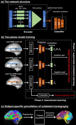 |
4 | An efficient deep learning framework for consistent superficial white matter tractography parcellation
Tengfei Xue1,2, Fan Zhang1, Chaoyi Zhang2, Yuqian Chen1,2, Yang Song3, Nikos Makris1, Yogesh Rathi1, Weidong Cai2, and Lauren Jean O’Donnell1
1Harvard Medical School, Boston, MA, United States, 2The University of Sydney, Sydney, Australia, 3University of New South Wales, Sydney, Australia
We propose a deep-learning-based framework, Superficial White Matter Analysis (SupWMA), which performs an efficient and consistent parcellation of 198 SWM clusters from whole-brain tractography. A point-cloud-based network is developed for our SWM parcellation task, and supervised contrastive learning enables more discriminative representations between plausible streamlines and outliers. SupWMA obtains highly consistent and accurate SWM parcellation results on a large tractography dataset with ground truth labels and on three independently acquired testing datasets from individuals across ages and health conditions.
|
||
3303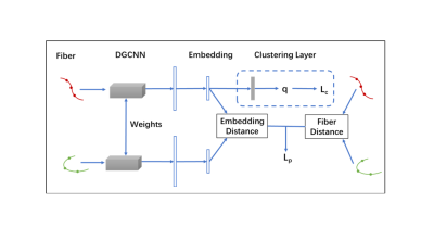 |
5 | Anatomically Informed Unsupervised Deep Learning for Fast and Effective White Matter Fiber Clustering
Yuqian Chen1,2, Chaoyi Zhang2, Yang Song3, Tengfei Xue1,2, Nikos Makris1, Yogesh Rathi1, Weidong Cai2, Fan Zhang1, and Lauren Jean O'Donnell1
1Harvard Medical School, Boston, MA, United States, 2The University of Sydney, Sydney, Australia, 3The University of New South Wales, Sydney, Australia
We propose a novel unsupervised deep learning framework for white matter fiber clustering. Self-supervised learning is adopted to enable joint deep embedding and cluster assignment. Anatomical information is incorporated into the neural network to improve anatomical coherence. In addition, outlier removal is performed to further improve cluster quality. Our method is evaluated on three datasets and showed superior performance in terms of cluster compactness, anatomical coherence and generalization across subjects compared to several state-of-the-art algorithms.
|
||
3304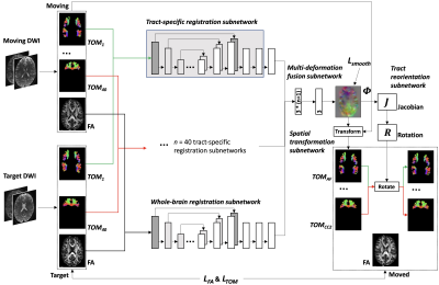 |
6 | A deep learning diffusion MRI registration method using joint whole-brain and tract-specific information
Fan Zhang1, William M Wells III1, and Lauren J O'Donnell1
1Harvard Medical School, Boston, MA, United States
We present a novel deep learning method, DDMReg, for accurate dMRI registration. In dMRI registration, the goal is to align brain anatomical structures while ensuring local fiber orientations consistency with the underlying white matter anatomy. DDMReg is an unsupervised method for deformable dMRI registration, without the need of non-rigidly pre-registered images and the corresponding deformation field as ground truth. We propose a novel registration architecture that leverages not only whole-brain information but also tract-specific fiber orientation information. We perform comparisons with four state-of-the-art methods on several independently acquired datasets. Experimental results show that DDMReg obtains highly improved registration performance.
|
||
3305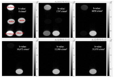 |
7 | Validation of the Multicomponent Diffusion Analysis using L1-norm regularized NNLS for Ultra-high b-value Diffusion-weighted MRI protocols
Jin Gao1,2, Weiguo Li2,3, Richard Magin3, and Danilo Erricolo1
1Electrical and Computer Engineering, University of Illinois at Chicago, Chicago, IL, United States, 2Research Resources Center, University of Illinois at Chicago, Chicago, IL, United States, 3Biomedical Engineering, University of Illinois at Chicago, Chicago, IL, United States
Multiple components analysis as a convenient tool to model multiexponential relaxations has been well developed in the applications of T2-weighted MRI. Nevertheless, this analysis is rarely used in diffusion-weighted MRI data and the lack of systematic studies makes the situation worse. Building on our previous studies, to validate the performance of the analysis becomes critical to carry out the ultra-high b-value DWI technique in vivo or translate to the clinical applications. We, therefore, proposed this phantom study to validate the stability and robustness of the multiple components analysis applied in the ultra-high b-value DWI technique.
|
||
3306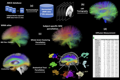 |
8 | Consistent White Matter Parcellation in Adolescent Brain Cognitive Development (ABCD): A ~10k Harmonized Diffusion MRI Study
Fan Zhang1, Suheyla Cetin-Karayumak1, Steve Pieper1, Yogesh Rathi1, and Lauren J O'Donnell1
1Harvard Medical School, Boston, MA, United States
We present a large-scale harmonized dMRI study where we have performed successful white matter(WM) tractography parcellation across ~10k subjects from the ABCD study. We first assess the effects of data harmonization on WM parcellation, followed by an evaluation of WM parcellation using the entire harmonized dataset. We show that after harmonization, more anatomical tracts are identified and their FA values are closer to the harmonization reference data. We also demonstrate highly successful WM parcellation, where overall 99.9% of tracts(73 per subject) are identified. The parcellated WM tracts, as well as their diffusion measures, will be made available to the public.
|
||
3307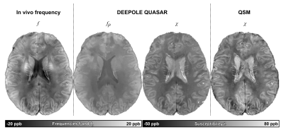 |
9 | In vivo validation and quantification of non-susceptibility frequency contrast obtained with DEEPOLE QUASAR
Thomas Jochmann1, Jens Haueisen1, Dejan Jakimovski2,3, Robert Zivadinov2,3, and Ferdinand Schweser2,3
1Department of Computer Science and Automation, Technische Universität Ilmenau, Ilmenau, Germany, 2Buffalo Neuroimaging Analysis Center, Department of Neurology at the Jacobs School of Medicine and Biomedical Sciences, University at Buffalo, The State University of New York, Buffalo, NY, United States, 3Center for Biomedical Imaging, Clinical and Translational Science Institute, University at Buffalo, The State University of New York, Buffalo, NY, United States Sources of MRI phase contrast include magnetic susceptibility, chemical exchange, and tissue microstructure. The novel DEEPOLE QUASAR method disentangles frequency sources and yields separate maps for magnetic susceptibility and non-susceptibility frequency shifts. In this work, we validated the method in vivo and performed the first quantitative study of non-susceptibility frequency in the human brain. We found substantial non-susceptibility contributions in WM and GM. The quantification of non-susceptibility in the human brain provides new ground for theories on the origins of frequency contrast. |
||
3308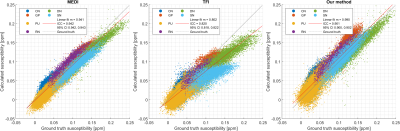 |
10 | Feature and voxel fidelity constraints improve the accuracy of direct inversion quantitative susceptibility mapping in deep gray matter
Jorge Campos Pazmiño1, Véronique Fortier1,2, and Ives Levesque1,2,3
1Medical Physics Unit, McGill University, Montreal, QC, Canada, 2Medical Imaging, McGill University Health Centre, Montreal, QC, Canada, 3Research Institute of the McGill University Health Centre, Montreal, QC, Canada We have designed a novel QSM algorithm that addresses some of the limitations of existing techniques that combine the background removal and dipole inversion steps in a single step. We propose that the solution to the direct inversion problem can be aided by an iterative k-space algorithm and the inclusion of a priori information that represents feature-based and voxel-fidelity-based constraints. The considered approach, when compared with other techniques, resulted in a more accurate depiction of the susceptibility in high susceptibility deep gray matter (dGM) structures without sacrificing performance in regions like the cortex of the brain. |
||
3309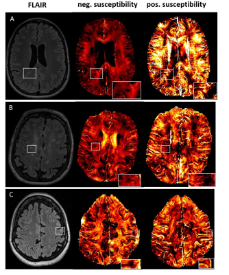 |
11 | Iron and Myelin Content in Multiple Sclerosis Lesions using Magnetic Susceptibility Source Separation
Reza Rahmanzadeh1,2, Po-Jui Lu1,2, Hyeong-Geol Shin 3, Matthias Weigel1,2, Thanh D. Nguyen4, Yi Wang4, Francesco La Rosa 5,6, Meritxell Bach Cuadra 5,6, Ernst-Wilhelm Radue1, Jens Kuhle2, Ludwig Kappos2, Jongho Lee 3, and Cristina Granziera1,2
1Translational Imaging in Neurology Basel, Department of Medicine and Biomedical Engineering, University Hospital Basel and University of Basel, Basel, Switzerland, 2Neurologic Clinic and Policlinic, Switzerland, Departments of Medicine, Clinical Research and Biomedical Engineering, University Hospital Basel and University of Basel, Basel, Switzerland, 3Laboratory for Imaging Science and Technology, Department of Electrical and Computer Engineering, Seoul National University, Seoul, Korea, Republic of, 4Department of Radiology, Weill Cornell Medical College, New York, NY, United States, 5Signal Processing Laboratory (LTS5), Ecole Polytechnique Fédérale de Lausanne (EPFL), Lausanne, Switzerland, 6Radiology Department, Center for Biomedical Imaging (CIBM), Lausanne University and University Hospital, Lausanne, Switzerland
Most multiple sclerosis (MS) white matter lesions (WMLs) appears hyperintense in quantitative susceptibility mapping (QSM). In the present study, we investigated the comparative contribution of myelin loss and iron deposit to QSM hyperintensity using a susceptibility source separation algorithm disentangling QSM- positive and QSM-negative susceptibility sources. Our results show that in most MS WMLs, demyelination is the source of QSM hyperintensity.
|
||
 Back to Meeting Home
 Back to the Program-at-a-Glance
The International Society for Magnetic Resonance in Medicine is accredited by the Accreditation Council for Continuing Medical Education to provide continuing medical education for physicians.
 Back to Meeting Home
 Back to the Program-at-a-Glance
 View the Presentation