Online Gather.town Pitches
MR Contrasts I
Joint Annual Meeting ISMRM-ESMRMB & ISMRT 31st Annual Meeting • 07-12 May 2022 • London, UK

| Booth # | ||||
|---|---|---|---|---|
3205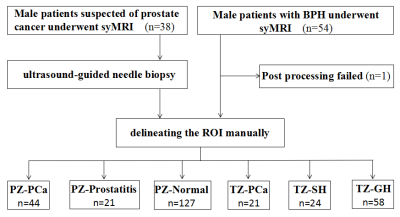 |
1 | Role of quantification of relaxation time from synthetic MRI in diagnosis and grading of prostate cancer
Suixing Zhong1, Ya Zhang1, Yingying Ding1, Lisha Nie2, Jing Tan1, Conghui Ai1, Yan Jin1, Hongbo Wang1, Huimei Zhang1, Miaomiao Li1, Rong Zhu1, and Shangwei Gu1
1Department of Radiology, Yunnan Cancer Hospital, Third Affiliated Hospital of Kunming Medical University, Kunming, China, 2MR Research, GE Healthcare, Beijing, China
This study aims to investigate the value of quantitative relaxation time derived from synthetic MRI (SyMRI) in the diagnosis and grading of prostate cancer (PCa). The results of this study showed that the diagnostic efficiencies of SyMRI in distinguishing PCa from most benign prostate lesions and different Gleason grades of PCa were similar to that of DWI, and the combined index of SyMRI and DWI can obtain higher diagnostic efficiencies. In addition, SyMRI has the advantages of short scanning time, unified parameters and simple operation, so it has a good prospect of clinical application.
|
||
3206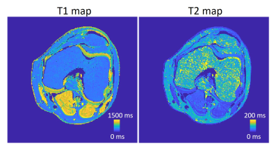 |
2 | Accurate and simultaneous acquisition of T1, T2, PD weighted, and MT weighted images using SPGR and DESS
Yi-Cheng Hsu1, Patrick Liebig2, and Ying-Hua Chu1
1Siemens Healthineers Ltd., Shanghai, China, 2Siemens Healthcare GmbH, Erlangen, Germany
We proposed a method to simultaneously acquire T1, T2, PD weighted, and MT weighted images. The scan time is the same as conventional SPGR T1 and DESS T2 mapping but with higher accuracy. This method will benefit diagnosis on muscular dystrophy, inflammation, fibrosis, and cartilage integrity.
|
||
3207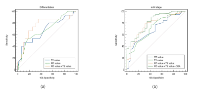 |
3 | Investigation of synthetic magnetic resonance imaging applied in the evaluation of the prognostic factors of rectal adenocarcinoma Video Permission Withheld
lidi ma1, shanshan lian1, huimin liu1, tiebao meng1, weilong zeng1, rui zhong1, linchang zhong1, long qian2, and chuanmiao xie1
1Sun Yat-sen University Cancer Center, guangzhou, China, 2MR Research, GE Healthcare, Beijing, China, guangzhou, China
The clinical significance of synthetic MRI in rectal adenocarcinoma remains unclear. This study aimed to explore the of quantitative parameters derived from SyMRI clinical stage according to “DISTANCE” criteria and differentiation of rectal adenocarcinoma. Our preliminary study demonstrated that quantitative T2 and PD values obtained by SyMRI might be used for noninvasive evaluation of prognostic factors of rectal adenocarcinoma. Furthermore, combining the two quantitative relaxation metrics further improved their diagnostic performance of mrN stage in rectal adenocarcinoma.
|
||
3208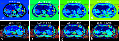 |
4 | T1 mapping for liver with EOB enhanced MRI: comparison of look-locker inversion recovery and B1 inhomogeneity-corrected method
Chenhui Li1, Liling Long1, Huiting Zhang2, and Dominik Nickel3
1The First Affiliated Hospital of Guangxi Medical University, Nanning, China, 2MR Scientific Marketing, Siemens Healthineers, Wuhan, China, 3MR Application Predevelopment, Siemens Healthcare GmbH, Erlangen, Germany
Two different T1 mapping sequences, based on the inversion recovery SNAPSHOT-FLASH sequence (LLIR-T1) and on B1 inhomogeneity-corrected variable flip angle (VFA-T1) acquisitions, were compared in a clinical setting. Results showed that the LLIR-T1 method had better image quality, and higher contrast between tumor and healthy liver tissue.c had a strong correlation with VFA-T1 method whether at different time point or regions. But LLIR-T1 had lower T1 in healthy liver lobe and higher T1 in tumor compared with VFA-T1.
|
||
3209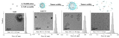 |
5 | Modular Graphene Quantum Dot Nano-transformers: An Efficient and Versatile Vehicle for Localized Tumor Imaging and Treatment Video Permission Withheld
Yuqi Yang1, Baolong Wang1, and Xin Zhou1
1Key Laboratory of Magnetic Resonance in Biological Systems, State Key Laboratory of Magnetic Resonance and Atomic and Molecular Physics, National Center for Magnetic Resonance in Wuhan, Wuhan Institute of Physics and Mathematics, Innovation Academy for Precision Measurement Science and Technology, Chinese Academy of Sciences–Wuhan National Laboratory for Optoelectronics, Wuhan, China
A versatile nano-vehicle, quantum dots nano-transformers, smartly transforms its shape in acidic tumor microenvironment to prolong the tumor retention, and then disassembles and releases the encapsulated photosensitizers to “turn on” the imaging contrast. The total drug dose in repeated photodynamic therapy is reduced because one injection is enough for four times of light irradiation.
|
||
3210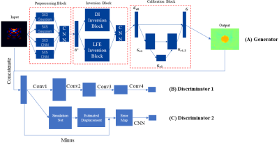 |
6 | A Model-based Neural Network for shear modulus estimation of MRE
Runke Wang1, Yu Chen1, Suhao Qiu1, and Yuan Feng1
1School of Biomedical Engineering, Shanghai Jiao Tong University, Shanghai, China
Algorithms providing accurate estimation of shear modulus and identification of tissue boundary are always desired for magnetic resonance elastography (MRE). In this study, we proposed a model-based neural network (MNN) embedding classic direct inversion (DI) and local frequency estimation (LFE). Convolution layers with Inception-like structure were applied for preprocessing, while postprocessing was implemented with a U-net structure. Additionally, DI and LFE algorithms were encapsulated as layers transforming wave images to shear modulus maps. The network performance was tested using a phantom with an inclusion. Compared with conventional algorithms, MNN provided modulus estimation with 5-fold higher contrast-to-noise ratio with clear boundaries.
|
||
3211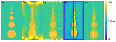 |
7 | A traveling-wave expansion based subspace search (TWESS) for shear modulus estimation using magnetic resonance elastography Video Permission Withheld
Shengyuan Ma1 and Yuan Feng1
1School of Biomedical Engineering, Shanghai Jiao Tong University, Shanghai, China
We proposed a traveling-wave expansion based subspace search (TWESS) algorithm for modulus estimation of MRE. Based on a traveling wave decomposition model, we explore the orthogonal characteristics of the signal subspace and the noise subspace. Eigenvalue decomposition of the covariance matrix of the wave data were used to search the wavenumber and retrieve the local modulus. Results showed the proposed method could provide better delineation of the inclusion boundaries, even when signal-to-noise ratio of the image was low.
|
||
3212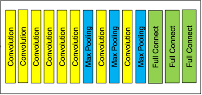 |
8 | Initial study of elastic modulus estimation method using deep learning in MR elastography
Takanori Aoki1 and Mikio Suga1,2
1Graduate School of Science and Engineering, Chiba University, Chiba, Japan, 2Center for frontier Medical Engineering, Chiba University, Chiba, Japan
MRE is a method for non-invasively and quantitatively estimating the stiffness of biological tissues. Although many estimation methods have been proposed, such as LFE, DI, and MMDI, there is a problem that both quantitativeness and spatial resolution cannot be achieved. In this study, we developed an elastic modulus estimation method by deep learning and compared its robustness to noise, quantitativeness, and spatial resolution with existing methods by numerical simulation. Compared with the conventional method (LFE), the proposed method has the same level of noise robustness, higher spatial resolution, and better quantitative performance in the case of high elastic modulus.
|
||
3213 |
9 | Frequency-dependent Correlation between Mechanical and Structural Properties of the Human Brain
Suhao Qiu1, Linghan Kong1, Runke Wang1, and Yuan Feng1
1School of Biomedical Engineering, Shanghai Jiao Tong University, Shanghai, China
The relationship between mechanical properties and microscopic structures of brain is of interest to both neuroscience and clinical communities. Using magnetic resonance elastography (MRE), we proposed and verified an empirical model to establish a linear correlation between shifted apparent diffusion coefficient (sADC) and shear modulus. A total of 43 healthy volunteers were included for MRE and diffusion weighted imaging (DWI) scanning. Results demonstrated that, in the parietal lobe, a strong correlation exists between shear modulus and sADC.
|
||
3214 |
10 | A combined deep learning and radiomics model predicts Ki-67 expression in HCC using MRI and multifrequency MR elastography
Xumei Hu1, Ruokun Li2, Jiahao Zhou2, Jing Guo3, Ingolf Sack3, Weibo Chen4, He Wang5, Fuhua Yan2, and Chengyan Wang1
1Human Phenome Institute,Fudan University, Shanghai, China, 2Department of Radiology, Ruijin Hospital, Shanghai Jiao Tong University School of Medicine, Shanghai, China, 3Department of Radiology, Charité–Universitätsmedizin Berlin, Berlin, Germany, 4Philips Healthcare, Shanghai, China, 5Institute of Science and Technology for Brain-inspired Intelligence, Fudan University, Shanghai, China
In this study, a combined deep learning and radiomics (DLR) approach using six different network architectures was tested and compared for the prediction of high Ki-67 expressions in patients with hepatocellular carcinoma (HCC). The model was based primarily on data from MRI and tomoelastography, a multifrequency MR elastography technique. Xception delivered the best performance and recognized seven prominent features among which four were obtained from tomoelastography. Our findings demonstrated that biomechanical properties, especially viscosity and the fluid behavior of the tumor, are crucial imaging features that are important for imaging-based cancer diagnostics.
|
||
3215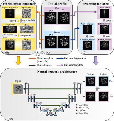 |
11 | Ultrafast water-fat separation using deep learning-based single-shot MRI
Xinran Chen1, Wei Wang1, Jianpan Huang2, Jian Wu1, Lin Chen1, Congbo Cai1, Shuhui Cai1, and Zhong Chen1
1Department of Electronic Science, Xiamen University, Xiamen, China, 2Department of Biomedical Engineering, City University of Hong Kong, Hong Kong, China
Water-fat separation is a powerful tool in diagnosing many diseases and many efforts have been made to reduce the scan time. Spatiotemporally encoded (SPEN) single-shot MRI, as an emerging ultrafast MRI method, can accomplish the fastest water-fat separation since only one shot is required. However, the SPEN water/fat images obtained by the state-of-the art methods still have some shortcomings. Here, a deep learning approach based on U-Net was proposed to obtain SPEN water/fat images simultaneously with improved spatial resolution, better fidelity and reduced reconstruction time. The efficiency of our method is demonstrated by numerical simulations, and in vivo rat experiments.
|
||
3216 |
12 | In Vivo Relaxation-Diffusion Correlation Revealed MRI Distribution Spectra of the Human Brain Using Genetic Algorithm Optimized Acquisitions
Lixian Wang1,2, Baogui Zhang1,2,3, Huilou Liang1,2, Yue Wu1,2, Jing An4, Yan Zhuo1,2,5, Rong Xue1,2,6, and Fangrong Zong7
1State Key Laboratory of Brain and Cognitive Sciences, Beijing MRI Center for Brain Research, Institute of Biophysics, Chinese Academy of Sciences, Beijing, China, 2University of Chinese Academy of Sciences, Beijing, China, 3Brainnetome Center, Institute of Automation, Chinese Academy of Sciences, Beijing, China, 4Siemens Shenzhen Magnetic Resonance Ltd., Shenzhen, China, 5CAS Center for Excellence in Brain Science and Intelligence Technology, Chinese Academy of Sciences, Beijing, China, 6Beijing Institute for Brain Disorders, Beijing, China, 7Institute of Biophysics, Chinese Academy of Sciences, Beijing, China Multidimensional MRI experiments have shown unique significance in resolving the correlations between physical parameters and relaxation properties. This study showed T1 Relaxation-Diffusion correlation imaging spectra of an in vivo human brain and a multicomponent phantom with optimized acquisitions. The acquisition paradigm was obtained from genetic algorithm (GA) by maximizing the singular values of the corresponding kernel functions. We detected significant differences in these spectra inversed from different components, which might help to identify tissue microstructure at the sub-voxel resolution. Our study offered a high probability of applying multidimensional MRI techniques in diagnosing neurological diseases. |
||
3217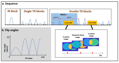 |
13 | Water- fat separated MR Fingerprinting (MRF) with simultaneous B1+ and B0 estimation
Jinmin Xu1, Huafeng Liu1, and HuiHui Ye1
1State key Laboratory of Modern Optical Science and Engineering, Zhejiang University, Hangzhou, China
A novel water-fat separated MRF approach was proposed to minimize the known biases introduced by the B1+, B0 and water and fat partial volume. By incorporating the water-fat separation approach in the framework, the dictionary size could be greatly reduced with the known B0 and FF map. Multiple maps of parameters including B1+, B0, FF and T1 and T2* of water and fat can be acquired within 14s for one slice.
|
||
3218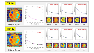 |
14 | Development of self-calibrating B1 correction for three-dimensional variable flip angle T1 mapping
Nagomi Fukuda1, Yuki Kanazawa2, Hiroaki Hayashi3, Yuki Matsumoto2, Masafumi Harada2, Motoharu Sasaki2, and Akihiro Haga2
1Institute of Biomedical Sciences, Tokushima University Graduate School, Tokushima, Japan, 2Graduate school of Health Science, Tokushima University, Tokushima, Japan, 3Faculty of Health Sciences, Institute of Medical, Pharmaceutical and Health Sciences, Kanazawa University, Kanazawa, Japan
We determine the accuracy of our B1 correction method using a cylindrical digital phantom and a digital brain phantom. Comparing the B1 correction accuracy of each sample, the relative uncertainties of each T1 value were less than 5%, and when long TR, the accuracy of B1 correction was almost equal to short TR. In addition, our method enabled us to correct the slice direction accurately. The proposed method can correct B1 inhomogeneity with high accuracy to in-plane and slice direction without additional scanning
|
||
3219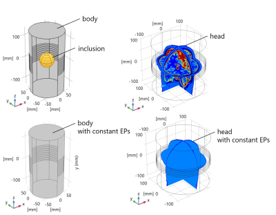 |
15 | Helmholtz Decomposition-Based Method for EPT with Approximation of Unmeasurable Magnetic Field Components Using Homogeneous Simulation Model
Naohiro Eda1, Motofumi Fushimi1, and Takaaki Nara1
1The University of Tokyo, Tokyo, Japan
We previously reported a method for magnetic resonance electrical properties tomography (MREPT) based on Helmholtz decomposition at ISMRM 2021. However, the method causes a reconstruction error due to the assumption that the unmeasurable components of the magnetic field are zero. This paper extends our previous method by approximating the unmeasurable components using a homogeneous simulation model that has the same geometry as the object to be examined and has homogeneous electrical properties (EPs). The efficacy of the proposed method was validated through numerical simulations.
|
||
 Back to Meeting Home
 Back to the Program-at-a-Glance
The International Society for Magnetic Resonance in Medicine is accredited by the Accreditation Council for Continuing Medical Education to provide continuing medical education for physicians.
 Back to Meeting Home
 Back to the Program-at-a-Glance
 View the Presentation