Online Gather.town Pitches
MR Contrasts IV
Joint Annual Meeting ISMRM-ESMRMB & ISMRT 31st Annual Meeting • 07-12 May 2022 • London, UK

| Booth # | ||||
|---|---|---|---|---|
4733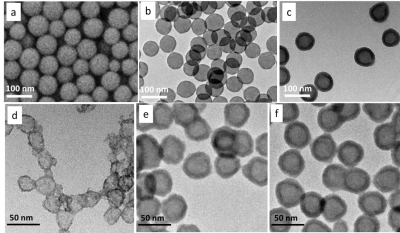 |
1 | The Development of Hollow Gd2O3 Nanospheres for T1 Contrast Agent
Hongyun Li1, Zhenxiong Wang2, Zhongping Zhang3, and Jie Fang1
1Nantong University, Nantong, China, 2Guangzhou First People's Hospital, Guangzhou, China, 3Philips Healthcare China, Guangzhou, China
Gd2O3 hollow nanospheres (HNS) with tuneable size and shell thickness was prepared. The thinnest shell of 30 nm Gd2O3 HNS achieved is 2.9 nm. This ultra-thin Gd2O3 HNS show effective T1 relaxivity in MRI with the highest r1 value of 3.77 mM-1s-1. Furthermore, the Gd2O3 HNS indicate strong chemical and physical stability which will minimize the toxicity caused by released free Gd3+ ions. The monodispersed and tuneable particle size will also facilitate the particle surface modification and blood circulation time design in bio-applications.
|
||
4734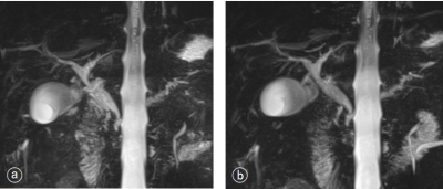 |
2 | The effect of contrast agent injection in MRCP under respiratory-triggered 3D-SPACE
Dayong Jin1, Xin Li1, Yue Qin1, Yinhu Zhu1, Liyao Liu1, Yifan Qian1, Juan Tian1, and Shaoyu Wang2
1Xi'an Daxing Hospital, Xi'an, China, 2Siemens Healthineers, Ltd., Xi'an, China
This study compared the image quality of MRCP before and after contrast agent injection on a 3T MRI scanner using the same respiratory-triggered 3D-SPACE sequence. Our results showed that enhancement of contrast-to-noise ratio (CNR) and background suppression will make the contour of pancreaticobiliary system more clear, which indirectly improves the imaging effect of MRCP. We performed MRCP scanning during the delayed waiting period for enhanced scans, which shortens the overall scan time for such patients. In summary, respiratory-triggered 3D-SPACE MRCP imaging with enhanced contrast injection significantly improved the imaging quality of the pancreaticobiliary duct and shorten the overall acquisition time. It is worthy of promotion and application in clinical practice.
|
||
4735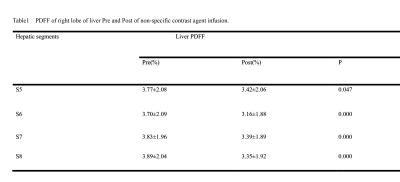 |
3 | Accuracy of fat fraction when using IDEAL-IQ with a liver nonspecific extracellular contrast agent
Lesheng Huang1, Hong yi Li1, Jun Chen1, Weiyin Vivian Liu 2, Kai li Cai1, Jing hua Jiang1, Wan chun Zhang1, Jia hui Tang1, Guang jun Tian3, Tao He1, Kai lian Yang1, Meng Hu1, Dong Zhang3, and Dan Li3
1Radiology, Guangdong Hospital of Traditional Chinese Medicine, Zhuhai, Zhu Hai, China, 2MR Research, GE Healthcare, Beijing, China, Beijing, China, 3Hepatology, Guangdong Hospital of Traditional Chinese Medicine, Zhuhai, Zhu Hai, China
To long-term track patients with liver disease is often carried out in clinics. It is also noted that nonspecific contrast agents change the relaxation time of tissue components, but few studies focus on the strong or weak FF alteration reflected by IDEAL-IQ derived measurements acquired before and after the contrast injection. In our study, we discovered significantly altered T1 values and fat fraction but not R2* even after the non-specific contrast agent achieved balanced condition at 3 minutes later of post-injection. Therefore, the liver non-specific contrast agent had an impact on the fat fraction measurements computed by IDEAL-IQ, and the utility of IDEAL-IQ should be performed before the injection of contrast agents to ensure the accuracy of fat fraction.
|
||
4736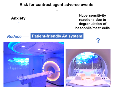 |
4 | A patient-friendly audiovisual MR system reduce adverse reaction to contrast agents
Keisuke Nitta1, Chiaki Tominaga1, Koji Matsumoto1, Hajime Yokota2, Yoshitada Masuda1, and Takashi Uno2
1Department of Radiology, Chiba University Hospital, Chiba, Japan, 2Diagnostic Radiology and Radiation Oncology, Graduate School of Medicine, Chiba, Japan
Patient anxiety could be a risk factor for adverse reaction to contrast agents. We hypothesized that a patient-friendly audiovisual system in the MR scanner room had a relaxing effect on patients and the occurrence of adverse reactions. As a result, the patient-friendly audiovisual system compared to the standard system reduced contrast agents reaction. Thus, this system may reduce adverse reaction in patients by providing a more patient-centered MRI examination environment.
|
||
4737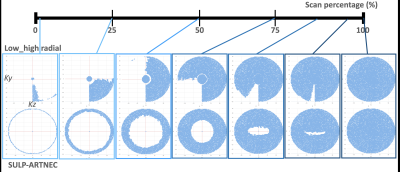 |
5 | Time-reversed CENTRA-PLUS profile ordering (SULP-ARTNEC) for improvement of image contrast in contrast-enhanced brain metastasis screening
Takayuki Sakai1, Hajime Yokota2, Masami Yoneyama3, Gabriele M Beck4, Daichi Murayama1, Shigehiro Ochi1, Tosiaki Miyati5, and Atsuya Watanabe6,7
1Radiology, Eastern Chiba Medical Center, Chiba, Japan, 2Diagnostic Radiology and Radiation Oncology, Graduate School of Medicine, Chiba University, Chiba, Japan, 3Philips Japan, Tokyo, Japan, 4Philips Healthcare, Best, Netherlands, 5Faculty of Health Sciences, Institute of Medical, Pharmaceutical and Health Sciences, Kanazawa University, Kanazawa, Japan, 6Orthopaedic Surgery, Eastern Chiba Medical Center, Chiba, Japan, 7General Medical Services, Graduate School of Medicine, Chiba University Graduate School of Medicine, Chiba, Japan
Shorter examination time might miss the appropriate timing for depicting the “delayed-enhanced” lesions, such as brain metastasis and multiple sclerosis plaques. We focused on the CENTRA-PLUS view-ordering framework and tried to modify the k-space profile ordering to enable completely timing-reversed (SLUP-ARTNEC). The purpose of this study is to demonstrate the feasibility of SULP-ARTNEC in the patients with brain metastasis. SLUP-ARTNEC is the unique approach in the post compressed sensing era that can improve the image contrast between delayed-enhanced brain metastasis lesions and background tissues in the contrast-enhanced studies without having the extra-waiting time, resulting in maintain the shorter examination time.
|
||
4738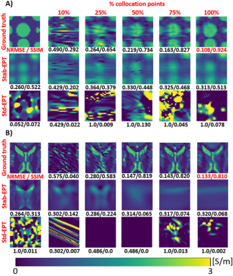 |
6 | Physics informed neural networks for phase-based magnetic resonance electrical properties tomography
Adan Jafet Garcia1, Shao Ying Huang2,3, Nevrez Imamoglu4, and Wenwei Yu1,5
1Medical systems, Chiba University, Chiba, Japan, 2Department of Surgery, National University of Singapore, Singapore, Singapore, 3Engineering Product Development, Singapore University of Technology and Design, Singapore, Singapore, 4Digital Architecture Research Center, National Institute of Advanced Industrial Science and Technology, Tokyo, Japan, 5Center for Frontier Medical Engineering, Chiba University, Chiba, Japan Magnetic Resonance Electrical Property Tomography (MREPT) could provide important contrast for non-calcified tumors. However, MREPT relies on numerical differentiation, which is noise sensitive and prone to artifacts near boundaries. In this work, physics-informed neural networks (PINN), NN empowered automatic differentiation is proposed to improve MREPT by mitigating artifacts and reducing noise sensitivity. Instead of calculating partial derivatives numerically, is obtained by backpropagation through PINNs. For clinical MREPT, reduction of ground-truth information to guide PINNs was investigated. Results show that above 25% collocation points, reconstruction can be made at 100 SNR. PINNs enable noise-robust and artifact-free MREPT from less ground-truth information. |
||
4739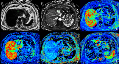 |
7 | Differentiation of liver benign, primary malignant and secondary malignant tumors using MR DCE and mDIXON-Quant imaging
Xue Ren1, Jiazheng Wang2, Liangjie Lin2, Lihua Chen1, Qingwei Song1, Renwang Pu1, Ying Zhao1, Tao Lin1, Qihao Xu1, and Ailian Liu1
1Department of Radiology, the First Affiliated Hospital of Dalian Medical University, Dalian, China, 2Philips Healthcare, Beijing, China
The quantitative MR dynamic contrast-enhanced (DCE) and mDIXON-Quant imaging were used in this study for differentiation between benign and malignant liver lesions. The combination of DCE and mDIXON-Quant showed significantly improved diagnostic efficacy between benign liver tumors and primary malignant tumors, as well as between primary and secondary malignant tumors.
|
||
 |
4740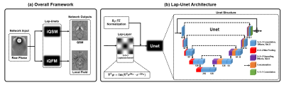 |
8 | QSM from the raw phase using an end-to-end neural network
Yang Gao1, Zhuang Xiong 1, Amir Fazlollahi2, Peter J Nestor2, Viktor Vegh3, G. Bruce Pike4, Stuart Crozier1, Feng Liu1, and Hongfu Sun1
1School of ITEE, University of Queensland, Brisbane, Australia, 2Queensland Brain Institute, University of Queensland, Brisbane, Australia, 3Centre for Advanced Imaging, University of Queensland, Brisbane, Australia, 4Departments of Radiology and Clinical Neurosciences, University of Calgary, Calgray, AB, Canada
Deep learning frameworks are emerging methods for solving QSM problems these days. However, most previous deep neural networks designed for QSM requires phase unwrapping and background field removal preprocessing procedures. This work presents a novel end-to-end network, namely Lap-Unet, for instant QSM and tissue field mapping from the raw phase in a single run. Comparative results find that the proposed method resulted in more accurate and robust reconstructions than previously established single- and multi-step methods, particularly for QSM of intracranial hemorrhages, which has been challenging due to fast signal decays.
|
|
4741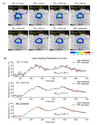 |
9 | Improved MR Temperature Imaging at 0.5T with Multi-echo Thermometry
Ziyi Pan1, Jianxiong Hu2, Hai Luo2, Simin Liu1, Sisi Li1, Ziyue Wu2, and Hua Guo1
1Center for Biomedical Imaging Research, Department of Biomedical Engineering, School of Medicine, Tsinghua University, Beijing, China, 2Marvel Stone Healthcare Co., Ltd., Wuxi, China
Low field MR-guided thermotherapy can provide some key advantages over the high-field alternative, including reduced cost, decreased susceptibility artifacts, and improved safety of interventional devices. However, both the accuracy and the speed of PRF temperature measurement suffer at the low field due to the reduced SNR, limited receive channels, and declined temperature-induced phase changes, making it unreliable for clinical MRgLITT treatments. In this study, we demonstrate that the multi-echo thermometry together with the view-sharing acceleration can be utilized to achieve high-quality PRF thermometry at 0.5T with satisfactory temperature measurement precision and temporal resolution.
|
||
4742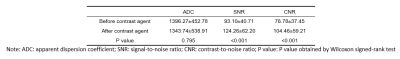 |
10 | Effects of Contrast Agents on Diffusion-Weighted Imaging and T2-Weighted Imaging for diagnosis of Breast tumor
Changxiang Wang1, Yongsheng Ao2, Lihua Qiu2, Lan Mu3, Junhui Huang1, Shuheng Zhang4, Shimin Yang4, Xin Liu1, Hairong Zheng1, and Na Zhang1
1Paul C. Lauterbur Research Center for Biomedical Imaging, Shenzhen Institute of Advanced Technology, Chinese Academy of Sciences, Shenzhen, China, 2Department of Radiology, The Second People’s Hospital of Yibin, Yibin, China, 3Department of Radiology,Pidu District People's Hospital, Chengdu, China, 4United Imaging Healthcare, Shanghai, China
Diffusion-Weighted Imaging (DWI) and T2-weighted imaging (T2WI) are two necessary sequences in breast MRI which of great significance for breast cancer. They are usually performed before contrast agent injection in the conventional breast MRI protocol. Recently, some studies have proposed simplified and fast breast scanning protocol, of which, DWI and T2WI are performed after contrast agent injection. This study evaluated the clinical value of DWI and T2WI after contrast agent in the diagnosis of breast cancer. The results showed that contrast injection has no effect on DWI, but it can improve the signal-to-noise ratio and contrast-to-noise ratio of T2WI.
|
||
 Back to Meeting Home
 Back to the Program-at-a-Glance
The International Society for Magnetic Resonance in Medicine is accredited by the Accreditation Council for Continuing Medical Education to provide continuing medical education for physicians.
 Back to Meeting Home
 Back to the Program-at-a-Glance
 View the Presentation