Online Gather.town Pitches
MR Contrasts II
Joint Annual Meeting ISMRM-ESMRMB & ISMRT 31st Annual Meeting • 07-12 May 2022 • London, UK

| Booth # | ||||
|---|---|---|---|---|
3929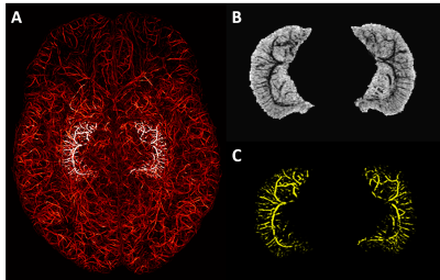 |
1 | Vascular Mapping of the Human Hippocampus Using USPIO
Sagar Buch1, Yongsheng Chen2, Pavan Jella3, Yulin Ge4, and Ewart Mark Haacke1
1Radiology, Wayne State University, Detroit, MI, United States, 2Neurology, Wayne State University, Detroit, MI, United States, 3Wayne State University, Detroit, MI, United States, 4Radiology, New York University School of Medicine, New York, NY, United States
In this work, we introduce the use of Ferumoxytol, an ultra-small superparamagnetic iron oxides (USPIO) agent, to increase the susceptibility in the veins and arteries to map the hippocampal microvasculature and to evaluate the fractional vascular density (FVD) in each of its subfields. We found that the hippocampal fissure, along with the fimbria, granular cell layer of the dentate gyrus and cornu ammonis layers (except for CA1), showed higher microvascular FVD than other parts of the hippocampus.
|
||
3930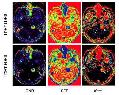 |
2 | Brain Tumour Studies using Low-Dose Gadolinium-Based Contrast Agent Dynamic Contrast Enhancement MRI
Xiaoping Zhu1, Daniel Lewis2, Ka-Loh Li1, and Alan Jackson1
1DIIDS, University of Manchester, Manchester, United Kingdom, 2Department of Neurosurgery, Salford Royal NHS Foundation Trust, Manchester, United Kingdom
Using one-fifth of standard GBCA dose, high spatial resolution (LDHS) whole brain pharmacokinetic maps was calculated and compared with those from dual temporal resolution (DTR) DCE, which combines high temporal resolution (LDHT) and full dose spatial resolution DCE. Results from Monte Carlo simulation suggested the measurement uncertainty and bias reduced to negligible whilst CNR of tumour concentration curves is greater than 3.1. The in-vivo LDHS maps have compatible accuracy with those from HT DCE, moreover revealing tumor heterogeneity to the quality DTR have achieved. Largely reduced dose enabled safer exams for patients undergoing repeated administration of GBCA.
|
||
3931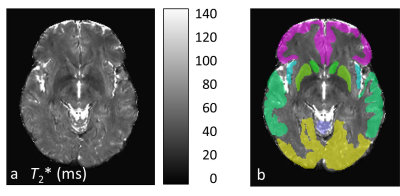 |
3 | 19-hour stability of T2* mapping of brain regions
Olaf Dietrich1, Johannes Levin2, and Jan Remi2
1Department of Radiology, LMU Ludwig Maximilian University of Munich, Munich, Germany, 2Department of Neurology, LMU Ludwig Maximilian University of Munich, Munich, Germany
The purpose of this study was to assess the temporal stability of T2* mapping results in the human brain of healthy volunteers. Three healthy male participants (36-45y) were each scanned 6 times over a period of 19 hours. Median T2* values were determined within the 9 regions of the MNI structural atlas. For each region, mean values, standard deviations (SD), and the coefficients of variation were calculated over the six acquisition time points. The results demonstrate very good intra-individual stability of T2* in different brain regions with coefficients of variations lower than 3 % in all evaluated regions and volunteers.
|
||
3932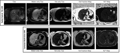 |
4 | A Free-Breathing Hybrid Technique for Simultaneous T2w, T1w, PDFF, and R2* Imaging at 0.55T
Mahesh Bharath Keerthivasan1, Mary Bruno2, Eddy Solomon3, Thomas Benkert4, David Grodzki4, Himanshu Bhat1, Kai Tobias Block2, and Hersh Chandarana2
1Siemens Medical Solutions USA, Malvern, PA, United States, 2Department of Radiology, New York University Grossman School of Medicine, New York, NY, United States, 3Department of Radiology, Weill Cornell Medical College, New York, NY, United States, 4Siemens Healthcare GmbH, Erlangen, Germany
Recently, there has been renewed interest in imaging at low field strengths. The increased T2* relaxation times at 0.55T and reduced chemical shift between fat and water requires the need for longer TEs for accurate fat fraction and R2* estimation, often resulting in longer acquisition times. In this work, we present a scan-efficient hybrid TSE-GRE technique for the simultaneous acquisition of T2- and T1-weighted images along with quantification of fat fraction and R2*. The use of a stack-of-stars trajectory allows SNR efficient free breathing imaging at 0.55T.
|
||
3933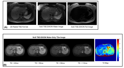 |
5 | Abdominal Water-Only T2 Estimation at 0.55T using a 3D Stack-of-Stars TSE-DIXON Technique
Mahesh Bharath Keerthivasan1, Mary Bruno2, Eddy Solomon3, David Grodzki4, Himanshu Bhat5, Kai Tobias Block2, and Hersh Chandarana2
1Siemens Medical Solutions USA, New York, NY, United States, 2Department of Radiology, New York University Grossman School of Medicine, New York, NY, United States, 3Department of Radiology, Weill Cornell Medical College, New York, NY, United States, 4Siemens Healthcare GmbH, Erlangen, Germany, 5Siemens Medical Solutions USA, Malvern, PA, United States
We explore the use of a stack-of-stars 3D TSE sequence along with multi-echo DIXON readouts for SNR-efficient, free-breathing water-only T2 estimation at 0.55T. A subspace constrained reconstruction is combined with a multi-step fat/ water separation algorithm to generate water-only T2-weighted images and T2 maps.
|
||
3934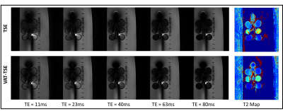 |
6 | Rapid Low SAR T2 Relaxometry near Metallic Implants
Mahesh Bharath Keerthivasan1, Jan Fritz2, Ran Schwarzkopf3, and Iman Khodarahmi2
1Siemens Medical Solutions USA, New York, NY, United States, 2Department of Radiology, New York University School of Medicine, New York, NY, United States, 3Department of Orthopedic Surgery, New York University School of Medicine, New York, NY, United States
Quantitative T2 mapping in the presence of metal implants is affected by susceptibility-related artifacts. We propose a low flip angle view angle tilting-enabled turbo-spin echo sequence along with spatiotemporal undersampling for low SAR scan efficient T2 mapping. The proposed technique would allow 2.5min T2 mapping with high temporal sampling of the T2 relaxation curve. Performance of the proposed technique is evaluated using retrospective under-sampling simulations and phantom experiments using a cobalt-chromium hip arthroplasty implant.
|
||
3935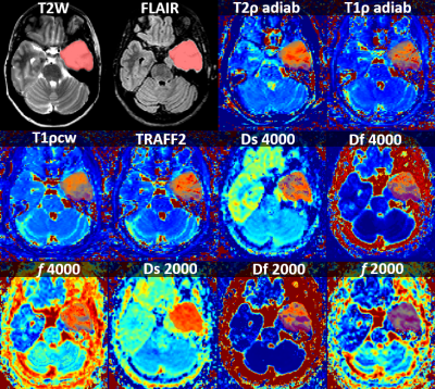 |
7 | Detection of IDH mutation in brain tumors using RAFF, continues wave T1rho, adiabatic T1rho, adiabatic T2rho and high b-value DWI
Harri Merisaari1,2, Aida Steiner1, Marko Pesola 1, Maria Gardberg 3, Janek Frantzén 4, Pekka Jokinen 4, Hannu Aronen 1, Timo Liimatainen 5, Heikki Minn 6, and Ivan Jambor1,7
1Department of Radiology, University of Turku, Turku, Finland, 2Turku Brain and Mind Center, University of Turku, Turku, Finland, 3Department of Pathology, Turku University Hospital, Turku, Finland, 4Department of Neurosurgery, Turku University Hospital, Turku, Finland, 5Department of Radiology, University of Oulu, Oulu, Finland, 6Department of Oncology and Radiotherapy, Turku University Hospital, Turku, Finland, 7Department of Radiology and Biomedical Imaging, Yale School of Medicine, New Haven, CT, United States
We evaluated the potential of Relaxation Along a Fictitious Field in second rotating frame (TRAFF2), continues wave T1rho (T1ρcw), adiabatic T1rho (T1ρadiab), adiabatic T2rho (T2ρadiab), DWI and anatomical MRI to differentiate isohydrogenase dehydrogenase (IDH) status between IDH wild type and IDH mutation in 22 patients with glioma. In voxel level analysis, DWI derived parameters using bi-exponential model (0-4000 s/mm2) provided improved performance in classification of IDH status of brain gliomas compared with TRAFF2, T1ρcw, T1ρadiab, T2ρadiab and anatomical MRI.
|
||
3936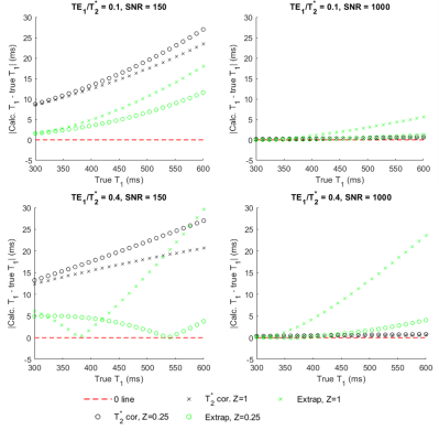 |
8 | Correction of Bias in Single Reference Variable Flip Angle T1-mapping with Simple Extrapolation Method
Michael Malmberg1, Henrik L Odéen1, and DENNIS L PARKER1
1University of Utah, Salt Lake City, UT, United States Single reference variable flip angle T1 mapping is subject to some remaining bias due to T2* effects between baseline and dynamic acquisitions. Previous work determined a computationally expensive correction for this bias. In this work, we present a much simpler extrapolation method to correct for T2* related bias in T1. This study provides an analysis through simulation of the benefits and drawbacks of this simpler extrapolation correction compared to the previous correction for bias on the SR-VFA T1 calculation. |
||
3937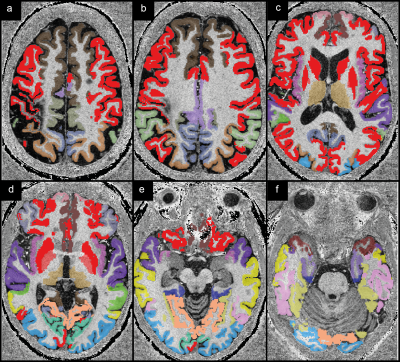 |
9 | Quantitative MRI biomarkers for cortical pathology in multiple sclerosis at 7 Tesla
Sina Straub1, Edris El-Sanosy2, Julian Emmerich1, Frederik L. Sandig2, Mark E. Ladd1,3,4, and Heinz-Peter Schlemmer2
1Medical Physics in Radiology, German Cancer Research Center (DKFZ), Heidelberg, Germany, 2Radiology, German Cancer Research Center (DKFZ), Heidelberg, Germany, 3Faculty of Physics and Astronomy, Heidelberg University, Heidelberg, Germany, 4Faculty of Medicine, Heidelberg University, Heidelberg, Germany
Although substantial cortical gray matter tissue damage has been revealed by advanced MRI methods and in histopathology studies, clinical assessment of MS still mainly focuses on white matter lesions. When cortical pathology is evaluated, predominantly structural markers are used. In this ultra-high field study, data for $$$T_1$$$, $$$T_2$$$, $$$R_2^*$$$, $$$T_1w-T_2w$$$-ratio, and susceptibility mapping were acquired in 21 patients and 17 healthy controls. $$$T_1$$$–weighted data were post-processed to obtain cortical gray matter and deep gray matter segmentations. Statistically significant differences were found in 31 out of 34 investigated cortical and in three out of four deep gray matter regions (p<0.05).
|
||
3938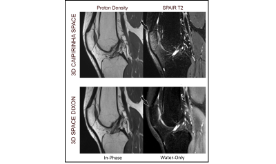 |
10 | A 3D SPACE-DIXON Technique for Fat Suppression in Knee Imaging
Mahesh Bharath Keerthivasan1, Xiaodong Zhong2, Mary Bruno3, Iman Khodarahmi4, and Jan Fritz4
1MR R&D Collaborations, Siemens Medical Solutions USA, New York, NY, United States, 2MR R&D Collaborations, Siemens Medical Solutions USA, Los Angeles, CA, United States, 3Department of Radiology, New York University Grossman School of Medicine, New York, NY, United States, 4Department of Radiology, NYU Grossman School of Medicine, New York, NY, United States
We present a 3D SPACE-DIXON technique with improved fat suppression performance for musculoskeletal 3D SPACE MRI. Phantom experiments were used to evaluate the fat suppression efficiency. Preliminary in vivo results indicate the clinical utility of this technique for proton density-weighted 3D MRI.
|
||
3939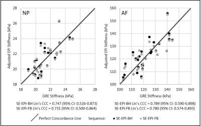 |
11 | Spin Echo-Echo Planar Imaging Sequence Validation for MR Elastography of Intervertebral Discs
Megan Co1, Huiming Dong2, Daniel J. Boulter2, Xuan V. Nguyen2, Safdar N. Khan2, Brian Raterman2, Brett Klamer3, Arunark Kolipaka1,2, and Benjamin A. Walter1,2,4
1Biomedical Engineering, The Ohio State University, Columbus, OH, United States, 2Radiology, The Ohio State University, Columbus, OH, United States, 3Center for Biostatistics, The Ohio State University, Columbus, OH, United States, 4Spine Research Institute, The Ohio State University, Columbus, OH, United States
Magnetic resonance elastography (MRE) is an imaging technique that can non-invasively assess shear properties of intervertebral discs (IVD), a potential biomarker for disease. This study validated the use of a spin echo-echo planar imaging (SE-EPI) sequence for MRE of the IVD compared against the standard gradient recalled echo (GRE) sequence. Volunteers were scanned with the GRE and two variants of the SE-EPI sequence: SE-EPI with breath holds (SE-EPI-BH) and SE-EPI with free breathing (SE-EPI-FB). SE-EPI-based MRE-derived stiffnesses are highly reproducible and repeatable and correlate with current standard GRE MRE-derived stiffness estimates while reducing scan times and potentially improving patient compliance.
|
||
3940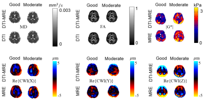 |
12 | Development of in vivo human brain DTI-MRE: Preliminary results
Shujun Lin1, Brad Sutton2, Richard Magin1, Aaron Anderson2, and Dieter Klatt1
1Biomedical Engineering, University of Illinois at Chicago, Chicago, IL, United States, 2Bioengineering, University of Illinois at Urbana-Champaign, Urbana, IL, United States
Simultaneous acquisition of diffusion tensor imaging (DTI) and magnetic resonance elastography (MRE) has been proven feasible in a pre-clinical study and in preliminary studies of in vivo human brain. However, the experimental parameters have to be optimized in order to prevent mutual interferences of DTI and MRE acquisitions. We identified in simulations two experimental parameter sets for in vivo human brain DTI-MRE that we classify as good and moderate and present a pilot study using these parameter sets. The experimental results indicate that both experimental parameter sets show good performance using motion-encoding gradients without flow compensation.
|
||
3941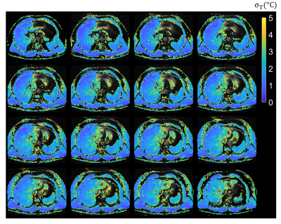 |
13 | Free-breathing PRF Thermometry of the Liver at 0.55T
Waqas Majeed1, Axel Joachim Krafft2, and Himanshu Bhat1
1Siemens Medical Solutions USA Inc., Malvern, PA, United States, 2Siemens Healthcare GmbH, Erlangen, Germany
Low field MRI offers advantages such as reduced cost and improved safety of implantable and interventional devices, and reduced RF energy deposition over high field alternatives. In this study we propose acquisition and analysis pipelines for free-breathing 2D and 3D liver thermometry and demonstrate the feasibility of MR thermometry of the liver at 0.55T.
|
||
3942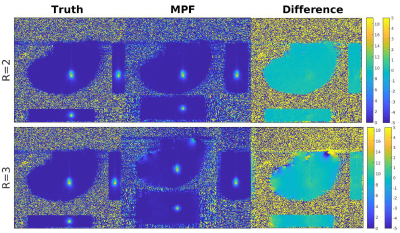 |
14 | Model Predictive Filtering and Green’s Function Heat Kernel for Real-Time Volumetric MRTI of Laser Interstitial Thermotherapy
Joshua Marchant1, Dennis L Parker2, and Henrik Odéen2
1Physics and Astronomy, University of Utah, Salt Lake City, UT, United States, 2Department of Radiology and Imaging Sciences, University of Utah, Salt Lake City, UT, United States Laser interstitial thermotherapy (LITT) is a clinical treatment modality performed under the guidance of magnetic resonance temperature imaging (MRTI). Current limitations in MRTI prevent real-time volumetric monitoring of the tissue regions being treated. In this work, we investigate the use of the model predictive filtering method (MPF), using a Green’s function heat kernel, to reconstruct large field-of-view MRTI data of phantom heating to validate their use for future real-time volumetric MRTI treatment monitoring. |
||
3943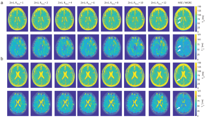 |
15 | Accelerated Simultaneous T2 and T2* Mapping of Multiple Sclerosis Lesions Using Compressed Sensing Reconstruction of 2in1-RARE-EPI Data Video Permission Withheld
Carl Julius Jacob Herrmann1, Ludger Starke1,2, Jason M. Millward1, Friedemann Paul3,4,5, Joseph Kuchling3,4,5, and Thoralf Niendorf1,3
1Berlin Ultrahigh Field Facility, Max Delbrück Center for Molecular Medicine in the Helmholtz Association, Berlin, Germany, 2Digital Health - Machine Learning Research Group, Digital Health Center, Hasso Plattner Institute, University of Potsdam, Potsdam, Germany, 3Experimental und Clinical Research Center (ECRC), a joint cooperation between the Charité Medical Faculty and the Max Delbrück Center for Molecular Medicine, Berlin, Germany, 4NeuroCure Clinical Research Center, Charité Medical Faculty, Berlin, Germany, 5Department of Neurology, Charité Medical Faculty, Berlin, Germany
We previously applied a radially-sampled RARE-EPI hybrid for simultaneous T2 and T2* mapping (2in1-RARE-EPI) in patients with multiple sclerosis, which reduced the scan time to 77% compared to Multi-Spin-Echo (MSE) and Multi-Gradient-Recalled-Echo (MGRE). Here we examine the potential for further acceleration utilizing a compressed sensing reconstruction of highly undersampled 2in1-RARE-EPI data. The accelerated T2 and T2* mapping is benchmarked against the MSE and MGRE references using regression and Bland-Altman plot analysis and the mean absolute percentage error. Our results show that an undersampling factor of 8-12 is feasible, achieving an acquisition time reduction to 23-17% compared to the references.
|
||
3944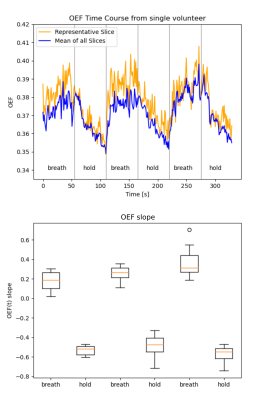 |
16 | Rapid T2’ quantification by 10-echo GESE-EPIK sequence with application to oxygen extraction fraction imaging
Fabian Küppers1,2, Seong Dae Yun1, and N. Jon Shah1,3,4,5
1Institute of Neuroscience and Medicine - 4, Forschungszentrum Jülich, Jülich, Germany, 2RWTH Aachen University, Aachen, Germany, 3Institute of Neuroscience and Medicine - 11, Forschungszentrum Jülich, Jülich, Germany, 4JARA-BRAIN - Translational Medicine, Aachen, Germany, 5Department of Neurology, RWTH Aachen University, Aachen, Germany
Owing to the advantages afforded by the simultaneous acquisition of T2/T2* in several practical applications, interest in this area remains high. In light of this, here we present an improved NLSQ fitting algorithm and a more detailed validation based of our previously introduced 10-echo GESE-EPIK sequence. The validation consists of two new phantoms, including a spectroscopic comparison to reference methods, and data from five in vivo subjects at 3T. In addition, GESE-EPIK is applied to OEF quantification during a breath-hold experiment to demonstrate its sensitivity to challenge-related changes.
|
||
3945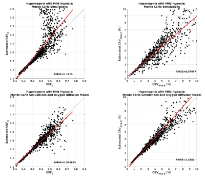 |
17 | Combining gradient and spin echo calibrated fMRI for mapping brain oxygen extraction fraction
Michael Germuska1,2, Antonio Maria Chiarelli 3,4, Hannah Chandler1, Ian Driver1, and Richard Wise3,4
1School of Psychology, Cardiff University, Cardiff, United Kingdom, 2School of Physics and Astronomy, Cardiff University, Cardiff, United Kingdom, 3Department of Neuroscience, University “G. d'Annunzio” of Chieti-Pescara, Chieti, Italy, 4Institute for Advanced Biomedical Technologies, University “G. d'Annunzio” of Chieti-Pescara, Chieti, Italy We propose a new calibrated fMRI approach for non-invasive measurement of the cerebral metabolic rate of oxygen of oxygen consumption and the deoxyhaemoglobin weighted blood volume. The method exploits the differential dependence of the gradient and spin echo BOLD signals on OEF. Unlike conventional calibrated approaches, we show that gradient-spin echo calibrated fMRI requires only a single respiratory modulation to determine OEF. The method is applied in-vivo using a repeated breath-holding protocol, and initial results show agreement with global measurements of oxygen saturation. |
||
3946 |
18 | Iterative MR-Electrical Properties Tomography using physics-based deep learning
Sabrina Zumbo1, Martina Teresa Bevacqua1, Ettore Flavio Meliadò2, Peter Stijnman2, Thierry Meerbothe2, Tommaso Isernia1, Cornelis A.T. van den Berg2, and Stefano Mandija2
1DIIES, Università Mediterranea di Reggio Calabria, Reggio di Calabria, Italy, 2Computational Imaging Group for MR Diagnostics & Therapy, Center for Image Sciences, Utrecht University, Utrecht, Netherlands
We introduce for the first time an iterative MR electrical properties tomography reconstruction method by exploiting a cascade of multi-layer convolutional neural networks (CNNs) able to learn spatial priors in an iterative fashion, alternated with physics-based gradient descent direction calculations. This method was tested on 2D simulated human brain data. The presented results demonstrate the feasibility of this methodology to reconstruct conductivity and permittivity maps at 128MHz. Ultimately, this method allows computational advantages compared to standard contrast source inversion electrical properties tomography (CSI-EPT), i.e. faster reconstructions, which will be extremely relevant when moving to 3D reconstructions.
|
||
 Back to Meeting Home
 Back to the Program-at-a-Glance
The International Society for Magnetic Resonance in Medicine is accredited by the Accreditation Council for Continuing Medical Education to provide continuing medical education for physicians.
 Back to Meeting Home
 Back to the Program-at-a-Glance
 View the Presentation