Online Gather.town Pitches
Molecular Imaging I
Joint Annual Meeting ISMRM-ESMRMB & ISMRT 31st Annual Meeting • 07-12 May 2022 • London, UK

| Booth # | ||||
|---|---|---|---|---|
3830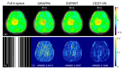 |
1 | Cross-domain Variational Network for Fast Chemical Exchange Saturation Transfer Imaging
Jianping Xu1, Tao Zu1, Yi-Cheng Hsu2, Yi Sun2, Dan Wu1, and Yi Zhang1
1Key Laboratory for Biomedical Engineering of Ministry of Education, Department of Biomedical Engineering, College of Biomedical Engineering & Instrument Science, Zhejiang University, Hangzhou, Zhejiang, China, 2MR Collaboration, Siemens Healthcare Ltd., Shanghai, China
Chemical exchange saturation transfer (CEST) is an emerging molecular imaging technique that can detect various biomolecules in vivo. However, the routine clinical application of CEST MRI is hammered by its long scan time due to the multiple saturation frames acquired. Here, a novel deep neural network modified from the variational network (VN) by utilizing cross-domain regularization structures, dubbed CEST-VN, is proposed for accelerated CEST imaging. In conjunction with multi-coil sensitivity encoding, the CEST-VN method demonstrated superior performance to the conventional parallel imaging and the original VN methods in healthy volunteers and glioma patients.
|
||
3831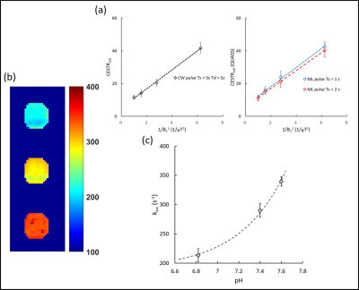 |
2 | Rapid Acquisition using Water pre-saturation for quantitive CEST imaging (RAW-qCEST): exchange rate quantitation at 3-Tesla Video Not Available
Wenxuan Chen1, Lele Ma2, Zhensen Chen3, and Xiaolei Song1
1Center for Biomedical Imaging Research, Department of Biomedical Engineering, Tsinghua University, Beijing, China, 2State Key Laboratory of Southwestern Chinese Medicine Resources, Chengdu University of Traditional Chinese Medicine, Chengdu, China, 3Institute of Science and Technology for Brain-Inspired Intelligence, Fudan University, Shanghai, China
We proposed a Rapid Acquisition using Water pre-saturation for quantitative CEST imaging (RAW-qCEST) sequence, which validated by phantom experiments at 3-Tesla. Raw-qCEST allowed rapid acquisition of multiple readouts with varied saturation powers within one TR, by adding a water pre-saturation module that suppress interference among readouts and a seconds-long CEST saturation module before each readout. Simulation and phantom experiments showed that RAW-qCEST could accurately determine pH-related exchange rates, calculated using a modified omega-plot analysis. Owing to its fast speed and quantitative capability, RAW-qCEST could be useful for imaging stroke and cancers, which will be investigated in future.
|
||
3832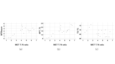 |
3 | New parameters on CEST imaging by multi pool model in gliomas in comparison with conventional IVIM and 11C-MET uptake on PET/CT
Yasukage Takami1, Naruhide Kimura1, Katsuya Mitamura1, Takashi Norikane1, Keisuke Miyake2, Tatsuya Yamasaki3, Kazuo Ogawa3, Mitsuharu Miyoshi4, Atsushi Nozaki4, and Yoshihiro Nishiyama1
1Department of Radiology, Faculty of Medicine, Kagawa University, Miki-cho, Kita-gun, Japan, 2Department of Neurological Surgery, Faculty of Medicine, Kagawa University, Miki-cho, Kita-gun, Japan, 3Kagawa University Hospital, Miki-cho, Kita-gun, Japan, 4GE Healthcare, Hino-shi, Japan
The purpose of this study was to evaluate the correlation among parameters on CEST imaging by multi pool model, IVIM and 11C-methionine (MET) uptake on PET/CT in gliomas. The maximum values of the parameters on CEST imaging, the minimum parameters (ADC, D, D*) and maximum f on IVIM were measured respectively. 11C-MET uptake was semiquantitatively assessed using tumor-to-contralateral normal brain tissue (T/N) ratio. Several strong correlations were observed among some of the parameters. These preliminary results suggest that parameters on CEST imaging by multi pool model and IVIM seems to correlate with 11C-MET uptake on PET/CT in gliomas.
|
||
3833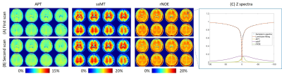 |
4 | Compressed sensing chemical exchange saturation transfer for simultaneous APT, ssMT, and rNOE imaging
Ying-Hua Chu1, Patrick Liebig2, Yifan Yuan3, He Wang4,5, and Yi-Cheng Hsu1
1Siemens Healthineers Ltd., Shanghai, China, 2Siemens Healthcare GmbH, Erlangen, Germany, 3Department of Neurosurgery, Huashan Hospital, Fudan University, Shanghai, China, 4Institute of Science and Technology for Brain-Inspired Intelligence, Fudan University, Shanghai, China, 5Human Phenome Institute, Fudan University, Shanghai, China We proposed a fast 3D CEST imaging method using compressed sensing SPACE and demonstrated its routine clinical application for simultaneous APT, ssMT, and rNOE imaging. The total scan time of CEST images with 58 z-spectra and an off-resonance map was 6 minutes and 49 seconds. We used low refocusing flip angle and high bandwidth to achieve a narrow point spread function, and the images could be reproduced between different scans. This method could be used for tumor grading and treatment response assessment at 3T. |
||
3834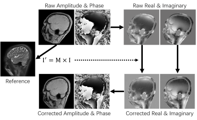 |
5 | Inclusion of B0 mapping images in motion correction for improved quality of CEST maps
Zhechuan Dai1, Yi-Cheng Hsu2, Yi Sun2, Dan Wu1, and Yi Zhang1
1Key Laboratory for Biomedical Engineering of Ministry of Education, Department of Biomedical Engineering, College of Biomedical Engineering & Instrument Science, Zhejiang University, Zhejiang, China, 2MR Collaboration, Siemens Healthcare Ltd., Shanghai, China
The inhomogeneity of the main magnetic field (B0) often results in degraded quality of CEST images. Currently, various B0 mapping methods have been used to correct the B0 inhomogeneity of CEST images. Motion inevitably happens between B0 mapping and CEST scans, which, however, is usually ignored in the current CEST processing pipeline. Here, we show that the inclusion of B0 mapping images into the motion correction process improved the quality of the CEST maps, and statistically significantly improved the uniformity indices. Thus, we suggest adding the motion correction of B0 mapping data step into the standard CEST processing pipeline.
|
||
3835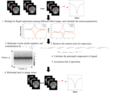 |
6 | Preliminary demonstration of a new motion-based noise reduction method for 3D CEST-MRI
Botao Zhao1 and Xiao-Yong Zhang1
1Institute of Science and Technology for Brain-Inspired Intelligence, Fudan University, Shanghai, China
Quantification of chemical exchange saturation transfer (CEST) imaging is a keystone for many clinical applications. Head motion and motion-caused noise produce non-negligible effects on the quantification in the whole brain 3D imaging. To solve the problem, we propose a new motion-based noise reduction method. Our preliminary results demonstrate that our method can reduce the noise efficiently and improve the amide proton transfer (APT) quantification results in the conditions of either with slight head motion or with huge head motion.
|
||
3836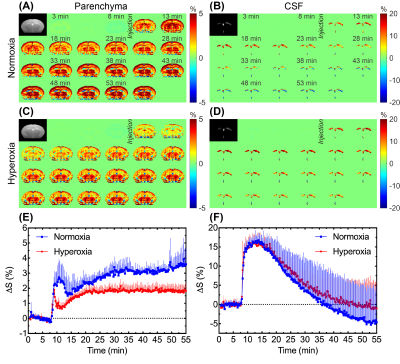 |
7 | Dynamic glucose enhanced MRI detects glucose-related responses in mouse brain under normoxia and hyperoxia
Jianpan Huang1, Zilin Chen1, Peter C. M. van Zijl2,3, Lok Hin Law1, Rohith Saai Pemmasani Prabakaran1,4, Se Weon Park1, Jiadi Xu2,3, and Kannie W. Y. Chan1
1Department of Biomedical Engineering, City University of Hong Kong, Hong Kong, China, 2F.M. Kirby Research Center for Functional Brain Imaging, Kennedy Krieger Research Institute, Baltimore, MD, United States, 3Russell H. Morgan Department of Radiology and Radiological Science, The Johns Hopkins University School of Medicine, Baltimore, MD, United States, 4Hong Kong Centre for Cerebro-Cardiovascular Health Engineering (COCHE), Hong Kong, China
Dynamic glucose enhanced (DGE) MRI can detect glucose-related events in the brain, however, the influence of oxygen levels on DGE signal remains unknown. Here, we investigated the DGE signal changes under normoxia and hyperoxia on mouse brain, using on-resonance variable delay multi-pulse (onVDMP) MRI. Significantly higher signal change under normoxia than that under hyperoxia was observed in parenchyma but not in cerebrospinal fluid (CSF). Without glucose infusion, a signal increment of about 3% was found in both parenchyma and CSF from hyperoxia to normoxia, interpreted as related to BOLD effect. These data provide insight into the origin of glucoCEST contrast.
|
||
3837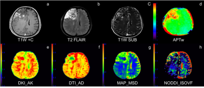 |
8 | Comparison of 3D amide proton transfer (APT) and four advanced diffusion models in gliomas IDH status decoding
Junjiao Hu1, Huiting Zhang2, Hu Guo3, Shan Jiang1, Weijun Situ1, Guang Yang4, and Jun Liu1
1Department of Radiology,The Second Xiangya Hospital, Central South University, Changsha, China, 2MR Scientific Marketing, Siemens Healthineers, Wuhan, China, 3MR Application, Siemens Healthineers, Changsha, China, 4Shanghai Key Laboratory of Magnetic Resonance, School of Physics and Electronic Science, Shanghai Key Laboratory of Magnetic Resonance, School of Physics and Electronic Science,East China Normal University, Shanghai, China
The purposed of study was to compare the diagnostic efficacy of APT imaging and four diffusion models, including DTI, DKI, NODDI, and MAP, in evaluating genotype IDH status, and to find the best imaging indicators for aiding accurate diagnoses and treatment decisions. Compared with IDH-mutant gliomas, IDH-wildtype gliomas had significantly higher mean and maximum of APTw, maximum axial, mean and radial diffusivity from DKI, maximum mean square diffusivity from MAP and maximum isotropic volume fraction from NODDI, as well as lower minimum APTw value.
|
||
3838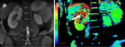 |
9 | Amide proton transfer weighted MRI in differentiating fat-poor angiomyolipoma from clearcell renal cell carcinoma Video Not Available
Qing Xu1, Weiqiang Dou2, and Jing Ye1
1Clinical Medical School of Yangzhou University, Northern Jiangsu People’s Hospital, Yangzhou, China, 2GE Healthcare, MR Research, Beijing, China
This study aimed to explore the feasibility of amide proton transfer weighted (APTw) imaging in discriminating fat-poor angiomyolipoma (AML) from clear cell renal cell carcinoma (ccRCC). 19 ccRCC patients and 9 fat-poor AML patients were recruited for APTw imaging. ccRCC group showed significantly higher MTRasym compared to fat-poor AML group (P < 0.05). High AUC of 0.827 in ROC analysis validated the efficacy of APTw imaging in differentiating fat-poor AML from ccRCC. With these findings, APTw imaging may be considered an effective method providing added clinical value in differentiating fat-poor AML from ccRCC.
|
||
3839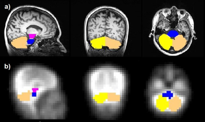 |
10 | Quantification of CMRO2 in Posterior Fossa Area Using Dynamic 17O-MRI Video Permission Withheld
Hao Song1, Ali Caglar Özen1, Stefan Schumann2, and Michael Bock1
1Dept. of Radiology, Medical Physics, Medical Center University of Freiburg, Faculty of Medicine, University of Freiburg, Freiburg, Germany, 2Dept. of Anesthesiology and Critical Care, Medical Center University of Freiburg, Faculty of Medicine, University of Freiburg, Freiburg, Germany Direct quantification of cerebral metabolic rate of oxygen consumption (CMRO2) is possible with dynamic 17O-MRI after inhalation of isotope-enriched 17O2. In this work, we investigated oxygen metabolism in posterior fossa tissue including cerebellum, pons and midbrain. The determined CMRO2 values in these areas were in good agreement with literature values obtained by 15O-PET studies. Our results demonstrate that dynamic 17O-MRI may provide a valuable tool for direct measurement of CMRO2 in posterior fossa tissue.
|
||
3840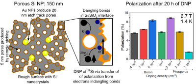 |
11 | Effect of doping on the dynamic nuclear polarization of porous silicon nanoparticles Video Permission Withheld
Konstantin Tamarov1, Gevin von Witte2,3, Viivi Hyppönen4, Jiri Jäntti1, Aaron Himmler3, Mohammed Albannay2, Matthias Ernst3, Sebastian Kozerke2, Vesa-Pekka Lehto1, Joakim Riikonen1, and Mikko I Kettunen4
1Department of Applied Physics, University of Eastern Finland, Kuopio, Finland, 2Institute for Biomedical Engineering, ETH Zurich, Zurich, Switzerland, 3Laboratory of Physical Chemistry, ETH Zurich, Zurich, Switzerland, 4A.I. Virtanen Institute, University of Eastern Finland, Kuopio, Finland
Porous Si nanoparticles (NPs) with different doping degree were prepared using low-load metal assisted catalytic etching and subjected to dynamic nuclear polarization at 3.4 T and 6.7 T. Thermal oxidation of Si was applied to form paramagnetic centers of dangling bond type in Si/SiO2 interface, which were used to polarize 29Si nuclei. The doping significantly affected the gained polarization and buildup times: high doping degree generally led to lower and faster polarization compared to the low doping. On the other hand, slight p-type or n-type doping was necessary to achieve the highest polarization of about 11 %.
|
||
3841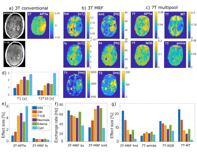 |
12 | Which CEST technique provides most insight into tumors – 3T APTw, 3T CEST-MRF or 7T multi-pool CEST?
Maria Sedykh1, Moritz Fabian1, Kai Herz2,3, Or Perlman4, Christian Farrar4, Angelika Mennecke1, Manuel Schmidt1, Arnd Dörfler1, and Moritz Zaiss1,2
1Neuroradiology, FAU Erlangen-Nuremberg, University Hospital, Erlangen, Germany, 2Magnetic Resonance Center, Max-Planck-Institute for Biological Cybernetics, Tübingen, Germany, 3Biomedical Magnetic Resonance, University of Tübingen, Tübingen, Germany, 4Radiology, Athinoula A. Martinos Center for Biomedical Imaging, Massachusetts General Hospital and Harvard Medical School, Charlestown, MA, United States
We report on only one glioblastoma patient, but who could be measured with several different CEST MRI methods: 3T APTw, 3T CEST fingerprinting, and 7T multi-pool CEST in order to reveal their relative performance and correlations. Coarse features can be observed in all methods, with MRF and 7T CEST being more versatile in non-active tumor parts. Isolation, separation and smarter combination of different CEST contrast is still needed to improve the diagnostic performance of CEST.
|
||
3842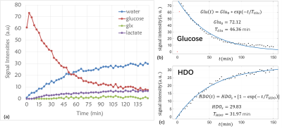 |
13 | Deuterium metabolic imaging for glioma in rat model using [2,3,4,6,6’-2H5]glucose Video Permission Withheld
Chao Zou1, Yingheng Ruan2, Feng Du1, Qikai Qin3, Qian Wan1, Jiawen Yuan1,4, Garth J. Thompson3, Xiaojun Yang2, Ye Li1, Xin Liu1, and Hairong Zheng1
1Shenzhen Institutes of Advanced Technology, Chinese Academy of Sciences, Shenzhen, China, 2Shenzhen Dingbang Bioscience Co., Ltd., Shenzhen, China, 3iHuman Institute, ShanghaiTech University, Shanghai, China, 4Southern University of Science and Technology, Shenzhen, China
Deuterium MRS(I) has emerged as a novel metabolic imaging method. However, the deuterium labeled substrate such as [6,6’-2H2]glucose is expensive, which becomes one of the obstacles for the clinical translation of this technique. Recently, we developed a cost-effective synthesis route for a new deuterated compound [2,3,4,6,6’-2H5]glucose. The cost of [2,3,4,6,6’-2H5]glucose is significantly lower than the [6,6’-2H2]glucose. In this study, we demonstrate that this new compound can measure the glycolytic flux through a rat glioma model.
|
||
3843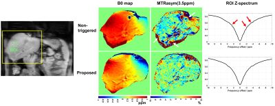 |
14 | Free-breathing abdominal CEST sequence using water presaturation and precise respiratory synchronization
Zhensen Chen1,2, Chuyu Liu3, Yishi Wang4, Weibo Chen5, Rui Guo6, He Wang1,7, and Xiaolei Song3
1Institute of Science and Technology for Brain-Inspired Intelligence, Fudan University, Shanghai, China, 2MOE Frontiers Center for Brain Science, Fudan University, Shanghai, China, 3Department of Biomedical Engineering, School of Medicine Tsinghua University, Beijing, China, 4Philips Healthcare, Beijing, China, 5Philips Healthcare, Shanghai, China, 6Department of Medicine (Cardiovascular Division), Beth Israel Deaconess Medical Center and Harvard Medical School, Boston, MA, United States, 7Human Phenome Institute, Fudan University, Shanghai, China
To date, chemical exchange saturation transfer (CEST) is rarely used in abdominal imaging of human body due to its high sensitivity to motion. In this study, we developed a free-breathing abdominal CEST sequence that uses a precise respiratory synchronization strategy and includes a water presaturation module. Preliminary in-vivo experiments showed that the proposed sequence outperformed non-triggered and breath-holding abdominal CEST in reducing the slice position inconsistency between the frequency offsets and motion-induced noise.
|
||
 Back to Meeting Home
 Back to the Program-at-a-Glance
The International Society for Magnetic Resonance in Medicine is accredited by the Accreditation Council for Continuing Medical Education to provide continuing medical education for physicians.
 Back to Meeting Home
 Back to the Program-at-a-Glance
 View the Presentation