Online Gather.town Pitches
RF Coils, Technologies & Sequences I
Joint Annual Meeting ISMRM-ESMRMB & ISMRT 31st Annual Meeting • 07-12 May 2022 • London, UK

Online Gather.town Pitches
RF Coils, Technologies & Sequences I
| Booth # | ||||
|---|---|---|---|---|
3220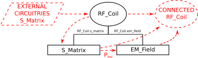 |
1 | CoSimPy: an Open-Source Python Library for MRI RF Coil EM/Circuit Cosimulation
Umberto Zanovello1, Frank Seifert2, Oriano Bottauscio1, Lukas Winter2, Luca Zilberti1, and Bernd Ittermann2
1Istituto Nazionale di Ricerca Metrologica (I.N.Ri.M.), Torino, Italy, 2Physikalisch-Technische Bundesanstalt (PTB), Braunschweig and Berlin, Germany
The Electromagnetic/Circuit cosimulation method represents an effective strategy to address those problems where a radiative structure has to interact with external supporting circuitries.This method proved to be particularly valuable for MRI RF coil design where the supporting circuitry optimisation is generally of particular concern. CoSimPy, an open-source library conceived for Electromagnetic/Circuit cosimulations, is presented. CoSimPy is written in Python following an Object-oriented programming. This improves its scalability and easily allows interested scientists to collaborate, integrating and improving the library with other methods. CoSimPy is available under https://github.com/umbertozanovello/CoSimPy together with a detailed documentation providing guidelines and examples.
|
||
3221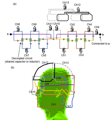 |
2 | Head Coil for Vertical-Field MRI using Loop/Dipole Parallel RF Coils Video Permission Withheld
Yosuke Otake1, Koichi Arai2, Takeshi Taniguchi2, Masayoshi Dohata2, Takahide Shimoda2, Kazuyuki Kato2, and Hisaaki Ochi1
1Innovative Technology Laboratory, FUJIFILM Healthcare Corporation, Tokyo, Japan, 2Radiation Diagnostic Systems Division, FUJIFILM Healthcare Corporation, Chiba, Japan
To improving the SNR and the g-factor in a vertical-field MRI, a multi-channel head coil has been developed. The coil consists of loop/dipole parallel RF coils (LDP) that improve the signal detection efficiency in the deep region of the subject in the vertical-field MRI. The performance of the coil was evaluated in a phantom experiment at 1.2T vertical-field MRI. The SNR and the g-factor of the coil using LDP were 1.2 and 1.2-2.9 times better than those of a conventional head coil, respectively. This technique will contribute to improve the performance of the vertical-field MRI.
|
||
3222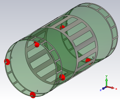 |
3 | Investigation of a transmit coil to reduce the magnetic field strength in the proximity of a MR system Video Permission Withheld
Daniel Durst1, Jürgen Nistler1, and Rainer Schneider1
1Siemens Healthcare GmbH, Erlangen, Germany
Birdcage coils used in MR imaging cause magnetic field strengths able to disturb electronic devices in the proximity to a MR system. Based on the principle of destructive superposition a configuration consisting of a main birdcage with additionally attached auxiliary birdcages is used to generate a field to reduce the magnetic field strength in the proximity of a MR system while maintaining the imaging quality. By electromagnetic simulations a reduction of up to 60 dB could be shown without drawbacks for the imaging performance if a distance of at least 25 cm between neighboring birdcages is retained.
|
||
3223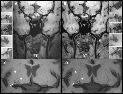 |
4 | A quadrature birdcage/48-channel receiver coil assembly for human brain imaging at 5T
Zidong Wei1,2,3, Qiaoyan Chen1,2, Qiang He3, Xiaoliang Zhang4, Xin Liu1,2, Hairong zheng1,2, and Ye Li1,2
1Paul C. Lauterbur Research Center for Biomedical Imaging, Shenzhen Institutes of Advanced Technology, Chinese Academy of Sciences, shenzhen, China, 2Key Laboratory for Magnetic Resonance and Multimodality Imaging of Guangdong Province, shenzhen, China, 3Shanghai United Imaging Healthcare, Shanghai, China, shanghai, China, 4Department of Biomedical Engineering, State University of New York at Buffalo, Buffalo, NY, United States
Ultra-high field magnetic resonance imaging (MRI) of human brain with high resolution has been increasing used for clinical and research applications. Due to RF transmit homogeneity and specific absorption issues, clinical use of ultra-high field MRI were limited. In this work, a local quadrature birdcage/48-channel receiver coil assembly was designed and evaluated on a novel whole body 5T MRI scanner. The coil at 5T showed improved SNR, higher parallel acceleration capability and improved detection in vessel wall imaging compared to a 32-channel coil at a 3T commercial scanner.
|
||
3224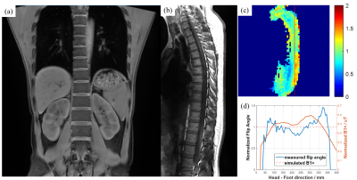 |
5 | Whole Spine Imaging at 5.0T Using An 8-channel Volume Transmit Coil and 48-channel Receive Array Video Not Available
Shao Che1,2,3, Fuyi Fang2, Kaida Dong2, Shihong Han2, and Ye Li1,3
1Lauterbur Research Center for Biomedical Imaging, Shenzhen Institute of Advanced Technology, Chinese Academy of Sciences, Shenzhen, China, 2United Imaging Healthcare, Shanghai, China, 3Key Laboratory for Magnetic Resonance and Multimodality Imaging of Guangdong Province, Shenzhen, China
Large field-of-view imaging of spine in ultra-high field 5.0T magnetic resonance imaging system is achieved with RF hardware design of an 8-channel volume transmit coil and a dedicated posterior spine array of 48 receive channels. The axial coverage and transmit homogeneity allow the use of refocusing pulses under specific-absorption ratio limits by safety regulations. Consequent high-resolution fast spin echo clinical images are acquired and compared with results on a commercial 3.0T scanner.
|
||
3225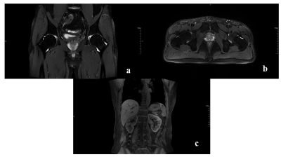 |
6 | A 24-channel ultra-light flexible receiver coil array for human body imaging at 5T Video Not Available
Yingchao Tan1,2,3, Qiaoyan Chen1,2, Rui Zhao3, Hai Lu3, Xianwen Fan3, Junyu Chen3, Xin Liu1,2, Hairong Zheng1,2, and Ye Li1,2
1Paul C. Lauterbur Research Center for Biomedical Imaging, Shenzhen Institutes of Advanced Technology, Chinese Academy of Sciences, Shenzhen, China, 2Key Laboratory for Magnetic Resonance and Multimodality Imaging of Guangdong Province, Shenzhen, China, 3Shanghai United Imaging Healthcare, Shanghai, China
To improve signal-to-noise ratio (SNR) and patient comfort, flexible coils need to be made of very light and flexible materials. In this study, a novel 24-channal ultra-light flexible receiver coil array was designed and constructed for human abdominal imaging at a 5T ultra-high field MRI system. The 24-channel ultra-light flexible coil showed much higher SNR and a better parallel acceleration capability compared to a 24-channel conventional receiver coil. High quality human abdominal images were acquired by using the 24-channel ultra-light flexible coil.
|
||
3226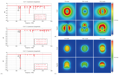 |
7 | Design of ultrahigh dielectric constant uHDC helmet for 3T and its intrinsic resonance modes
Xinlian Chen1, Navid P. Gandji2, Christopher T. Sica2, Gary W. Yang3, Parisa Lotfi Poshtgol2, Hannes M Wiesner4, Xiao-Hong Zhu4, Michael Lanagan5, Wei Chen4, Qing X Yang2, and Xuegang Xin1
1Department of Biomedical Engineering, South China University of Technology, GuangDong, China, 2Departments of Neurosurgery and Radiology, College of Medicine, Pennsylvania State University, Hershey, PA, United States, 3Departments of Computer Science and Engineering, Pennsylvania State University, Hershey, PA, United States, 4Center for Magnetic Resonance Research, University of Minnesota, Minneapolis, MN, United States, 5Department of Engineering Science and Mechanics, Pennsylvania State University, Hershey, PA, United States
This investigation characterize the intrinsic resonance modes (IRM) in a helmet made from utrahigh dielectric constant (uHDC) material and their interactions with RF field produced by conventional array coils using numerical simulation. The RF field of the three lower order modes, TE, TM1 and TM2, were investigated. TM1 mode produces uniform H-field in the transverse plane inside the helmet similar to that of a birdcage coil. TM2 mode produces a strong RF field but with a large gradient. Understanding of the IRMs of large uHDC structures such as helmets provides theoretical foundation for wider applications of uHDC materials RF engineering.
|
||
3227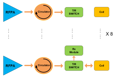 |
8 | High isolation 8-channel pTx system for UHF MRI Video Not Available
Jifeng Chen1, Ye Li1, Jun Luo1, Xin Liu1, Xu Chu2, and Hairong Zheng1
1Lauterbur Imaging Research Center, Shenzhen Institutes of Advanced Technology, Chinese Academy of Sciences, Shenzhen, China, 2United Imaging Healthcare, Shanghai, China
Ultra-high filed (UHF) whole-body MRI technique usually utilizes radio frequency power amplifier (RFPA) with high power rating [1,2]. It requires high isolation among transmission channels as well as between transmitting and receiving (T/R) circuits. It also requires high B1 homogeneity [3] and signal-to-noise ratio (SNR). On the premise of meeting the abovementioned requirements, this paper presents an 8-channel high power pTx architecture, which can support the whole-body MRI system with high isolation among transmission channels as well as between T/R circuits.
|
||
3228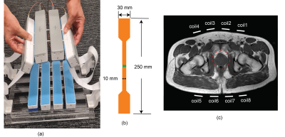 |
9 | Prostate imaging with Integrated Multi-modal Antenna with coupled Radiating dipoles (I-MARS) at 7T
Mingyan Li1, Jin Jin1,2, Ewald Weber1, Craig Engstrom 3, Aurelien Destruel1,4, Stuart Crozier1, and Feng Liu1
1School of Information Technology and Electrical Engineering, The University of Queensland, Brisbane, Australia, 2Siemens Healthineers, Brisbane, Australia, 3School of Human Movement and Nutrition Sciences, The University of Queensland, Brisbane, Australia, 4The Center for Magnetic Resonance in Biology and Medicine, Aix-Marseille University, Marseille, France
Prostate imaging is challenging at 7T due to its small size seated deeply inside the pelvic region. As such, both transmit receive performance of the RF coils are heavily challenged. We have recently developed a new paddle-shape Integrated Multi-modal Antenna with coupled Radiating structures (I-MARS) allowing an ensemble of eight elements to be placed tightly together among each other and closely to the imaging region. In the current work, we demonstrate optimized work-flow and in vivo imaging results using such an 8-element paddle I-MARS coil with an upgraded MAGNETOM 7T Plus system.
|
||
3229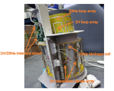 |
10 | A dual-nuclei 1H/23Na knee coil for high-resolution 1H and 23Na MR imaging at 7T Video Not Available
xing yang1, Xin Liu1, feng du1, Nan Li1, Hairong Zheng1, and Ye Li1
1Shenzhen Institutes of Advanced Technology,Chinese Academy of Sciences, Shenzhen, China
An ideal MRI study for early osteoarthritis should provide both structure imaging and functional imaging, which can show early morphologic degenerative changes and physiology changes of the cartilage. Most of previous dual-nuclei coils pushed the limits of non-proton nuclei sensitivity at the expense of 1H performance. To get high performance imaging for both nuclei, a novel dual-nuclei 1H/23Na coil array was developed. The bench test results show that all coils were sufficiently matched and decoupled, and the interference between sodium coils and proton coils was nearly negligible, thus can be expected to get high-resolution 1H imaging and quantitative 23Na imaging.
|
||
3230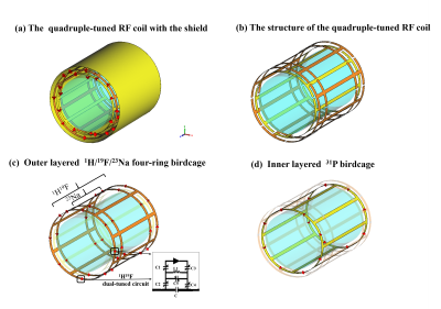 |
11 | Design of a double-layered quadruple-nuclear birdcage coil system for 1H / 19F / 23Na/31P MR imaging at 3T
Nan Li1,2, Feng Du1,2, Xiaoliang Zhang3, Xin Liu1,2, Hairong Zheng1,2, and Ye Li1,2
1Lauterbur Imaging Research Center, Shenzhen Institutes of Advanced Technology, Chinese Academy of Sciences, Shenzhen, China, 2Key Laboratory for Magnetic Resonance and Multimodality Imaging of Guangdong Province, Shenzhen, China, 3Department of Biomedical Engineering, State University of New York, Buffalo, NY, United States
The lower natural abundance and gyromagnetic ratios of non-hydrogen nuclei require the use of a highly efficient RF coil to optimize the quality of the MR image and spectroscopy. In this study, a new quadruple-tuned RF coil system capable of 1H / 19F / 23Na /31P imaging was proposed, including the inner high-pass birdcage coil (31P) and a triple-tuned outer four-ring birdcage coil (1H / 19F / 23Na). The feasibility and performance of this proposed RF coil were evaluated and validated by the numerical electromagnetic simulation in the scattering parameters and B1+field distributions.
|
||
3231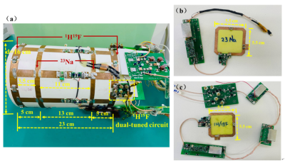 |
12 | Triple-nuclear RF coil system for Simultaneous acquisition of 1H / 19F / 23Na MR imaging at 3T
Nan Li1,2, Feng Du1,2, Xiaoliang Zhang3, Xin Liu1,2, Hairong Zheng1,2, and Ye Li1,2
1Lauterbur Imaging Research Center, Shenzhen Institutes of Advanced Technology, Chinese Academy of Sciences, Shenzhen, China, 2Key Laboratory for Magnetic Resonance and Multimodality Imaging of Guangdong Province, Shenzhen, China, 3Department of Biomedical Engineering, State University of New York, Buffalo, NY, United States
The lower natural abundance and gyromagnetic ratios of non-hydrogen nuclei require the use of a highly efficient RF coil to optimize the quality of the image. In this study, a novel triple-nuclear RF coil system capable of 1H / 19F / 23Na imaging was designed. The coil consists of a triple-tuned four-ring birdcage transmit coil and the tight fit local X-nuclei single-loop receive coils. A range of bench tests were performed by using the developed triple-nuclear coil. The performance evaluations showed that the coil system was appropriately tuned, matched and decoupled at 1H/19F/23NA frequencies when working simultaneously.
|
||
3232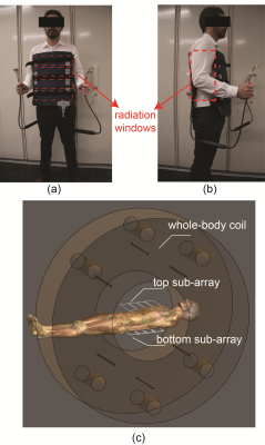 |
13 | An 8-element torso RF coil array for the Australian inline MRI-Linac: Initial imaging results
Mingyan Li1, Ewald Weber1, David Waddington2, Shanshan Shan2, Paul Liu2, Bin Dong3, Craig Freakley1, Stuart Crozier1, and Feng Liu1
1School of Information Technology and Electrical Engineering, The University of Queensland, Brisbane, Australia, 2ACRF Image X Institute, The University of Sydney, Sydney, Australia, 3Ingham Institute, Sydney, Australia
The hybrid MRI-Linac system demands dedicated radiolucent imaging coils to visualise and track tumours without interfering the Linac beams. A radiolucent whole-body transceive RF coil for the Australian inline MRI-Linac system has been previously developed and produced images with good quality. Recently, an 8-element torso RF coil array was manufactured to provide better SNR with fast imaging capability, which will facilitate real-time tumour tracking during radiotherapy. Initial imaging results acquired from the 1.0 T Australian MRI-Linac system on a phantom and a volunteer demonstrate the potential benefits of such a torso coil array for MRI-guided radiotherapies.
|
||
3233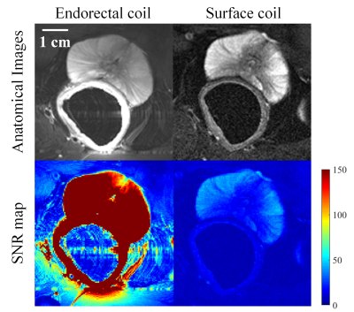 |
14 | A novel three-channel endorectal coil for prostate magnetic resonance imaging at 3T
Zhiguang Mo1, Xiaoliang Zhang2, Changjun Tie1, Huageng Liang3, Xiaoping Zhang3, Wen Xiao3, Qi Cao3, and Ye Li1
1Lauterbur Research Center for Biomedical Imaging, Shenzhen Institutes of Advanced Technology, Chinese Academy of Sciences, Shenzhen, Guangdong, 518055, China, Shenzhen, China, 2Department of Biomedical Engineering, State University of New York at Buffalo, NY, United States., Buffalo, NY, United Stats, Buffalo, NY, United States, 3Union Hospital, Tongji Medical College, Huazhong University of Science and Technology, Wuhan, China
We designed and fabricated a novel three-channel endorectal prostate coil and validated its feasibility of high-resolution magnetic resonance imaging (MRI) at 3T. A commercial 3T surface coil was used for comparison. Both phantom imaging and in-vivo prostate imaging show that the 3-channel endorectal coil provides an improved SNR than the external surface coil. Prostate images at resolution of 0.47 mm × 0.47 mm × 4.0 mm are acquired using the 3-channel endorectal coil to demonstrate the performance. With the improved SNR and multichannel design, the proposed endorectal coil could enable parallel imaging based fast imaging in prostate imaging studies.
|
||
3234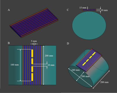 |
15 | Combining High Permittivity Pads with Periodic Passive Components in RF Coil Design at 7T
Haiwei Chen1, Zhuang Xiong1, Lei Guo1, Xiaotong Zhang2, and Feng Liu1
1the University of Queensland, Brisbane, Australia, 2Zhejiang University, Hangzhou, China Periodic passive components (PPC) such as metamaterials can improve SAR efficiency and B1 penetration. However, due to the shorter RF wavelength in MRI, the performance of PPC is confined by the limited number of unit cells. In this work, a novel PPC structure has been proposed to combine with a high permittivity pad (HPP), so a larger number of unit cells can be used. The designed structure is excited by a dipole coil and compared with the case when a single HPP is used. It is observed that more than 50% SAR reduction can be achieved. |
||
3235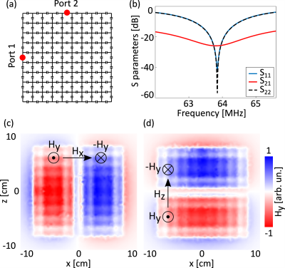 |
16 | Multichannel MRI receive coil based on artificial material Video Not Available
Eugene Koreshin1, Denis Burov1, and Pavel Seregin1
1School of Physics and Engineering, ITMO University, Saint-Petersburg, Russian Federation
Metamaterials and metasurfaces, in particular, are gaining increasing popularity in recent years. One of the novel applications of metasurfaces are receive coils for magnetic resonance imaging. The major drawback of using such structures is the lack of parallel MRI capabilities due to the single-channel acquisition regime. In this work, we will demonstrate that it is possible to design a multichannel metasurface.
|
||
3236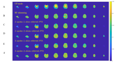 |
17 | Parallel transmission RF pulse design for simultaneous multi‐slab excitation at 7 Tesla: a simulation study
Xin Shao1, Xiaodong Ma2, Simin Liu1, Kamil Ugurbil2, Hua Guo1, and Xiaoping Wu2
1Center for Biomedical Imaging Research, Department of Biomedical Engineering, School of Medicine, Tsinghua University, Beijing, China, 2Center for Magnetic Resonance Research, Radiology, Medical School, University of Minnesota - Twin Cities, Minneapolis, MN, United States
It has been reported that combining ultrahigh field (UHF) with 3D imaging is beneficial for high-resolution imaging. Nevertheless, image quality may suffer from increased magnetic field inhomogeneity. Thus, we combined radiofrequency (RF) parallel transmission (pTx) with 3D simultaneous multi-slab (SMSlab) acquisition at 7T to get improved transmit field and optimal SNR efficiency concurrently. By simulation, it was proved that by using a 3-spoke RF pulses we could get 25% more uniform excitation magnitude than 2-spoke pulses (with almost the same RF peak power and identical pulse duration).
|
||
3237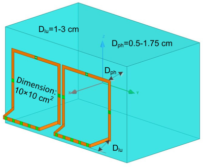 |
18 | Bumped Self-decoupled RF Coil
Ming Lu1, John C. Gore2,3, and Xinqiang Yan2,3
1College of nuclear equipment and nuclear engineering, Yantai University, Yantai, China, 2Vanderbilt University Institute of Imaging Science, Vanderbilt University Medical Center, Nashville, TN, United States, 3Department of Radiology and Radiological Sciences, Vanderbilt University Medical Center, Nashville, TN, United States
In this work, we propose a novel “bump” design to further improve the coil robustness versus loadings. For the loop-mode self-decoupled coil, the rotating magnetic field (composed of Bx and By) and electric field are mainly determined by the feed and arm conductors; the frequency shift that decreases coil robustness is mainly caused by the conductor with small-valued mode capacitors (referred to as mode conductor for short). Therefore, a bumped coil with a spaced mode conductor but unchanged arm and feed conductors potentially provides more robust tuning/matching performance without decreasing coil efficiency.
|
||
3238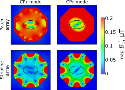 |
19 | Bore integrated coil for whole-body imaging at 7 Tesla based on patch antennas
Georgiy Solomakha1, Svetlana Egorova1, Alena Shchelokova1, Egor Kretov1, and Stanislav Glybovski1
1Department of Physics and Engineering, ITMO University, Saint Petersburg, Russian Federation
This work proposes a novel transmit radiofrequency coil integrated into the scanner bore for whole-body MRI for 7 Tesla MRI based on patch antennas. Numerical studies with a homogeneous phantom demonstrated 4.5-fold higher transmit efficiency in the phantom center compared to the state-of-the-art bore integrated stripline array.
|
||
 Back to Meeting Home
 Back to the Program-at-a-Glance
The International Society for Magnetic Resonance in Medicine is accredited by the Accreditation Council for Continuing Medical Education to provide continuing medical education for physicians.
 Back to Meeting Home
 Back to the Program-at-a-Glance
 View the Presentation