Online Gather.town Pitches
New Systems & Devices IV
Joint Annual Meeting ISMRM-ESMRMB & ISMRT 31st Annual Meeting • 07-12 May 2022 • London, UK

Online Gather.town Pitches
New Systems & Devices IV
| Booth # | ||||
|---|---|---|---|---|
5023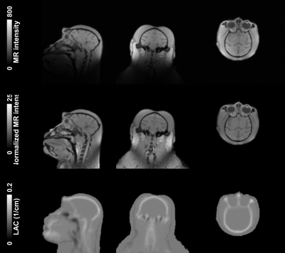 |
1 | Application of the template-based attenuation correction to simultaneous MR-PET brain imaging of non-human primates
Chi-Hyeon Yoo1, Jonathan DuBois2, Lu Wang3, Steven Liang4, David Izquierdo-Garcia1, and Hsiao-Ying Wey1
1Athinoula A. Martinos Center for Biomedical Imaging, Department of Radiology, Massachusetts General Hospital, Harvard Medical School, Charlestown, MA, United States, 2Biogen, Cambridge, MA, United States, 3Department of Nuclear Medicine and PET/CT-MRI Center, The First Affiliated Hospital of Jinan University & Institute of Molecular and Functional Imaging, Jinan University, Guangzhou, China, 4Division of Nuclear Medicine and Molecular Imaging, Department of Radiology, Massachusetts General Hospital, Harvard Medical School, Boston, MA, United States
The goal of this study was to generate a population-based template/atlas and apply the template/atlas-based attenuation correction (AC) method for simultaneous positron-emission-tomography/magnetic-resonance-imaging (PET/MRI) neuroimaging studies on non-human primates (NHPs). Population-based template/atlas of computed tomography image and anatomical MRI, were generated from ten NHPs (rhesus macaque). By using the generated template/atlas, the linear attenuation coefficient (LAC) of objects can be reliably derived, showing good differentiation for tissues, also skull and air. Comparison of the PET images reconstructed by the template/atlas-based and simple threshold-based AC has shown that the template/atlas-based AC can increase PET signal by avoiding overcompensation of attenuation.
|
||
5024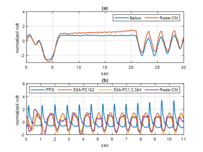 |
2 | Noncontact in-bore cardiopulmonary sensing using CW doppler radar
Wonje Lee1, Kanghyun Ryu1, Fraser Robb2, John Pauly3, Shreyas Vasanawala1, and Greig Scott3
1Radiology, Stanford University, Stanford, CA, United States, 2GE healthcare, Cleveland, OH, United States, 3Electrical Engineering, Stanford University, Stanford, CA, United States
Noncontact vital sensing applications have great potential to aid in patient care. We leverage a reconfigurable SDR to assess the feasibility of in-bore cardiopulmonary sensing using CW doppler radar that operates independently of the MRI system and data acquisition.
|
||
5025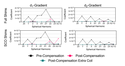 |
3 | Spherical Harmonic Active Shim Set Design for a Shoulder Cut-out MRI Platform
Eric J Lessard1, William B Handler1, and Blaine A Chronik1
1Western University, London, ON, Canada
This simulation and electromagnetic design work explored the development of an active second order spherical harmonic shim set for eddy current compensation in a shoulder cut-out head and neck imaging platform. While the small radius shoulder cut-out shims can be driven faster and stronger than the larger radius complete cylinder shims they suffer in homogeneity and eddy current compensation. This difference in compensation is minimized with the addition of two purpose designed eddy current compensation coils.
|
||
5026 |
4 | MC-ECLIPSE for arbitrary ROI shaping and whole brain shimming for 3D MRSI
Chathura Kumaragamage1, Peter B Brown1, Scott McIntyre1, Terence W Nixon1, Henk M De Feyter1, and Robin A de Graaf1
1Radiology and Biomedical Imaging, Yale University, New Haven, CT, United States
ECLIPSE, a pulsed second order gradient insert can provide robust lipid suppression over an elliptical ROI for applications in human brain MRSI. While ECLIPSE provides excellent axial slice coverage for brain shapes that closely resemble an ellipse, coverage can be compromised for other head shapes. The development of an ECLIPSE gradient coil in combination with a 54-channel multi-coil array (MC-ECLIPSE), that allows B0 field shaping for arbitrary ROI generation is presented. Simulation results demonstrate that near-perfect (99%) axial slice coverage is achievable with MC-ECLIPSE, for a wide range of highly asymmetrical brain and head shapes.
|
||
5027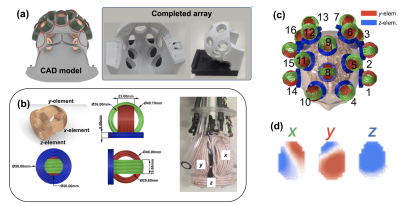 |
5 | Spatial encoding with a 48-ch Transcranial Magnetic Stimulation coil array: Application to diffusion imaging
Jason Peter Stockmann1,2, Lucia Navarro de Lara1, Mohammed Daneshzand1, Qinglei Meng1, Congyu Liao3, Hong-Hsi Lee1,2, Lawrence L Wald1,2, Berkin Bilgic1,2, Susie Huang1,2, and Aapo Nummenmaa1,2
1A. A. Martinos Center for Biomedical Imaging, Massachusetts General Hospital, Charlestown, MA, United States, 2Harvard Medical School, Boston, MA, United States, 3Lucas Center, Stanford University, Stanford, CA, United States
We show that multi-channel TMS (mTMS) probe arrays can be used to generate B-fields inside the head for spatial encoding in concurrent TMS/MRI studies. We assess the field-synthesis capability of a 48-ch mTMS array for diffusion imaging and spatial encoding more broadly. Proof-of-concept diffusion imaging experiments on a 3T scanner using a 3-axis TMS probe achieve spatially-varying b-values >500 s/mm2 at the depth of the peripheral gray matter using only 4A of current. We consider the potential for scaling up to 200A for studies of gray matter microstructure.
|
||
5028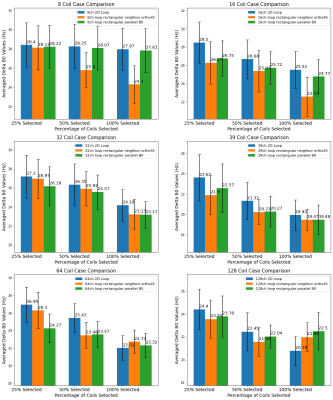 |
6 | Improving the performance of integrated RF-B0 shim array using 3D coil elements with orthogonal loops
Siyu Li1 and Danny JJ Wang1
1Laboratory of FMRI Technology (LOFT), Mark & Mary Stevens Neuroimaging and Informatics Institute, Keck School of Medicine, University of Southern California, Los Angeles, CA, United States B0 field inhomogeneity can reduce the effectiveness of RF pulses, result in signal voids, blurring and image artefacts. Integrated RF-shim coil arrays1,2,3 offer capabilities for concurrent parallel RF reception and local B0 shimming, and is superior to spherical harmonic shimming. However, its performance may still be limited due to the lack of flexibility for shimming and limited depth penetration for signal reception. We simulated the B0 shimming performance of a range of integrated RF-shim coil arrays with 3D orthogonal loop coil geometry, and compared the results with standard 2D loop coil arrays. |
||
5029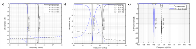 |
7 | A Proposed Method for Retuning an RF Coil by Changing the Shield Radius for Multinuclear Imaging
Kieffer Davieau1, Matthew Fox1,2, Blaine Chronik1,3, and Alexei Ouriadov3,4
1Medical Biophysics, Western University, London, ON, Canada, 2Lawson Health Research Institute, London, ON, Canada, 3Physics and Astronomy, Western University, London, ON, Canada, 4School of Biomedical Engineering, Western University, London, ON, Canada
This study investigates the feasibility of changing the shield radius of an RF coil to shift the resonant frequency from 1H to 19F for multinuclear imaging. The method proposed in this investigation was verified by changing the resonant frequency of a 129Xe RF Coil a total of 1.18 MHz by changing the shield radius, allowing it to be used in a 2.9T Siemens MRI and a 3.0T GE MRI. It was found that altering the shield radius had similar effects on the resonant frequency as using a variable tuning capacitor, allowing for one coil to be compatible with multiple scanners.
|
||
5030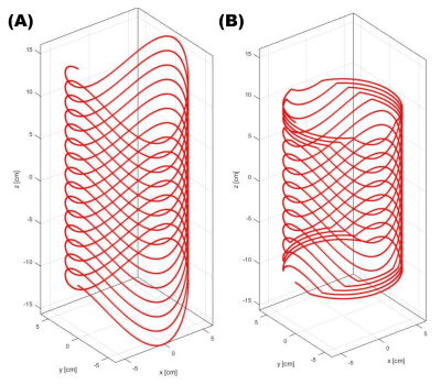 |
8 | A Truncated Twisted Solenoid RF Phase Gradient Transmit Coil for TRASE MRI
Christopher James Sedlock1, Aaron Purchase1, Boguslaw Tomanek1, and Jonathan Sharp1
1University of Alberta, Edmonton, AB, Canada TRASE is an MR imaging sequence that achieves k-space encoding through the use of phase gradients in the RF transmit field. The twisted solenoid is the most efficient RF transmit coil used for TRASE encoding; however, the geometry results in a long coil with a relatively short imaging volume. We introduce a new truncated design for the twisted solenoid to increase the usable imaging volume relative to the coil’s size. Simulation studies conducted indicate the truncated design can create similar imaging volumes to the untruncated version whilst significantly reducing the coils length by as much as a half. |
||
5031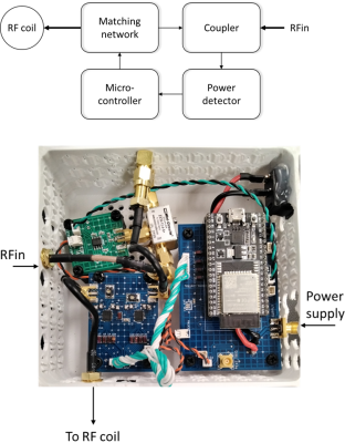 |
9 | Mobile application based monitoring system for RF coils at 7T Magnetic Resonance Imaging
Sri Kirthi Kandala1, Alberto Fuentes2, Gregory H Turner2, and Sung-Min Sohn1
1Arizona State University, Tempe, AZ, United States, 2Barrow-ASU Center for Preclinical Imaging, Phoenix, AZ, United States
Like most modern electronic devices, a mobile application based Radio Frequency (RF) coil monitoring system will make it easy to carry out any real-time changes to the RF coil. This work focuses on developing a mobile application based RF monitoring system, that allows the user to gather real-time RF coil characteristics and control the coil. It gives the user a chance to make changes when necessary. The opportunity to wirelessly implement such changes without physical contact with the coil, using a mobile application via Bluetooth is a step in the direction towards building next-generation RF coils for MRI.
|
||
5032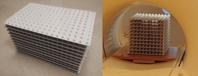 |
10 | Distortion Measurements Using a Large-volume, Lightweight, Modular MRI Phantom With Solid Signal Sources
Mariko Gardiner1, Keith Wachowicz2,3, and Nicola De Zanche2,3
1University of Alberta, Edmonton, AB, Canada, 2Oncology, University of Alberta, Edmonton, AB, Canada, 3Medical Physics, Cross Cancer Institute, Edmonton, AB, Canada
Phantoms that cover large volumes are required to characterize geometrical distortion for image-guided interventions such as MR-guided radiation therapy (linac-MR). Liquid-filled phantoms of appropriate size can, however, be impractically heavy. Our phantom uses silicone rubber spheres arranged on a three-dimensional grid, resulting in a lightweight, portable structure. Displacements of each bead relative to the known grid positions are calculated in Matlab or 3D Slicer from 3D bSSFP scans. Design files and image processing code are available online.
|
||
5033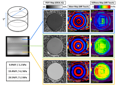 |
11 | Combined Stiffness and PDFF Phantom for Quality Assurance of Quantitative Magnetic Resonance Elastography and Fat Fraction Imaging
David Rutkowski1, Alejandro Roldán-Alzate2,3,4, José Guerrero-González1, Diego Hernando1,3,5, Scott B Reeder1,3,4,5,6,7, and Jean H Brittain1
1Calimetrix, Madison, WI, United States, 2Mechanical Engineering, University of Wisconsin, Madison, WI, United States, 3Radiology, University of Wisconsin, Madison, WI, United States, 4Biomedical Engineering, University of Wisconsin, Madison, WI, United States, 5Medical Physics, University of Wisconsin, Madison, WI, United States, 6Medicine, University of Wisconsin, Madison, WI, United States, 7Emergency Medicine, University of Wisconsin, Madison, WI, United States
MRI proton density fat fraction (PDFF) and MR elastography (MRE) methods are frequently used to evaluate patients with known or suspected NAFLD or NASH. Reference standards (phantoms) are needed for quality assurance of these methods. Currently available phantoms mimic only a single tissue property. A phantom that simultaneously mimics PDFF and tissue stiffness would more realistically mimic NAFLD/NASH tissue and would enable robust and convenient quality assurance. In this work, a chemical formulation and phantom configuration were developed for a multi-layer combined PDFF-MRE phantom, in which each layer simultaneously mimics different PDFF and stiffness values.
|
||
5034 |
12 | Modular, 3D Printed, Open-source Phantoms for Small Animal Systems
Mariko Gardiner1, Anthony G. Tessier2, Keith Wachowicz2,3, and Nicola De Zanche2,3
1University of Alberta, Edmonton, AB, Canada, 2Medical Physics, Cross Cancer Institute, Edmonton, AB, Canada, 3Oncology, University of Alberta, Edmonton, AB, Canada
Phantoms for pre-clinical systems have not been standardized, and none in the literature permit the wide variety of measurements possible with the ACR phantoms. We present a low-cost, open-source, modular phantom made of 3D-printed and off-the-shelf parts that mimics the functionality of the ACR phantoms on an appropriate scale. Standard, inexpensive manufacturing techniques are sufficient to create the fine features required at this scale. Configuration of the phantom can be customized to suit each site’s needs. Possible measurements include image uniformity, distortion and SNR; slice location, thickness, and angle; radial, linear, and low-contrast resolution.
|
||
5035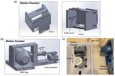 |
13 | Low-cost MR-compatible pneumatic respiratory motion simulator for development of MR-guided therapies
Kisoo Kim1, Peter Jones2, Chris J Diederich2, and Eugene Ozhinsky1
1Radiology & Biomedical Imaging, University of California, San Francisco, San Francisco, CA, United States, 2Radiation Oncology, University of California, San Francisco, San Francisco, CA, United States
We developed a low-cost and simple MR-compatible respiratory motion simulator to support proof-of-concept studies of MR monitoring approaches with respiration-induced organ motion. The motion system integrates pneumatic control via an actuator subsystem outside the MRI and coupled via plastic tubing to a compressible bag for distention and retraction within the MRI motion subsystem and phantom within the MRI scanner. The proposed respiratory motion simulator provided consistent periodic respiratory motion with displacement in the range 3.7-9.0 mm. The respiratory simulator could be easily assembled with off-the shelf and 3D-printed parts, based on open-source design models.
|
||
5036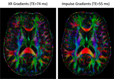 |
14 | Evaluation of High Resolution Diffusion MRI on the Next-Generation 7T scanner
An T Vu1,2, Alexander JS Beckett3,4, Salvatore T Torrisi3,4, Sinyeob Ahn5, Michael Koehler6, Peter Dietz6, and David A Feinberg3,4
1Radiology and Biomedical Imaging, University of California, San Francisco, San Francisco, CA, United States, 2Radiology, San Francisco Veteran Affairs Health Care System, San Francisco, CA, United States, 3Helen Wills Neuroscience Institute, University of California, Berkeley, Berkeley, CA, United States, 4Advanced MRI Technologies, Sebastopol, CA, United States, 5Siemens Medical Solutions USA, Inc., Melvern, PA, United States, 6Siemens Healthcare, Erlangen, Germany
The newly developed head gradient (200 mT/m Gmax, 900 T/m/s SR) on the new NexGen 7T system provides many potential benefits for high-resolution diffusion imaging. Faster, stronger gradients can significantly reduce diffusion encoding times and TE for up to a 3-fold improvement SNR while also reducing TR and total scan time. We demonstrate these benefits in high resolution diffusion imaging applications with b-values of up to 10,000 s/mm2 at 7T.
|
||
 |
5037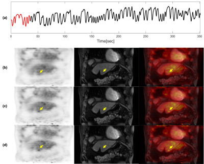 |
15 | MR-Assisted PET Respiratory Motion Correction Using Deep-Learning Based Short-Scan Motion Fields
Sihao Chen1, Tyler J. Fraum1, Cihat Eldeniz1, Joyce Mhlanga1, Weijie Gan1, Thomas Vahle2, Uday B. Krishnamurthy3, David Faul4, H. Michael Gach1, Michael M. Binkley1, Ulugbek S. Kamilov1, Richard Laforest1, and Hongyu An1
1Washington University in St. Louis, Saint Louis, MO, United States, 2Siemens Healthcare GmbH, Erlangen, Germany, 3Siemens Medical Solutions USA, Saint Louis, MO, United States, 4Siemens Medical Solutions USA, Malvern, PA, United States
Respiratory motion causes signal blurring and image artifacts. Simultaneous PET/MRI allows for MR-assisted motion correction (MoCo) in PET imaging, leading to improved PET images for detection and evaluation of lesions. In this study, we proposed and examined a PET MoCo approach using motion vector fields (MVFs) from a deep-learning reconstructed MRI scan. MRI-based MVFs were derived from either 2000 spokes (MoCo2000, 5-6 minutes acquisition time) using a Fourier transform reconstruction or 200 spokes (MoCoP2P200, 30-40 seconds acquisition time) using a deep-learning Phase2Phase (P2P) reconstruction and then incorporated into PET MoCo reconstruction.
|
|
 Back to Meeting Home
 Back to the Program-at-a-Glance
The International Society for Magnetic Resonance in Medicine is accredited by the Accreditation Council for Continuing Medical Education to provide continuing medical education for physicians.
 Back to Meeting Home
 Back to the Program-at-a-Glance
 View the Presentation