Online Gather.town Pitches
New Systems & Devices II
Joint Annual Meeting ISMRM-ESMRMB & ISMRT 31st Annual Meeting • 07-12 May 2022 • London, UK

Online Gather.town Pitches
New Systems & Devices II
| Booth # | ||||
|---|---|---|---|---|
3859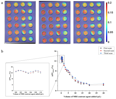 |
1 | Quantification of the impact of Gadolinium agent on amide-proton-transfer weighted MRI: an ex vivo and in vivo study Video Permission Withheld
Qian Yang1, Zhou Liu1, Yin Wu2, Long Qian3, and Dehong Luo1
1National Cancer Center/National Clinical Research Center for Cancer/Cancer Hospital & Shenzhen Hospital,Chinese Academy of Medical Sciences and Peking Union Medical College, Shenzhen, China, 2Paul C. Lauterbur Research Center for Biomedical Imaging, Shenzhen Institute of Advanced Technology, Chinese Academy of Sciences, Shenzhen, China, 3MR Research, GE Healthcare, Beijing, China
The clinical value of Amide proton transfer (APT) imaging in brain tumors has been intensively studied and validated. However, whether and how much Gadolinium-based (Gd) agent in clinical dosage has impact on the APT-weighted signal in clinical scenarios remains controversial. Hence, this study designed an in vitro study using a phantom with a gradient dosage of Gd-based agent and then validated the result in 6 patients with brain tumors. The result revealed that APT effect was significantly influenced by Gd contrast agent both in the phantom in vitro study and in vivo study.
|
||
3860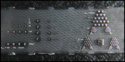 |
2 | Resolution phantoms for MR microimaging with 4µm≤ps≤256µm based on anisotropic tube channels (PSF + MTF) fabricated by Deep X-Ray-Lithography Video Permission Withheld
Andreas Georg Berg1,2 and Martin Börner3,4
1Center for Medical Physics and Biomedical Engineering, MR-Physics, Medical University of Vienna, Vienna, Austria, 2High field MR-Center, Medical University of Vienna, Vienna, Austria, 3Karlsruhe Nano-Micro facility (KNMFi), Karlsruhe Institute of Technology (KIT), Eggenstein-Leopoldshafen, Germany, 4Institute of Microstructure Technology (IMT), Karlsruhe Institute of Technology (KIT), Eggenstein-Leopoldshafen, Germany
The most important quality control criterions in Magnetic Resonance Imaging (MRI) are represented by the contrast-to-noise-ratio (CNR) and the spatial resolution. This report presents the design, a prototype object, an exemplary MRI evaluation, qualitative results and quantitation of the spatial resolution up to microscopic scale beyond pixel-size characterization. The phantom is manufactured using Deep X-Ray Lithography (DXRL) at the Synchrotron radiation source KARA (Karlsruhe Research Accelerator) for obtaining highly anisotropic tube type cavities for getting the Point-Spread-Function (PSF) and Modulation Transfer Function (MTF) in MR-µ-imaging ranging from d=256µm down to d≈4 µm.
|
||
3861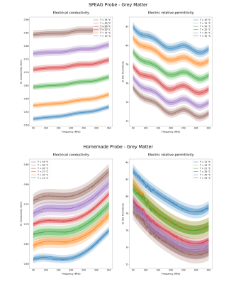 |
3 | Temperature Dependence of the Electric Properties in MR Phantoms
Umberto Zanovello1, Wolfgang Kilian2, Adriano Troia1, Bernd Ittermann2, Alessandro Arduino1, and Luca Zilberti1
1Istituto Nazionale di Ricerca Metrologica (I.N.Ri.M.), Torino, Italy, 2Physikalisch-Technische Bundesanstalt (PTB), Braunschweig and Berlin, Germany
Phantom realization represents an important activity for the development and validation of advanced MR-based quantitative imaging techniques such as Magnetic Resonance Fingerprinting or Electrical Properties Tomography. In this regard, phantoms represent a reliable ground truth with the requirement that the sought properties are properly characterised in the variety of conditions at which the phantoms will be finally deployed. In this abstract, measurements of the electrical properties of two different gel-based tissue-mimicking materials are presented as a function of their temperature. A linear dependence with positive (negative) slope between temperature and electrical conductivity (relative permittivity) is observed.
|
||
3862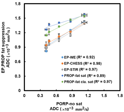 |
4 | Quantitatively Evaluating the Effect of Fat on ADC Measurement in EP-DWI and PROPELLER-DWI Using Fat-Water Phantom
Jo-Hua Peng1, Chun-Jung Juan1,2,3, Yi-Jui Liu4, Ruey-Hwang Chou5, Hing-Chiu Chang6, Chang-Hsien Liou2,7, Szu-Hsien Chou1,2, Yen-Ting Wu1,2, Bai-Wen Wu2, and Hsu-Hsia Peng1
1Department of Biomedical Engineering and Environmental Sciences, National Tsing Hua University, Hsinchu, Taiwan, 2Department of Medical Imaging, China Medical University Hsinchu Hospital, Hsinchu, Taiwan, 3Department of Radiology, School of Medicine, China Medical University, Taichung, Taiwan, 4Department of Automatic Control Engineering, Feng Chia University, Taichung, Taiwan, 5Institute of Cancer Biology, China Medical University, Taichung, Taiwan, 6Department of Biomedical Engineering, Chinese University of Hong Kong, Sha Tin, Hong Kong, 7Institute of Nuclear Engineering and Science, National Tsing Hua University, Hsinchu, Taiwan
ADC estimations are still known to be influenced by the content of fat. Fat suppression is normally applied in order to avoid the ADC measurement with fat contamination. The aim of this study was to evaluate the fat effect on ADC measurement by EP-DWI and PROP-DWI combined with different fat suppression techniques using a fat-water phantom with different fat fractions. Our results suggest that the ADC measurement is affected by the fat fraction and the MR sequences but not the fat suppression methods in EP-DWI.
|
||
3863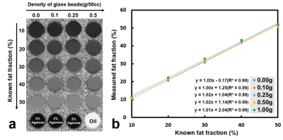 |
5 | An Innovative MRI phantom Allowing Evaluation of the Influence of Fat Fraction on the Measurement of ADC under Different Glass Bead Densities
Jo-Hua Peng1, Chun-Jung Juan1,2,3, Yi-Jui Liu4, Ruey-Hwang Chou5, Hing-Chiu Chang6, Chang-Hsien Liou2,7, Szu-Hsien Chou1,2, Yen-Ting Wu1,2, Bai-Wen Wu2, and Hsu-Hsia Peng1
1Department of Biomedical Engineering and Environmental Sciences, National Tsing Hua University, Hsinchu, Taiwan, 2Department of Medical Imaging, China Medical University Hsinchu Hospital, Hsinchu, Taiwan, 3Department of Radiology, School of Medicine, China Medical University, Taichung, Taiwan, 4Department of Automatic Control Engineering, Feng Chia University, Taichung, Taiwan, 5Institute of Cancer Biology, China Medical University, Taichung, Taiwan, 6Department of Biomedical Engineering, Chinese University of Hong Kong, Sha Tin, Hong Kong, 7Institute of Nuclear Engineering and Science, National Tsing Hua University, Hsinchu, Taiwan
Although a single-function phantom for either ADC or fat content is popular, it is crucial to develop a multiple-function phantom to simulate the real situation of human tissue in vivo. The aim of our study was to design a novel phantom containing different fat fractions and cellularity to evaluate the effect of fat content on ADC measurement in vivo as in human tissue environment. In conclusion, the influence of fat contents and glass beads density on ADC values was examined in our dual-function phantom, which can be helpful to improve the accuracy of ADC quantification in clinical practice.
|
||
 |
3864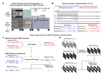 |
6 | Deep Learning Driven EMI Prediction and Elimination for RF Shielding-Free MRI at 0.055T and 1.5T
Yujiao Zhao1,2, Linfang Xiao1,2, Yilong Liu1,2, Alex T. L. Leong1,2, and Ed X. Wu1,2
1Laboratory of Biomedical Imaging and Signal Processing, The University of Hong Kong, Hong Kong, China, 2Department of Electrical and Electronic Engineering, The University of Hong Kong, Hong Kong, China
All clinical MRI scanners require bulky and enclosed RF shielding rooms to prevent external electromagnetic interference (EMI) signals during data acquisition, and quality electronics inside shielding room (i.e., with minimal EMI emission). A deep learning EMI cancellation strategy is presented to model, predict and remove EMI signals from acquired MRI signals, eliminating the need for RF shielding. We demonstrated that this method worked robustly for various EMI sources from both external environments and internal scanner electronics, producing final image SNRs highly comparable to those obtained using a fully enclosed RF shielding cage in 0.055T and 1.5T experiments.
|
|
3865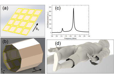 |
7 | Design of a Metamaterial-inspired Flexible Structure for MRI Field Enhancement at 3T: a Numerical Approach
Morteza Teymoori1 and Arda Deniz Yalçınkaya2
1Institute of Biomedical Engineering, Boğaziçi University, Istanbul, Turkey, 2Department of Electric and Electronics Engineering, Boğaziçi University, Istanbul, Turkey MR imaging has a significant role in medical diagnostics. Imaging quality depends on SNR levels. Higher B0 field and consequently higher RF-field intensity increase SNR but it also elevates the total body SAR. This can be alleviated by local magnetic field enhancement. Metamaterial based structures have shown promising applications in this regard. Here we report a new metamaterial inspired resonant structure for local field enhancement in 3T MRI machine. We show field enhancement capabilities of our design in planar and cylindrical forms, each of which can be used based on the anatomical site under investigation. |
||
3866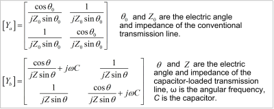 |
8 | Miniaturized and Reproducible Phase Shifters using Capacitor-Loaded Transmission Lines for Add-On RF Shimming at High Field
Ming Lu1, Yue Zhu2,3, John Gore2,3,4, William A. Grissom2,3,4, and Xinqiang Yan2,3
1College of nuclear equipment and nuclear engineering, Yantai University, Yantai, China, 2Vanderbilt University Institute of Imaging Science, Vanderbilt University Medical Center, Nashville, TN, United States, 3Department of Radiology and Radiological Sciences, Vanderbilt University Medical Center, Nashville, TN, United States, 4Department of Biomedical Engineering, Vanderbilt University, Nashville, TN, United States
Add-on RF shimming devices, including phase shifters, could provide additional degrees of freedom for RF transmit chains to improve field uniformity and reduce SAR. At the Larmor frequencies of MRI, however, the transmission line length for realizing large phase shifts becomes unrealistically long. We introduce a capacitor-loaded transmission line design to build a miniaturized microstrip line phase shifter for RF shimming at high fields. The fabricated phase shift boards had a miniaturized size of 3 x 4 cm2, exhibited a low loss of < 0.26 dB, and cost much less than those made from other materials.
|
||
3867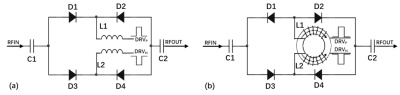 |
9 | A Balanced RF Switch for Ultra-Wideband System with Microsecond Dead Time
JunCheng Xu1, Shouquan Yao1, Ming Shen1, Yiqiao Song2,3, Yu Jiang1, and Jianqi Li1
1Shanghai Key Laboratory of Magnetic Resonance, School of Physics and Electronic Science, East China Normal University, shanghai, China, 2Athinoula A. Martinos Center for Biomedical Imaging, Department of Radiology, Massachusetts General Hospital, Charlestown, MA, United States, 3John A. Paulson School of Engineering and Applied Sciences, Harvard University, Cambridge, MA, United States
This work demonstrated a fully balanced strcture of wideband RF switch. The switch utilizes PIN diodes to form two balanced paths which transmit the RF signals in common mode and the drive signal of PIN diode in differential mode.The common-mode choke can be used to block AC signal and feed DC bias. It can also be used to suppress the transient voltage due to the drive signal. The switch achieved low insertion loss and high isolation within a bandwidth between 10MHz-100MHz. The switch also suppressed the transient voltage to less than 3.3mV with a very short dead time.
|
||
3868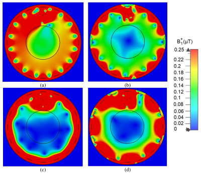 |
10 | Wireless power transfer on non-main eigenmodes within birdcage coil
Oleg I. Burmistrov1, Egor I. Kretov1, Dmitrii S. Dashkevich1, and Pavel S. Seregin1
1School of Physics and Engineering, ITMO university, Saint-Petersburg, Russian Federation
In this work, we investigate a concept of a wireless power transfer system for 1.5T MRI scanner. Our system exploits additional eigenmodes of a birdcage body coil that are not used during the scanning process. Such a solution could be implemented in existing hardware with a minor change in MRI components and at the same time minimize the interference between the power transfer system and imaging functions of the scanner.
|
||
3869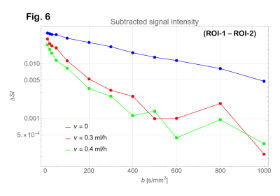 |
11 | Intravoxel incoherent motion imaging of a small-vessel MRI phantom: Preliminary study.
Hajime Tamura1, Hideki Ota2, Tatsuo Nagasaka3, Ryuichi Mori3, Yuki Ichinoseki3, and Kenichi Funamoto4
1Department of Medical Physics, Tohoku University, Graduate school of medicine, Sendai, Japan, 2Department of Advanced MRI Collaborative Research, Tohoku University, Graduate school of medicine, Sendai, Japan, 3Department of Radiology, Tohoku University hospital, Sendai, Japan, 4Institute of Fluid Science, Tohoku University, Sendai, Japan
IVIM signal intensities could be compared with the ‘ground truth’ by use of a micro-vessel phantom in this study. In IVIM imaging, images with b = 0 are often included in the analysis, but this study indicated that images with b = 0 should be excluded. In this study, IVIM analysis showed a tendency to overestimate D* and underestimate f.
|
||
3870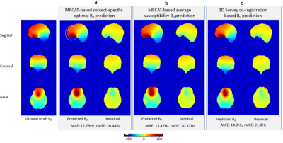 |
12 | Analytical B0 shimming from 3D survey scans: feasibility and impact on EPI image quality
Sharun S Thazhackal1, Ashvin Srinivasan1, Suja Saraswathy1, Manivannan Jayapalan2, Umesh Rudrapatna2, and Jaladhar Neelavalli2
1Philips Research, Bangalore, India, 2Philips Healthcare, Bangalore, India
The inhomogeneity in the main magnetic field (B0) leads to SNR loss and image artifacts. B0 inhomogeneity increases with the field strength, thus leading to more pronounced B0 inhomogeneity at 3T and beyond. However repeated B0 map acquisition for shimming and for image reconstruction is time consuming. We show that subject-specific B0 and the corresponding shim settings can be predicted accurately using pseudo-CT generated from MR images – potentially obviating the need for a B0 scan. We propose an alternative approach based on image co-registration techniques that yields comparable results using survey images, without the need of any additional scans.
|
||
3871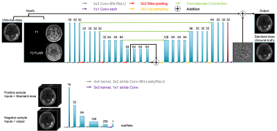 |
13 | Diagnostic Ultra-low-dose 18F-PI-2620 Tau PET/MRI with Generative Adversarial Network-based Enhancement
Kevin T Chen1,2, Robel Tesfay3, Mary Ellen I Koran2, Jiahong Ouyang2, Sara Shams2, Tie Liang2, Mehdi Khalighi2, Elizabeth Mormino2, and Greg Zaharchuk2
1National Taiwan University, Taipei, Taiwan, 2Stanford University, Stanford, CA, United States, 3Meharry Medical College, Nashville, TN, United States
With the focal and lower uptake in tau PET imaging compared to other tracers such as amyloid, we aim to use multimodal simultaneous PET/MR imaging combined with training a generative adversarial network (GAN) to enhance ultra-low-dose 18F-PI-2620 tau PET images. We showed that the deep learning-enhanced images greatly reduced image noise as compared to the ultra-low-dose images, outperformed the ultra-low-dose images metrics-wise, and were able to be read clinically for regional uptake patterns of tau accumulation similarly as the full-dose images.
|
||
3872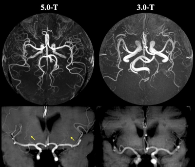 |
14 | High-Resolution 3D-TOF MR Angiography in intracranial arteries and branches: A Initial Experience on Comparison between 3.0-T and 5.0-T Video Not Available
Zhang Shi1, Jiang Lin1, Yunfei Zhang2, Yongming Dai2, and Mengsu Zeng1
1Zhongshan Hospital of Fudan University, Shanghai, China, 2Central Research Institute, United Imaging Healthcare, Shanghai, China
3D-Time-of-Flight MR Angiography (3D TOF-MRA) of the intracranial vessels is a first-line and noninvasive tool for the evaluation of patients with cerebrovascular disease. However, it is still challenged at 3.0-T MR imaging because peripheral segments of the main arteries can often not be sufficiently visualized. The expected remarkable increase in visualizations of intracranial arteries segments and vessel brancher provided by ultra-high field 5.0-T MRI may be valuable for addressing above issues. Our prospective analysis demonstrated that the 5.0-T TOF-MRA provides higher improvement in visualization of distal large arteries and small vessel branches than that at 3.0-T.
|
||
 Back to Meeting Home
 Back to the Program-at-a-Glance
The International Society for Magnetic Resonance in Medicine is accredited by the Accreditation Council for Continuing Medical Education to provide continuing medical education for physicians.
 Back to Meeting Home
 Back to the Program-at-a-Glance
 View the Presentation