Online Gather.town Pitches
New Systems & Devices I
Joint Annual Meeting ISMRM-ESMRMB & ISMRT 31st Annual Meeting • 07-12 May 2022 • London, UK

Online Gather.town Pitches
New Systems & Devices I
| Booth # | ||||
|---|---|---|---|---|
3239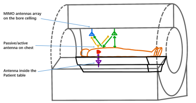 |
1 | High Accuracy Heartbeat Detection with 433MHz CW-Doppler Radar for MRI Application
Yuxin Wu1, John Pauly2, and Greig Scott2
1Department of Electronic Engineering, Tsinghua University, Beijing, China, 2Department of Electrical Engineering, Stanford University, Stanford, CA, United States
This work assesses heartbeat detection by continuous-wave (CW) Doppler radar using low and high carrier frequencies of 433MHz and 5GHz as options for non-contact vitals sensing in MRI. The experimental results show that using the amplitude of received complex signals in low frequency 433MHz-detection can perform as well as 5GHz-detection due to near field interactions within a 20cm range, with similar heartbeat detection efficacy.
|
||
3240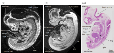 |
2 | MR microscopy of human embryo at high isotropic spatial resolution
Kazuyuki Makihara1, Kazuki Kunieda1, Shigehito Yamada2, Katsumi Kose3, and Yasuhiko Terada1
1Graduate School of Science and Technology, University of Tsukuba, Tsukuba, Japan, 2Congenital Anomaly Research Center, Kyoto University Graduate School of Medicine, Kyoto, Japan, 3MRI simulations Inc., Minato-ku, Japan
High-resolution MR microscopy is advantageous for the human embryo study, but had not been realized because of many challenges. In this study, we demonstrated high-resolution MR microscopy of a human embryo specimen with 18 µm isotropic resolution. To achieve the higher spatial resolution than the previous system, we improved the hardware and optimized the pulse sequence. The MR images of the embryo showed detailed anatomical structures and were similar in appearance to the stained images obtained from an embryo specimen at the same developmental stage.
|
||
3241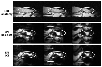 |
3 | Design of a 5-channel local B0 shimming coil for rat brain imaging at 3T
Qiaoyan Chen1,2, Chao Luo1,2, Xiaoliang Zhang3, Xin Liu1,2, Hairong Zheng1,2, and Ye Li1,2
1Paul C. Lauterbur Research Center for Biomedical Imaging, Shenzhen Institutes of Advanced Technology, Chinese Academy of Sciences, Shenzhen, China, 2Key Laboratory for Magnetic Resonance and Multimodality Imaging of Guangdong Province, Shenzhen, China, 3Department of Biomedical Engineering, State University of New York at Buffalo, New York, NY, United States
To improve B0 magnetic field homogeneity and minimize interferences on RF coils, local shimming coils with a few channel number can be applied. In this study, we designed and constructed a 5-channel local B0 shimming coil for the rat brain. There was a marginal SNR loss within 5% after converting the local shimming coil into a 3-channel RF coil. The reduction of B0 inhomogeneity for the rat brain with respect to the basic set was about 34%. A large portion of the distortion in EPI images was recovered and some image artifacts were eliminated after using the local shimming coil.
|
||
3242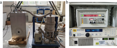 |
4 | Development of a Benchtop 7T cryogen-free animal MRI system
Yaohui Wang1, Qiuliang Wang1, Jigang Zhao1, Yang Liu1, Yijie Zheng1, Zhifeng Chen2, and Feng Liu3
1Institute of Electrical Engineering, Chinese Academy of Sciences, Beijing, China, 2USC Stevens Neuroimaging and Informatics Institute, Keck School of Medicine, University of Southern California, Los Angeles, CA, United States, 3School of Information Technology and Electrical Engineering, The University of Queensland, Brisbane, Australia
A small-size, light-weight, cryogen-free 7T animal MRI system is being developed in IEE CAS. The magnet system has been completed and acceptable magnetic field strength and homogeneity indices have been achieved. The gradient assembly with passive shimming is going into the final assembly stage and the RF coil is ready for tunning. The integration of the whole system will be very soon and a mouse experiment will be conducted then. It is expected the ultimate product will largely boost the scientific research activities that are unapproachable to advanced MRI scanners.
|
||
3243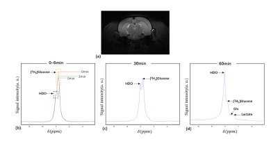 |
5 | Deuterium metabolism imaging of rat brain at 9.4T using a double-nuclear transceiver
Feng Du1,2, Jiawen Yuan1,3, Nan Li1,2, Chao Zhou1,2, Qian Wan1,2, Qikai Qin4, Garth J. Thompson4, Qiong Ye5, Xiaoliang Zhang6, Xin Liu1,2, Hairong Zheng1,2, and Ye Li1,2
1Shenzhen Institutes of Advanced Technology, Chinese Academy of Sciences, Shenzhen, China, 2Key Laboratory for Magnetic Resonance and Multimodality Imaging of Guangdong Province, Shenzhen, China, 3Southern University of Science and Technology, Shenzhen, China, 4iHuman Institute, ShanghaiTech University, Shanghai, China, 5High Magnetic Field Laboratory, Hefei Institutes of Physical Science, Chinese Academy of Sciences, Hefei, China, 6Department of Biomedical Engineering, State University of New York, Buffalo, NY, United States
Deuterium metabolic imaging (DMI) is an emerging technique, which can measure the metabolism in the brain non-invasively after intake of deuterated glucose. In this work, we construct a versatile and dedicated 1H/2H dual-nuclear transceiver for a 9.4T pre-clinical research system. The performance of the transceiver was evaluated by measuring the in vivo 2H spectra data. The metabolites of water, glucose, Glx and lactate after glucose infusion were quantified in healthy and glioma rats using the constructed double-nuclear transceiver.
|
||
3244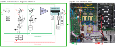 |
6 | A 2kW non-magnetic RF power amplifier with negative feedback for 5T MRI proton imaging
Jun Luo1,2,3, Shengping Liu1, Jifeng Chen2, and Ye Li2,3
1Chongqing University of Technology, Chongqing, China, 2Lauterbur Imaging Research Center, Shenzhen Institutes of Advanced Technology, Chinese Academy of Sciences, Shenzhen, China, 3Key Laboratory for Magnetic Resonance and Multimodality Imaging of Guangdong Province, Shenzhen, China
Parallel transmission technology has attracted much attention because of its advantages of uniform excitation and artifact correction in ultra-high field. A 2kW non-magnetic prototype RF amplifier for 5T multi-channel parallel transmission (pTx) is presented. It is equipped with analogic feedback loops and provides a high linearity up to peak output power. It maintains excellent fidelity performance even without a circulator.
|
||
3245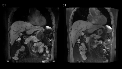 |
7 | Abdominal imaging at 5.0 Tesla: Preliminary results
Liyun Zheng1,2, Chun Yang3, Ruofan Sheng3, Yongming Dai2, and Mengsu Zeng1,3
1Shanghai Institute of Medical Imaging, Shanghai, China, 2United Imaging Healthcare, Shanghai, China, 3Department of Radiology, Zhongshan Hospital, Fudan University, Shanghai, China Higher magnetic field strength leads to an increase of the SNR. On the other hand, the increase of magnetic field strength implies larger magnetic susceptibility inhomogeneities. Recently, 5.0T whole body magnetic resonance imaging system has been introduced into research and clinical settings. In this study, we evaluated the feasibility of abdominal imaging at 5T and compared the image quality and potential artifacts with 3T. Our results indicated that 5T MRI is able to acquire artifact-free anatomical images of the abdomen. Compared to 3T MRI, abdominal MRI at 5T resulted in comparable image quality and better contrast ratios. |
||
3246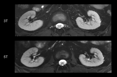 |
8 | Renal MRI at 5T: A feasibility and quantitative study
Liyun Zheng1,2, Chun Yang3, Ruofan Sheng3, Yongming Dai2, and Mengsu Zeng1,3
1Shanghai Institute of Medical Imaging, Shanghai, China, 2United Imaging Healthcare, Shanghai, China, 3Department of Radiology, Zhongshan Hospital, Fudan University, Shanghai, China Compared to clinical field strengths, MRI at ultra-high magnetic fields allows higher SNR. However, imaging at 7T remains challenging, especially in renal MRI. This study investigated the feasibility of renal MRI at 5T, with a brand new 5T MR scanner. Compared to 3T examination, 5T renal MRI demonstrated higher SNR and improved corticomedullar discrimination with diagnostic image quality. Functional imaging, including DWI and T2* mapping, was also feasible at 5T. Therefore, in vivo 5T renal MRI may better elucidate the renal diseases with both anatomical and functional imaging, compared to conventional clinical MRI scanners. |
||
3247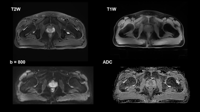 |
9 | Multiparametric MRI method development for clinical prostate imaging using 5T MR imaging
Liyun Zheng1,2, Chun Yang3, Ruofan Sheng3, Yongming Dai2, and Mengsu Zeng1,3
1Shanghai Institute of Medical Imaging, Shanghai, China, 2United Imaging Healthcare, Shanghai, China, 3Department of Radiology, Zhongshan Hospital, Fudan University, Shanghai, China Moving to higher magnetic field strength (> 3 T) may have clinical advantages because of an intrinsic increase of the signal-to-noise ratio (SNR), while heterogeneity in the transmit radiofrequency (RF) field or B1 can cause signal voids throughout images. This study aimed to evaluate the development of prostate MRI at 5T by providing assessment of image quality on clinical sequences, including T1W, T2W and DWI images. According to current results about image quality, present of artifacts, visibility of anatomical structures of T2W images, and geometric distortion of DWI images, clinical prostate imaging is feasible at 5T MRI. |
||
3248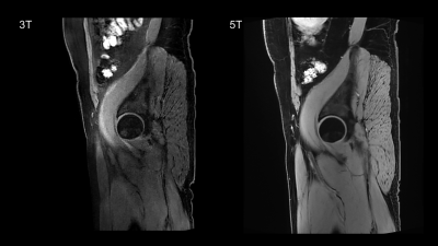 |
10 | Morphological 5T MRI of hip in comparison to 3T: An initial study
Liyun Zheng1,2, Chun Yang3, Ruofan Sheng3, Yongming Dai2, and Mengsu Zeng1,3
1Shanghai Institute of Medical Imaging, Shanghai, China, 2United Imaging Healthcare, Shanghai, China, 3Department of Radiology, Zhongshan Hospital, Fudan University, Shanghai, China Imaging the hip cartilage using magnetic resonance imaging (MRI) is more challenging compared with other joints. The utilize of higher magnetic field strengths (>3T) is able to improve the assessment of cartilage structures in the hip. In this study we evaluated morphological 5T hip MRI in healthy subjects and compared image quality at 5T with 3T MRI. The results revealed that morphological 5T hip MRI in healthy subjects was superior to 3T MRI in image quality, visibility of the anatomical structures and contrast ratios. |
||
3249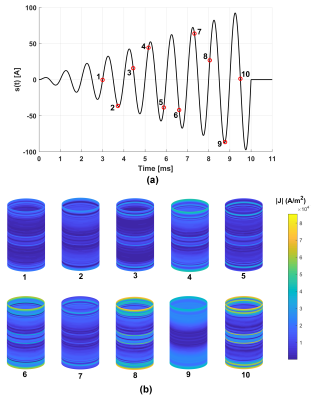 |
11 | Transient Computations of Eddy-Currents Induced by an Amplitude-Modulated Sinusoidal Gradient Pulse
Sadeq S Alsharafi1, Haile Baye Kassahun1, Ahmed M Badawi1, and AbdEl-Monem M El-Sharkawy1
1Systems and Biomedical Engineering, Cairo University, Giza, Egypt
Eddy-currents are generated in MRI scanners’ metallic structures due to the rapid switching of gradient coils. They may result in image distortions and induce acoustic-noise particularly for fast/spiral sequences. Transient eddy-currents computations can be used to understand the extent of such effects. In this work, a numerical framework was devised to compute transient eddy-currents induced by an amplitude-modulated sinusoidal pulse for a longitudinal gradient coil configuration. Stream functions representation of eddy-currents, Multilayer Integral Method, and an excitation current with amplitude-modulated sinusoidal function are used to solve the circuit equation and efficiently compute transient eddy-currents generated in the cryostat.
|
||
3250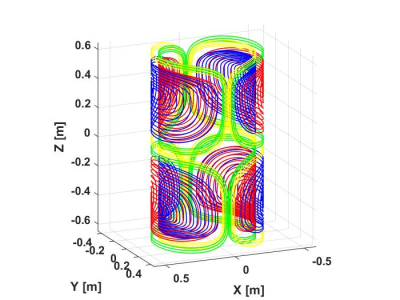 |
12 | Actively Shielded Two-channel Transverse MRI Gradient Coil Numerical Design Using Discrete Wire Method
Haile Baye Kassahun1, Sadeq S Alsharafi1, Ahmed M Badawi1, and AbdEl-Monem M El-Sharkawy1
1Systems and Biomedical Engineering, Cairo University, Cairo, Egypt
A two-channel, self-shielded, cylindrical, transverse gradient coil is numerically designed. The four quadrants of both the inner and outer cylinders were each divided into two sections. All symmetric inner sections of the primary and corresponding shielding coil turns are assigned to the first channel where the outer enclosing sections belong to the second channel. Quasi-elliptic curves are used for coil tracks design. Achieving a comparable target gradient field, the DC dissipated power of a two-channel coil is lower compared to a conventional coil designed with similar dimensions and number of turns by at least 20%, thereby reducing ohmic losses.
|
||
3251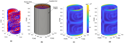 |
13 | Eddy-Currents Computations Using A Single-Layer Stream-Function Method
Sadeq s Alsharafi1, Haile Baye Kassahun1, Ahmed M Badawi1, and AbdEl-Monem M El-Sharkawy1
1Systems and Biomedical Engineering, Cairo University, Giza, Egypt
Eddy-currents induced by MRI gradient coils due to rapid switching result in undesirable thermal effects, field distortions and acoustic-noise. Efficient numerical computations are needed to analyze eddy-currents for complex configurations. The skin-depth adds an extra computational burden for numerical eddy-current computations since meshing in the skin-depth direction may be needed to achieve reliable computations during harmonic analysis. Here, we use a single-layer stream-function method (SSM) applying resistance compensation and compare our results to a multi-layer integral method (MIM) approach. We show that SSM method achieves accurate comparable results relative to both MIM and Ansys albeit at higher computational effeciency.
|
||
 Back to Meeting Home
 Back to the Program-at-a-Glance
The International Society for Magnetic Resonance in Medicine is accredited by the Accreditation Council for Continuing Medical Education to provide continuing medical education for physicians.
 Back to Meeting Home
 Back to the Program-at-a-Glance
 View the Presentation