Online Gather.town Pitches
Safety, Low Field & Interventional II
Joint Annual Meeting ISMRM-ESMRMB & ISMRT 31st Annual Meeting • 07-12 May 2022 • London, UK

Online Gather.town Pitches
Safety, Low Field & Interventional II
| Booth # | ||||
|---|---|---|---|---|
4367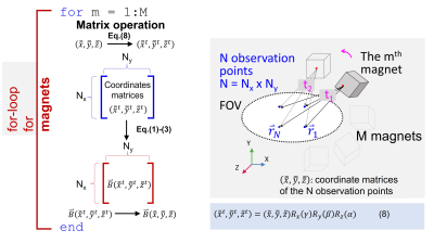 |
1 | A Simulator, “MagTetris”, for Fast Calculation of Magnetic Field and Force for Permanent Magnets
Tingou Liang1, Tie Qiu1, and Shaoying Huang1
1Singapore University of Technology and Design, Singapore, Singapore
We propose a magnet simulator (named “MagTetris”) for fast calculation of both magnetic field and magnetic force generated by multiple cuboid magnets in arbitrary configurations. The accuracy of “MagTetris” is examined by comparing the calculated results to the FEM-based simulations and the experimental results through a 2-magnet experiment. The average differences between the FEM-based simulations and the “MagTetris” calculations are within 6% for field and 2% for force, while those between the measurements and the “MagTetris” are within 10% for field and 4.5% for force. For the calculation speed, “MagTetris” is 123 times faster compared to the FEM-based commercial software.
|
||
4368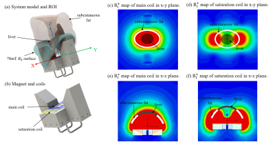 |
2 | RF Coil design for improving human liver fat quantification in a portable single-side MR system
Shiwei Yang1, Xiao Chen2, Shuen Chen1, Hai Luo2, Yue Zhao2, Ziyue Wu2, and Zhiyong Zhang1
1School of Biomedical Engineering, Shanghai Jiao Tong University, Shanghai, China, Shanghai, China, 2Wuxi Marvel Stone Healthcare Co. Ltd., Wuxi, Jiangsu, China, Wuxi, China
Earlier diagnosis of non-alcoholic fatty liver disease (NAFLD) becomes important to prevent the disease progression. Recently, a low-cost portable MR system was developed as a point-of-care screening tool for in vivo liver fat quantification. However, the subcutaneous fat may confound the live fat quantification, particularly in the NAFLD susceptible population. In this work, we propose a novel RF coil design composed of a main target coil sandwiching a set of “saturation” coil to improve human liver fat quantification. We demonstrate the capability and effectiveness of the novel RF design in phantom experiments as well as in-vivo liver scans.
|
||
4369 |
3 | Simulation of RF heating for patients with metallic gamma knife stereotaxic headframe in MRI
Weiman Jiang1, Fan Yang1, and Kun Wang1
1GE Healthcare, Beijing, China
In this study, electromagnetic simulation was used to calculate RF heating for the patients with metallic gamma knife stereotaxic headframe in MRI. The relationship between headframe structure parameters and patient SAR was investigated. Two effective SAR derating methods of adjusting the headframe structure parameters were proposed, which can significantly decrease 10 g local SAR from 253 W/kg to 68 W/kg and head SAR from 4.2 W/kg to 2.0 W/kg. This study provides the efficient and reliable methods to ensure safety of the patients with metallic gamma knife headframe in MRI.
|
||
4370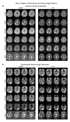 |
4 | A Feasibility Study of 0.055T MRI for Neuroimaging and Comparison with Clinical 3T MRI
Alex T. L. Leong1,2, Yilong Liu1,2, Yujiao Zhao1,2, Linfang Xiao1,2, Henry K. F. Mak3, Tsang Anderson4, Gary K. K. Lau5, Gilberto K. K. Leung4, and Ed X. Wu1,2,6
1Laboratory of Biomedical Imaging and Signal Processing, The University of Hong Kong, Hong Kong SAR, China, 2Department of Electrical and Electronic Engineering, The University of Hong Kong, Hong Kong SAR, China, 3Department of Diagnostic Radiology, Li Ka Shing Faculty of Medicine, The University of Hong Kong, Hong Kong SAR, China, 4Department of Surgery, Li Ka Shing Faculty of Medicine, The University of Hong Kong, Hong Kong SAR, China, 5Department of Medicine, Li Ka Shing Faculty of Medicine, The University of Hong Kong, Hong Kong SAR, China, 6School of Biomedical Sciences, Li Ka Shing Faculty of Medicine, The University of Hong Kong, Hong Kong SAR, China
Magnetic resonance imaging is a key diagnostic tool in modern healthcare, yet its accessibility is low with the vast majority of clinical MRI scanners being placed in highly specialized radiology departments, large centralized imaging centers, and housed on ground floors of hospitals and clinics. In this study, we deployed our recently developed 0.055T brain ultra-low-field MRI scanner to demonstrate preliminary feasibility in diagnosing tumor and stroke cases with direct comparisons to 3T clinical MRI results. The development of such ULF MRI technologies will enable patient-centric and site-agnostic MRI scanners to fulfill the unmet clinical needs across various global healthcare sites.
|
||
4371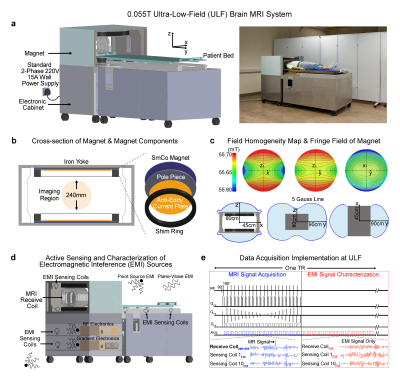 |
5 | A Shielding-free Ultra-low-field 0.055T Brain MRI Scanner for Accessible Healthcare
Ed X. Wu1,2,3, Yilong Liu1,2, Alex T. L. Leong1,2, Yujiao Zhao1,2, Linfang Xiao1,2, Henry K. F. Mak4, Tsang Anderson5, Gary K. K. Lau6, and Gilberto K. K. Leung5
1Laboratory of Biomedical Imaging and Signal Processing, The University of Hong Kong, Hong Kong SAR, China, 2Department of Electrical and Electronic Engineering, The University of Hong Kong, Hong Kong SAR, China, 3School of Biomedical Sciences, Li Ka Shing Faculty of Medicine, The University of Hong Kong, Hong Kong SAR, China, 4Department of Diagnostic Radiology, Li Ka Shing Faculty of Medicine, The University of Hong Kong, Hong Kong SAR, China, 5Department of Surgery, Li Ka Shing Faculty of Medicine, The University of Hong Kong, Hong Kong SAR, China, 6Department of Medicine, Li Ka Shing Faculty of Medicine, The University of Hong Kong, Hong Kong SAR, China
In this study, we describe an ultra-low-field brain MRI scanner that operates using a standard AC power outlet and is low cost to build. Using a permanent 0.055 Tesla Samarium-cobalt magnet and deep learning for cancellation of electromagnetic interference, it requires neither magnetic nor radiofrequency shielding cages. We also successfully implement four standard clinical neuroimaging protocols (T1W, T2W, FLAIR like, and DWI) and demonstrate human brain imaging on this system. This proof-of-concept work will advance the development of a new class of MRI technologies to democratize MRI for low and middle income countries and increase MRI accessibility in healthcare.
|
||
4372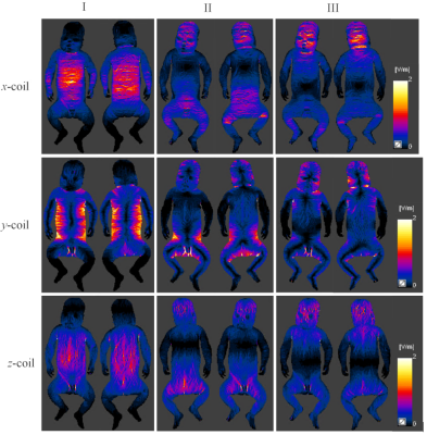 |
6 | Exposure of infants in baby gradient coils Video Permission Withheld
Fangfang Tang1, Luca Giaccone2, Fabio Freschi1,2, Stuart Crozier1, and Feng Liu1
1School of Information Technology and Electrical Engineering, The University of Queensland, Brisbane, Australia, 2Department of Energy, Politecnico di Torino, Torino, Italy
In pediatric magnetic resonance imaging, infants are exposed to rapid, time-varying gradient magnetic fields, leading to electric fields induced in the body of infants and potential safety risks (e.g., peripheral nerve stimulation). In this work, the induced electric fields by the small x, y, z gradient coils in an infant model were numerically evaluated at different model positions. It was found that the induced electric fields in most tissues exceeded the basic restrictions of the ICNIRP 2010 guidelines.
|
||
4373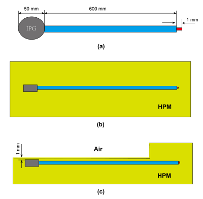 |
7 | Preliminary Study of the Uncertainty for RF-induced Heating Assessment of Subcutaneous and Partially Implanted Devices during MRI Exposure
Aiping Yao1, Minjuan Ma1, Yunfeng Pei1, and Longjun Tang2
1Lanzhou University, Lanzhou, China, 2Shanghai NeuraZing Co., Ltd, Shanghai, China
This work intends to provide a preliminary evaluation of the uncertainty produced by current Tier 3 approach during the assessment of subcutaneous and partially implanted devices. The transfer function of a 600 mm long DBS and guidance wire for Interventional MRI are derived under homogeneous and heterogeneous tissue environments, respectively. The resulting power deposition using different transfer function are evaluated and compared. The results show that using current Tier 3 approach may lead to potential large underestimation of the power deposition for subcutaneous and partially implanted devices, methods which can overcome or reduce these uncertainty are needed.
|
||
4374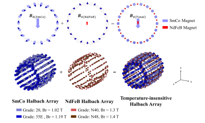 |
8 | A Temperature-Insensitive Halbach Magnet Array Design for Ultra-Low-Field MRI
Ziming Huang1,2, Alex T. L. Leong1,2, and Ed X. Wu1,2
1Laboratory of Biomedical Imaging and Signal Processing, The University of Hong Kong, Hong Kong, China, 2Department of Electrical and Electronic Engineering, The University of Hong Kong, Hong Kong, China
Halbach permanent magnet array offers lightweight and mobility but its field is sensitive to temperature, thus hindering its robust point-of-care MRI applications. This work presents a temperature-insensitive Halbach magnet array design for ultra-low-field MRI. The proposed array is of adequate bore diameter and reasonable weight (~50kg), and provides a 30 mT B0 within a 20 cm imaging field of view (FOV). Numerical simulation is conducted to test the proposed design, demonstrating a drastic reduction in temperature-induced B0 drifting and inhomogeneity.
|
||
 Back to Meeting Home
 Back to the Program-at-a-Glance
The International Society for Magnetic Resonance in Medicine is accredited by the Accreditation Council for Continuing Medical Education to provide continuing medical education for physicians.
 Back to Meeting Home
 Back to the Program-at-a-Glance
 View the Presentation