Online Gather.town Pitches
Safety, Low Field & Interventional I
Joint Annual Meeting ISMRM-ESMRMB & ISMRT 31st Annual Meeting • 07-12 May 2022 • London, UK

Online Gather.town Pitches
Safety, Low Field & Interventional I
| Booth # | ||||
|---|---|---|---|---|
 |
3575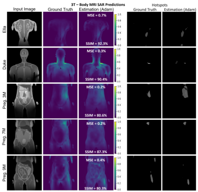 |
1 | MRSaiFE: towards the real-time prediction of SAR in 3T and 7T MR RF coils - a feasibility study with 10 body models
Sayim Gokyar1, Isabelle Saniour1, Fraser Robb2, Arthur Nghiem3, Wolfgang Kainz4, Akshay S. Chaudhari5, and Simone Winkler1
1Radiology, Weill Cornell Medicine, New York, NY, United States, 2General Electric Healthcare, Aurora, OH, United States, 3Biomedical Engineering, University of Minnesota, Minneapolis, MN, United States, 4Center for Devices and Radiological Health, U.S. Food and Drug Administration, Silver Spring, MD, United States, 5Radiology, Stanford University, Stanford, CA, United States
Significant RF power deposition in the body causing local specific absorption rate (SAR) in the form of hotspots is an important safety concern at 3T (128 MHz) and, even more so, at 7T (298 MHz). In this work, we expand the proof-of-concept of artificial intelligence based real-time MRI safety prediction software (MRSaiFE) to 10 body models. We show that SAR patterns can be predicted with a mean squared error (MSE) of less than 1% and a structural similarity index of above 90% for 7T brain and above 85% for 3T body MRI.
|
|
3576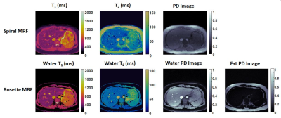 |
2 | Assessment of MRF for Simultaneous T1 and T2 Quantification and Water-Fat Separation in the Liver at 0.55T
Yuchi Liu1, Jesse Hamilton1,2, Yun Jiang1,2, and Nicole Seiberlich1,2
1Department of Radiology, University of Michigan, Ann Arbor, MI, United States, 2Department of Biomedical Engineering, University of Michigan, Ann Arbor, MI, United States This study aims to assess the feasibility of performing MRF in the liver and examine the feasibility of water-fat separation using rosette MRF at low field. MRF sequences using spiral and rosette trajectories were implemented on a 0.55T scanner. T1 and T2 quantification in the liver was achieved in a single breath-hold of <15s with in-plane resolution of 1.56×1.56mm2 and slice thickness of 5mm. In addition, water-fat separation was achieved along with T1 and T2 quantification with no time penalty using rosette MRF. |
||
3577 |
3 | RF coil modeling with mechanical components: initial investigation
Xin Chen1 and Michael Steckner1
1Canon Medical Research USA Inc, Mayfield Village, OH, United States
Electromagnetic modeling has been widely used to study MRI RF coils, but the coil model usually only consists of conductors and lumped elements. The impact of mechanical components (such as coil housing, patient table) has not yet been thoroughly investigated. This work showed that including mechanical parts in the model lead to noticeable changes of total B1 and E fields (up to 15% and 32% increase respectively in the central axial slice), peak SAR (22%), and coil input power (67%) calculations.
|
||
3578 |
4 | Study of B1 and SAR Field Distribution for loop coil with different bending angles at 3T Video Permission Withheld
Tripta Sharma1 and Xiaoliang Zhang1
1Biomedical Engineering, University at Buffalo, Buffalo, NY, United States
The aim is to investigate the RF field behavior of flexing loop coils as resonant elements in flexible RF arrays. Numerical modeling and simulation of a mechanically flexible single channel RF coil at different bending angles for 3T MRI are implemented to determine the best bending angle that will generate highest B1 magnetic field inside imaging area with least flux cancellation at center. Results include B1 magnetic and SAR field distribution of the coronal, sagittal and transverse planes of phantom at 9 bent angles. It also includes the comparative quantitative analysis of magnetic field at different biological samples.
|
||
3579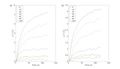 |
5 | Extension of Simple Safety Framework to Realistic Configurations
Jagjit Sidhu1, Pallab Bhattacharyya1, Ken Sakaie1, and Mark Lowe1
1Cleveland Clinic, Cleveland, OH, United States
Implanted electrodes in patients used to treat movement disorders can induce unsafe heating in scanners above 1.5T. However, this patient population will likely benefit strongly from having the option of high field and ultra-high field scanners. To remedy this situation, we demonstrate a proof-of-concept that by carefully selecting the weights of the B1 shim on a Siemens 3T Prisma scanner, negligible temperature rise can be obtained using a T2-TSE sequence. We also show that when no temperature rise is obtained, the image quality is better and more homogeneous.
|
||
3580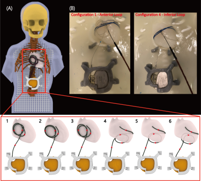 |
6 | A small loop makes a big difference: Modifying trajectory of epicardial leads substantially reduces RF heating of CIEDs in children during 1.5T MRI
Fuchang Jiang1, Bhumi Bhusal2, Bach Nguyen2, Michael Monge3, Gregory Webster4, Daniel Kim2, Giorgio Bonmassar5, Andrada R. Popsecu6, and Laleh Golestanirad1,2
1Department of Biomedical Engineering, McCormick School of Engineering, Northwestern University, Evanston, IL, United States, 2Department of Radiology, Feinberg School of Medicine, Northwestern University, Chicago, IL, United States, 3Division of Cardiovascular-Thoracic Surgery, Ann & Robert H. Lurie Children's Hospital of Chicago, Chicago, IL, United States, 4Division of Cardiology, Ann & Robert H. Lurie Children's Hospital of Chicago, Chicago, IL, United States, 5A. A. Martinos Center for Biomedical Imaging, Massachusetts General Hospital, Boston, MA, United States, 6Division of Medical Imaging, Ann & Robert H. Lurie Children’s Hospital of Chicago, Chicago, IL, United States
Infants with congenital heart defects, inherited arrhythmia syndromes, and congenital disorders of cardiac conduction often require cardiac implantable electronic devices (CIEDs). Some infants receive a CIED within hours, or even minutes, of birth. The presence of an epicardial CIED is a relative contraindication for cardiac magnetic resonance imaging (MRI) due to the risk of RF heating. We present results of phantom experiments and electromagnetic simulations showing that a simple surgical modification in the trajectory of epicardial leads can reduce RF heating more than 40-fold during MRI at 1.5 T.
|
||
3581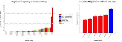 |
7 | Magnetic Susceptibility of Common Metals and Alloys Used in Medical Devices
Grant M. Baker1, Eric D. Anttila1, Erick Smith1, Andrew Robison1, and David C. Gross1
1MED Institute Inc., West Lafayette, IN, United States
The goal of this study was to characterize the magnetic susceptibility and accompanying magnetically induced force, torque, and image artifact of metals and alloys that are commonly used in medical devices. These values were reported for 46 metals and alloys, with an overall trend that magnetically induced force, torque, and image artifact increased with increasing susceptibility of a material. The results of this study can inform medical device design for the development of safer and better performing devices, especially in the growing area of MR guided procedures.
|
||
3582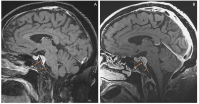 |
8 | Compact 3T brain MRI for patients with abandoned leads of cardiac implantable electronic devices
Lydia J Bardwell Speltz1,2, Yunhong Shu1, Robert E Watson1, Joshua D Trzasko1, Erin Gray1, Maria Halverson1, Joseph Arant1, John Huston III1, Thomas KF Foo3, and Matt A Bernstein1
1Department of Radiology, Mayo Clinic, Rochester, MN, United States, 2Mayo Clinic Graduate School of Biomedical Sciences, Mayo Clinic, Rochester, MN, United States, 3GE Global Research, Niskayuna, NY, United States
Patients with abandoned pacemaker leads require special attention during MR exams due to the risk of lead tip heating from RF energy deposition. The electromagnetic fields on the compact 3T (C3T) scanner fall off rapidly caudal to the head, and therefore present a reduced risk of lead tip heating during brain imaging. We compared the images from a whole-body 1.5T scanner and a high-performance C3T scanner. The C3T images show substantial improvements in image quality and greater cortical detail. This work establishes the feasibility of C3T brain MRI with a 32-channel receive coil for patients with abandoned leads.
|
||
3583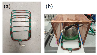 |
9 | Design of an asymmetric spiral RF coil for RF spatial encoding
Yonghyun Ha1, Kartiga Selvaganesan1, Kasey Hancock1, Gigi Galiana1, and R. Todd Constable1
1Department of Radiology and Biomedical Imaging, Yale University, New Haven, CT, United States Gradients in B1 can be useful as a source of spatial encoding for low-field MRI. Here, an asymmetric spiral surface coil is introduced that generates an RF gradient around the coil. Simulations have shown that the shape of the B1 field can be controlled by adjusting the spacing between the wires. The measured B1 field of the asymmetric spiral coil shows that the RF gradient is generated near the coil as shown in the simulation results. |
||
3584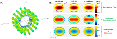 |
10 | Shimming a Halbach Magnet for TRASE MRI on the International Space Station
Aaron R. Purchase1, Christopher Sedlock1, Gordon E. Sarty2, Jonathan C. Sharp1, and Boguslaw Tomanek1
1Oncology, University of Alberta, Edmonton, AB, Canada, 2Biomedical Engineering, University of Saskatchewan, Saskatoon, SK, Canada
Astronauts experience detrimental loss to bone and muscle during long-term space flight. MRI is desired to monitor bone and muscle loss, but current portable systems remain unsuitable for the International Space Station (ISS). We built a 25 kg dipolar Halbach magnet dedicated to the transmit array spatial encoding (TRASE) method over a cylindrical region of interest (ROI), which satisfies the requirements for MRI on the ISS. Still, the natural axial gradient is not well suited to slice selection because it has a quadratic-type profile producing unwanted aliasing. We propose a technique to implement a linear axial B0 gradient for encoding.
|
||
3585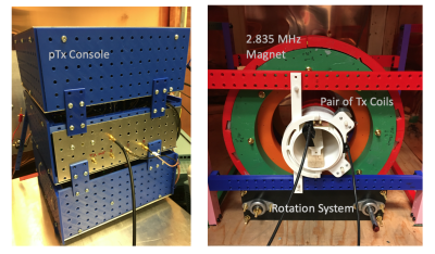 |
11 | A Parallel-Transmit Halbach Magnet TRASE MRI System
Jonathan C. Sharp1, Aaron Purchase1, Christopher Sedlock1, and Boguslaw Tomanek1
1Oncology, University of Alberta, Edmonton, AB, Canada
We have designed and constructed a low-field TRASE RF imaging system. In TRASE k-space encoding is achieved by refocusing with RF phase gradients. The system includes a motorized rotatable magnet, twin RF power amplifiers, two geometric decoupled truncated twisted solenoid RF transmit coils (1D radial TRASE encoding), and a multi-channel transmit SDR console. The magnet is a 2.835 MHz 8-ring Halbach design. All components are custom made. An inherent axial gradient of the inhomogeneous magnet function as slice selection. The unique feature of this imaging configuration is the simplicity. Two RF channels and magnet rotation provide multi-slice projection reconstruction imaging.
|
||
 Back to Meeting Home
 Back to the Program-at-a-Glance
The International Society for Magnetic Resonance in Medicine is accredited by the Accreditation Council for Continuing Medical Education to provide continuing medical education for physicians.
 Back to Meeting Home
 Back to the Program-at-a-Glance
 View the Presentation