Online Gather.town Pitches
Body: Liver/GI/Chest II
Joint Annual Meeting ISMRM-ESMRMB & ISMRT 31st Annual Meeting • 07-12 May 2022 • London, UK

Online Gather.town Pitches
Body: Liver/GI/Chest II
| Booth # | ||||
|---|---|---|---|---|
4183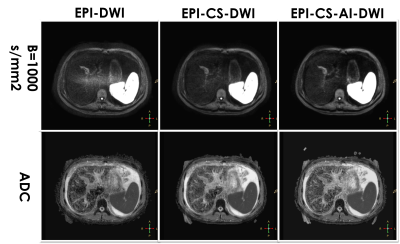 |
1 | Liver DWI using deep learning constrained compressed sensing: A primary study compared to conventional DWI and compressed sensing-based DWI
Ting Duan1, Zhen Zhang2, Hanyu Jiang2, Yidi Chen2, Xiaoyong Zhang3, and Bin Song2
1Radiology, West China Hospital, Sichuan University, Chengdu, China, 2West China Hospital, Sichuan University, Chengdu, China, 3Clinical Science, Philips Healthcare, Chengdu, China, Chengdu, China
Compressed sensing AI-based DWI substantially reduced noise artifacts and improved the signal-to-noise ratio and lesion contrast-to-noise ratio compared with conventional DWI and Compressed sensing based DWI, without any penalty for scan parameters. This technique may prove of value in better diagnostic liver imaging.
|
||
4184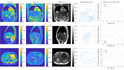 |
2 | Motion-resolved and Free-breathing Liver MRF
Cao Peng1, Zuojun Wang1, Chenyang Liu2, Tian Li2, Edward S. Hui3, and Jing Cai2
11. Department of Diagnostic Radiology, The University of Hong Kong, Hong Kong, Hong Kong, 23. Department of Health Technology & Informatics, The Hong Kong Polytechnic University, Hong Kong, Hong Kong, 3The Hong Kong Polytechnic University, Kowloon, Hong Kong
We developed a motion-resolved and free-breathing liver magnetic resonance fingerprinting (MRF) protocol. The deformation maps were obtained from the first singular image of MRF data. The reconstruction method enforced the consistency of the MRF data with the deformation maps by adding the deformation maps to the encoding matrix. MRF was tested on four healthy volunteers. We demonstrated a motion-resolved MRF with a nominal frame rate of 2.5 Hz for free-breathing liver imaging.
|
||
4185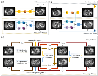 |
3 | Dual-domain self-supervised network for removing motion artifact related to Gadoxetic acid-enhanced MRI Video Not Available
Qingjia Bao1, Feng Pan2, Chongxin Bai3, Kewen Liu3, Zhao Li1, Peng Sun4, Jiazheng Wang4, Linkun Zhong5, Aodong Xiao6, Lian Yang2, and Chaoyang Liu1
1State Key Laboratory of Magnetic Resonance and Atomic and Molecular Physics, Wuhan Institute of Phys, Wuhan, China, 2Department of Radiology, Union Hospital, Tongji Medical College, Huazhong University of Science and Technology, Wuhan, China, 3Wuhan University of Technology School of Information Engineering, Wuhan, China, 4Philips Healthcare, Beijing, China, 5Wuhan University of Arts and Science, Wuhan, China, 6Henan University of Science and Technology, Luoyang, China
We proposed a dual-domain self-supervised motion artifacts disentanglement network (DSMAD-Net) for the liver's gadoxetic acid-enhanced arterial phase images. The motion correction is converted to the image-to-image translation problem by assuming that motion-free images and motion-corrupted images belong to different domains. Specifically, image-to-image translation within the same domain is designed to constrain auto-encoders to learn the feature representation by utilizing the input images as supervision information. Moreover, the cross-domain translation explores the cycle consistency in the absence of paired motion-free and motion-corrupted images. Experimental results demonstrate that our method remarkably removes artifacts in the gadoxetic acid-enhanced arterial phase images.
|
||
4186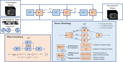 |
4 | Dynamic MRI Reconstruction of the Whole Liver with High Acceleration using Low Rank Tensor and Weighted Group Sparsity
Bei Liu1, Huajun She1, Yufei Zhang1, Zekang Ding1, Zhijun Wang1, and Yiping P. Du1
1School of Biomedical Engineering, Shanghai Jiao Tong University, Shanghai, China
We propose an algorithm for dMRI reconstruction from highly under-sampled k-space data acquired during free breathing. Stack-of-star GRE radial sequence with self-navigator is used to acquire the data. We explore spatial and temporal redundancy for the reconstruction by using weighted group sparsity, weighted sparsity, and low-rank tensor. Additionally, a tensor total variation is used to ensure spatial and temporal smoothness. By applying a weighting function to the sparsity and group sparsity, the subtle structural sparsity features in the sparse domain can be better utilized. The proposed algorithm has the potential to be used in clinical applications such as MR-guided surgery.
|
||
4187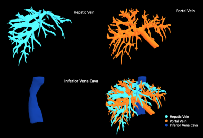 |
5 | Automatic segmentation and reconstruction of liver vascular structure on MRI with a deep learning model
Mengsi Li1, Yanjie Hong2, Zhixuan Yu2, Dandan Zheng2, and Jin Wang1
1Department of Radiology, The Third Affiliated Hospital, Sun Yat-Sen University, Guangzhou, China, 2Shukun (Beijing) Technology Co., Ltd, Beijing, China The accurate segmentation of liver vascular structure is one of the key components of automated radiological diagnosis. Unfortunately, accurate vessel segmentation in clinical practice usually relies on the manual delineation by radiologists on each slice, which is extremely tedious and time-consuming. We developed and evaluated a new liver vessel segmentation and reconstruction workflow, which consist of a 3D Res-Net, a 2D Dense-Net and a 3D U-net model, allows for automated extraction of the portal vein, hepatic vein and inferior vena cava on 3D contrast enhanced portal venous phase MR images. |
||
 |
4188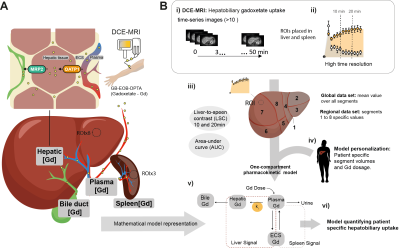 |
6 | In silico liver resection to estimate global and regional hepatobiliary function
Christian Simonsson1,2,3, Wolf Claus Bartholomä2,3, Anna Lindhoff Larsson4, Markus Karlsson2, Bengt Norén2, Gunnar Cedersund1,3, Per Sandström4, Nils Dahlström2,3, and Peter Lundberg2,3
1Department of Medical Engineering, Linköping University, Linköping, Sweden, 2Departments of Radiation Physics, Radiology, Department of Medical and Health Sciences, Linköping University, Linköping, Sweden, 3Center for Medical Image Science and Visualization (CMIV), Linköping University, Linköping, Sweden, 4Department of Surgery, Department of biomedical and clinical sciences, Linköping University, Linköping, Sweden
In some cases, treatment of severe liver diseases requires resective surgery. This brings serious complications if the remnant tissue fails to match the requirement of liver function. Therefore, a pre-operative risk assessment is vital. However, usually the assessment only investigates global liver function. For a more precise assessment, we investigate the possibility of using DCE-MRI in combination with pharmacokinetic modeling to quantify both global- and regional liver function. Also, we show a novel eight-compartment hepatic model, capable of performing an in-silico resection. This framework might in the future be used as a tool for a more precise pre-operative assessment.
|
|
4189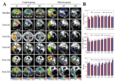 |
7 | Assessment of fibrotic liver regeneration with IVIM imaging: an experimental study in a rat model Video Permission Withheld
Shuangshuang Xie1, Caixin Qiu1, Yajie Sun1, Jinxia Zhu2, Robert Grimm3, and Wen Shen1
1Tianjin First Central Hospital, Tianjin, China, 2MR Collaboration, Siemens Healthcare Ltd., Beijing, China, 3MR Application development, Siemens Healthcare GmbH, Erlangen, Germany
This study investigated the feasibility of IVIM to evaluate fibrotic liver regeneration and function recovery after partial hepatectomy in a rat model. In fibrotic rats, significant increase in D values and decrease in D* and PF values followed with recovery to pre-PH levels were shown. PF values correlated well with Ki-67 indices, D* and PF values correlated well with ALT, AST, and TBil values. IVIM may be an noninvasive and promising approach to monitor liver regeneration and functional recovery after PH.
|
||
4190 |
8 | Comparison and Optimization b-Values combination of liver Diffusion-Weight Imaging in hepatic stiffness discrimination
Jie Yuan1, Wenli Tan1, Songhua Zhan1, Zhigang Gong1, and Yongming Dai2
1Shuguang Hospital Affiliated to Shanghai University of Traditional Chinese Medicine, Shanghai, China, 2United Imaging Healthcare, Shanghai, China
DWI can indirectly reflect the changes of tissue microstructure by detecting the direction and extent of water molecule diffusion. In liver fibrosis, collagen fibers and cell membranes may block the diffusion of water molecules, resulting in changes in the diffusion signal. B-value selection is closely related to the image quality of DWI. Previous studies have found the correlation between tissue water diffusivity and tissue elasticity in the liver. However, there are no widely accepted ADC values on the b-value combination for the stiffness of liver fibrosis. Therefore, the study of b-value combination optimization is of important clinical interest.
|
||
4191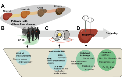 |
9 | NAFLD and Hepatic Inflammation: Multi-modal MR-Investigations of Disease Progression
Christian Simonsson1,2,3, Markus Karlsson2,3, Patrik Nasr3,4, Ralph Sinkus 5,6, Simone Ignatova7, Nils Dahlström2,3, Mattias Ekstedt3,4, Stergios Kechagias3,4, and Peter Lundberg2,3
1Department of Biomedical Engineering (IMT), Linköping University, Linköping, Sweden, 2Department of Radiation Physics, Radiology, Department of Medical and Health Sciences, Linköping University, Linköping, Sweden, 3Center for Medical Image Science and Visualization (CMIV), Linköping University, Linköping, Sweden, 4Department of Gastroenterology and Hepatology, Department of Health, Medicine and Caring Sciences, Linköping University, Linköping, Sweden, 5Imaging Sciences & Biomedical Engineering, Kings College, London, United Kingdom, 6LVTS, INSERM U1148, Paris, France, 7Department of Clinical Pathology and Clinical Genetics, Department of Biomedical and Clinical Sciences, Linköping University, Linköping, Sweden
Several MR-biomarkers characterizing different aspects of NAFLD progression, especially for the early onset, and late stages, are available. However, there is a need to combine and characterize, not only the endpoints but also the intermediate stages of the disease, in particular the early stages of hepatic inflammation. Using an extensive multi-modal MRI protocol, we have investigated correlations between different MR-biomarkers with histopathology measurements of hepatic inflammation, fat accumulation and fibrosis. The long-term aim of this research is to characterize each patient in the early to intermediate stages of disease progression, with a higher degree of precision.
|
||
4192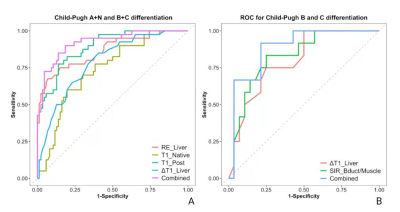 |
10 | A comparative study of multi-parameters from Gd-EOB-DTPA-enhanced MRI in the assessment of liver function in patients with cirrhosis. Video Not Available
Yexin He1, Jia Shao1, Jinxia Guo2, Qiang Gao1, and Cheng Xu1
1SHANXI PROVINCIAL PEOPLE'S HOSPITAL, Taiyuan, China, 2GE Healthcare, Beijing, China
Gd-EOB-DTPA-enhanced MRI was used to quantitatively assess liver function benefiting the follow-up of patients with chronic liver disease and the appropriate plan for liver cancer treatment. A systematic multi-parametric analysis with non-enhanced and Gd-EOB-DTPA-enhanced T1 weighted and T1 mapping MRI were conducted to assess the liver function in patients with cirrhosis. Independent factor for differentiation of A+N(normal and Child-Pugh A) and B+C (Child-Pugh B and C) were found to be RE_Liver, T1_Native, T1_Post and ΔT1_Liver, while for differentiation of Child-Pugh B and C were found to be ΔT1_Liver and SIR_Bdult/Muscle. Both combined model with these factors obtained better diagnostic performance.
|
||
 |
4193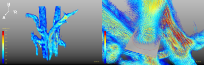 |
11 | Noninvasive Diagnosis of Portal Hypertension and Detecting High Risk Gastroesophageal Varices in Chronic Liver Disease Patients with 4D Flow MRI
Jiachen Ji1, Hanyu Wei1, Yunduo Li1, Mengmeng Zhang2, Changchun Liu2, Bo Jin3, Xiaolong Qi4, Wen Shen5, Jianming Cai2, and Rui Li1
1Biomedical Engineering, Center for Biomedical Imaging Research, Tsinghua Univ., Beijing, China, 2Radiology, Department of Radiology, The Fifth Medical Centre of Chinese PLA General Hospital, Beijing, China, 3First Liver Cirrhosis Diagnosis and Treatment Center, The Fifth Medical Center of Chinese PLA General Hospital, Beijing, China, 4CHESS Center, Institute of Portal Hypertension, The First Hospital of Lanzhou University, Lanzhou, China, 5Department of Radiology, First Center Hospital of Tianjin, Tianjin, China
Chronic liver disease patients commonly suffer from portal hypertension (PH), which could lead to gastroesophageal varices. Noninvasive methods to diagnose PH and to detect high risk varices are urgently needed. 4D Flow MRI is an advanced technique which could provide visualization and quantification of blood flow. In the study, we observed significant hemodynamic differences in portal vein between PH patients and controls. Similar differences were observed between patients with high risk and low risk varices. The performance of diagnosing PH/detecting high risk varices using the single hemodynamic parameter and the logistic regression model was acknowledged by the ROCs.
|
|
4194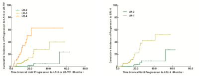 |
12 | Longitudinal evolution of liver imaging reporting and Data system Version 2018 category 2, 3, and 4 observations in cirrhosis on MRI Video Not Available
Fei Xing1 and Xiance Zhao2
1the Third People’s Hospital of Nantong, Nantong, China, 2Philips Healthcare, Shanghai, China
The purpose of this study was to evaluate the category modifications and prognosis of LI-RADS 2,2 and 4 observations in cirrhosis on MRI. We retrospectively analyzed 105 LR-2 , 97 LR-3, and 43 LR-4 observations following-up datas. Follow-up duration and category modifications that progressed, remained stable, or decreased were recorded for each observation. Our study showed that LR-2, LR-3 and LR-4 lesions in liver cirrhosis patients present with different natural disease courses and outcomes, the lesions with a higher LI-RADS category have a higher cumulative incidence of progression to LR-5/-TIV lesion, thus demonstrating an increasing risk of developing HCC.
|
||
4195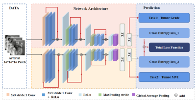 |
13 | Jointly grading and microvascular invasion predicting of hepatocellular carcinoma based on multi-task deep learning
Yanyan Xie1, Lijuan Zhang2, Guangyi Wang3, and Wu Zhou1
1School of Medical Information Engineering, Guangzhou University of Chinese Medicine, Guangzhou, China, 2Shenzhen Institutes of Advanced Technology, Chinese Academy of Sciences, Shenzhen, China, 3Department of Radiology, Guangdong General Hospital, Guangzhou, China
The pathological grade and microvascular invasion (MVI) of hepatocellular carcinoma (HCC) are two key factors related to the patient's prognosis. Previous studies usually predict these two factors separately based on medical images. In this study, we propose an end-to-end multi-task deep learning network to simultaneously predict the MVI and grading information. Specifically, we are the first to demonstrate that these two tasks are related and can promote each other in the framework of multi-task deep learning. Experimental results of HCC in Contrast-enhanced MR demonstrate the effectiveness of the proposed method, outperforming the single task learning.
|
||
4196 |
14 | Image Quality of Deep-Learning Reconstructed Near-isotropic (3D) Enhanced MR enterography with LAVA HyperSense in Crohn disease patients Video Permission Withheld
Jung Hee Son1, Yedaun Lee1, Ho-Joon Lee1, Jisook Yi1, Joonsung Lee2, and Ersin Bayram3
1Radiology, Heaundae Paik Hospital, Busan, Korea, Republic of, 2GE Healthcare, Seoul, Korea, Republic of, 3GE Healthcare, Houston, TX, United States
In this study, we evaluate image quality of high resolution contrast-enhanced T1-weighted MR enterography with deep learning reconstruction. Use of DLRecon for near-isotropic CE-T1WI MRE provides improved image quality, and increased SNR.
|
||
4197 |
15 | Differentiation between rectal mucinous and common adenocarcinoma by amide proton transfer weighted and intravoxel incoherent motion imaging
Juan Li1, cheng jingliang1, and Lin Liangjie 2
1Department of MRI, The First Affiliated Hospital of Zhengzhou University, Zhengzhou, China, 2Philips Healthcare, Beijing, China
Rectal mucinous adenocarcinoma (MC) is a typical subtype of rectal adenocarcinomas, which has a poor prognosis and it is not sensitive to neoadjuvant chemoradiotherapy thus associated with significantly different individualized treatments as those for rectal common adenocarcinoma (AC). The amide proton transfer (APT) weighted and intravoxel incoherent motion (IVIM) imaging were investigated in this study for differential diagnosis between MC and AC. Results indicated that APT signal intensity and the D value by IVIM can be used in discriminating between MC and AC, slightly inferior to the performance of apparent diffusion coefficient by conventional diffusion weighted imaging.
|
||
|
4198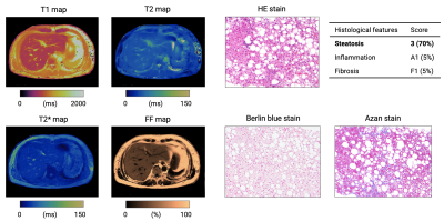 |
16 | MR fingerprinting for liver tissue characterization in a single breath-hold scan: A histopathological correlation study
Shohei Fujita1,2, Katsuhiro Sano1, Gastao Cruz3, Yuki Fukumura4, Hideo Kawasaki1, Issei Fukunaga1, Masami Yoneyama5, Koji Kamagata1, Osamu Abe2, Kenichi Ikejima6, Rene Botnar3, Claudia Prieto3, and Shigeki Aoki1
1Department of Radiology, Juntendo University, Tokyo, Japan, 2Department of Radiology, The University of Tokyo, Tokyo, Japan, 3Department of Biomedical Engineering, School of Biomedical Engineering and Imaging Sciences, King’s College London, London, United Kingdom, 4Department of Human Pathology, Juntendo University, Tokyo, Japan, 5MR Clinical Science, Philips Japan, Tokyo, Japan, 6Department of Gastroenterology, Juntendo University, Tokyo, Japan
Magnetic resonance fingerprinting (MRF) has been proposed for simultaneous T1, T2, T2*, and fat-fraction mapping of the liver, which is suitable for the diagnosis and monitoring of liver diseases. We evaluated the repeatability of liver MRF measurements and correlated it with reference standard imaging in patients with diffuse liver disease. Furthermore, we validated MRF-derived measurements against histological grading of liver biopsies. Liver MRF provided repeatable multi-parametric maps with high agreement with measurements of separate conventional quantitative mapping as well as histological grading of liver biopsies. This approach may enable objective and non-invasive hepatic tissue characterization in a single breath-hold scan.
|
|
 Back to Meeting Home
 Back to the Program-at-a-Glance
The International Society for Magnetic Resonance in Medicine is accredited by the Accreditation Council for Continuing Medical Education to provide continuing medical education for physicians.
 Back to Meeting Home
 Back to the Program-at-a-Glance
 View the Presentation