Online Gather.town Pitches
Body: Liver/GI/Chest I
Joint Annual Meeting ISMRM-ESMRMB & ISMRT 31st Annual Meeting • 07-12 May 2022 • London, UK

Online Gather.town Pitches
Body: Liver/GI/Chest I
| Booth # | ||||
|---|---|---|---|---|
3364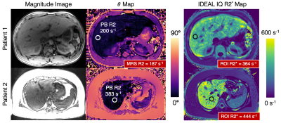 |
1 | Whole Liver Phase-Based R2 Mapping in Liver Iron Overload within a Breath-hold
Ruvini Navaratna1,2, Daiki Tamada2, Diego Hernando1,2, and Scott B Reeder1,2,3,4,5
1Medical Physics, University of Wisconsin-Madison, Madison, WI, United States, 2Radiology, University of Wisconsin-Madison, Madison, WI, United States, 3Emergency Medicine, University of Wisconsin-Madison, Madison, WI, United States, 4Medicine, University of Wisconsin-Madison, Madison, WI, United States, 5Biomedical Engineering, University of Wisconsin-Madison, Madison, WI, United States
There is an unmet need for non-invasive methods that provide rapid and accurate quantification of liver iron concentration (LIC) over a wide range of iron overload severity. Current R2 mapping techniques suffer from long acquisition times and limited spatial coverage. Recently, a phase-based R2 mapping technique for rapid whole-liver R2 quantification within a single breath-hold has been introduced. However, the feasibility of this method to quantify liver iron overload has not been demonstrated. In this work, we optimize and validate phase-based R2 mapping to quantify R2 in phantom experiments, healthy volunteers, and demonstrate feasibility in patients with iron overload.
|
||
3365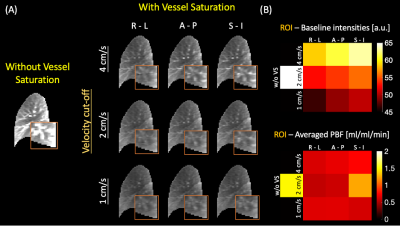 |
2 | Lung perfusion at 0.55T using ASL: Feasibility and Initial Results
Ziwei Zhao1, Nam G. Lee2, Sophia X. Cui3, and Krishna S. Nayak1,2
1Electrical and Computer Engineering, University of Southern California, Los Angeles, CA, United States, 2Biomedical Engineering, University of Southern California, Los Angeles, CA, United States, 3Siemens Medical Solutions USA, Inc., Los Angeles, CA, United States
Lung perfusion is challenging because of the complex anatomy and pulsatile blood flow. At 1.5T and 3T, a major challenge is the low signal from parenchyma due to the short T2*. Here, we demonstrate feasibility of lung ASL perfusion imaging on a high-performance 0.55T system. Experiments were performed using FAIR labeling with snapshot bSSFP imaging and quantified using Buxton’s GKM. In four healthy volunteers, pulmonary blood flow (PBF) ranges from 0.62 to 0.99 ml-blood/ml-tissue/min, which are within the expected range (0.51 to 1.52 ml/ml/min) and agree with the total average PBF estimated by a phase contrast method.
|
||
3366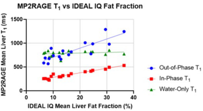 |
3 | Fat-Compensated T1 Mapping of Non-Alcoholic Fatty Liver Disease with MPRAGE and MP2RAGE
Andrew Duffy1, Andreu Costa2, Sharon Clarke2,3, Ashley Stueck4, Magnus McLeod5, and James Rioux2,3
1Faculty of Medicine, Dalhousie University, Halifax, NS, Canada, 2Diagnostic Radiology, Dalhousie University, Halifax, NS, Canada, 3Biomedical Translational Imaging Centre, Nova Scotia Health Authority, Halifax, NS, Canada, 4Pathology, Dalhousie University, Halifax, NS, Canada, 5General Internal Medicine, Dalhousie University, Halifax, NS, Canada Fat is a confounding factor in the use of T1 measurements as a non-invasive method of assessing non-alcoholic fatty liver disease. A Dixon-based water/fat separation was used to produce T1 maps based on the separated water signal in MPRAGE and MP2RAGE sequences. A correlation inconsistent with biophysical processes was observed between uncorrected MP2RAGE T1 values and fibrosis stage, but using the fat compensated T1 values corrects the correlation. The same effect was seen to a lesser extent with MPRAGE T1 values. Further investigation and validation with a larger cohort may provide a more reliable biomarker of liver fibrosis within NAFLD. |
||
3367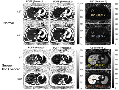 |
4 | Reproducibility of Chemical Shift-Encoded MRI Fat Quantification in Liver Across Field Strengths in Patients with Iron Overload
Zihan Wang1,2, Scott B Reeder, MD, PhD1,2,3,4,5, and Diego Hernando, PhD2,3
1Biomedical Engineering, University of Wisconsin-Madison, Madison, WI, United States, 2Radiology, University of Wisconsin-Madison, Madison, WI, United States, 3Medical Physics, University of Wisconsin-Madison, Madison, WI, United States, 4Medicine, University of Wisconsin-Madison, Madison, WI, United States, 5Emergency Medicine, University of Wisconsin-Madison, Madison, WI, United States
The purpose of this study is to assess the reproducibility of CSE-MRI methods to measure proton density fat fraction (PDFF) in liver in patients with iron overload. The PDFF measurements of 31 patients with known or suspected iron overload and 10 healthy volunteers were imaged at both 1.5T and 3.0T. Patients were separated into four groups with increasing liver iron concentration (LIC). The reproducibility across field strengths was quantified within each group and reported below.
|
||
3368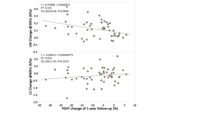 |
5 | Longitudinal assessment of nonalcoholic steatohepatitis with MRI and MRE – changes of imaging biomarkers and their relationships
Zheng Zhu1, Jiahui Li1, Usman Yaqoob2, Alina M Allen2, Terry Therneau3, Kevin J Glaser1, Safa Hoodeshenas1, Sudhakar K Venkatesh1, Armando Manduca1, Vijay H Shah2, Richard L Ehman1, and Meng Yin1
1Radiology, Mayo Clinic, Rochester, MN, United States, 2Gastroenterology and Hepatology, Mayo Clinic, Rochester, MN, United States, 3Biomedical Statistics and Informatics, Mayo Clinic, Rochester, MN, United States
We measured the longitudinal change of MRE-assessed liver stiffness (LS), loss modulus (LM) and MRI-assessed proton density fat fraction (PDFF) in a preclinical NASH model of 25 rats and 52 patients with NAFLD to assess disease severity, as well as the direction of progression/regression. Our pilot studies suggest that ΔLM and ΔPDFF are promising for monitoring treatment effects in a short-term period, while ΔLS is recommended for stratifying significant fibrosis that needs treatment in long-term follow-ups.
|
||
3369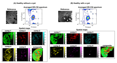 |
6 | Probing Liver Microstructure in-vivo Using Diffusion-Relaxation Correlation Spectroscopic Imaging (DR-CSI)
Daeun Kim1, Brian P Lee2, Junzhou Chen1,3, Kevin King1, Justin P Haldar4,5, Norah A Terrault2, and Zhaoyang Fan1,5,6
1Department of Radiology, University of Southern California, Los Angeles, CA, United States, 2Department of Medicine, Division of Gastroenterology and Liver Diseases, University of Southern California, Los Angeles, CA, United States, 3Department of Bioengineering, University of California, Los Angeles, Los Angeles, CA, United States, 4Department of Electrical and Computer Engineering, University of Southern California, Los Angeles, CA, United States, 5Department of Biomedical Engineering, University of Southern California, Los Angeles, CA, United States, 6Department of Radiation Oncology, University of Southern California, Los Angeles, CA, United States
Diffusion-relaxation correlation spectroscopic imaging (DR-CSI) is an advanced microstructure imaging approach that can resolve sub-voxel tissue compartments and quantify their fractions. In this work, we investigated the feasibility of DR-CSI with an optimized experiment design for in-vivo liver imaging. Our study showed that DR-CSI can measure multiple sub-voxel compartments in the liver and provide consistent component fraction maps in healthy livers. An initial test on a subject with chronic hepatitis B also demonstrated the potential of DR-CSI to identify and characterize pathological changes in liver parenchyma. Further studies on variable liver diseases are underway.
|
||
3370 |
7 | Radiomics for noninvasive prediction of HCC immunophenotyping
Enamul Bhuiyan1,2, Octavia Bane1,2, Paul Kennedy1,2, Sema Yildiz2, Muhammed Shareef2, M. Isabel Fiel3, Stephen Ward3, Myron Schwartz4, Thomas Marron5, Miriam Merad6, and Bachir Taouli1,2
1BioMedical Engineering and Imaging Institute (BMEII), Icahn School of Medicine at Mount Sinai, New York, NY, United States, 2Department of Diagnostic, Molecular and Interventional Radiology, Icahn School of Medicine at Mount Sinai, New York, NY, United States, 3Department of Pathology, Molecular and Cell Based Medicine, Icahn School of Medicine at Mount Sinai, New York, NY, United States, 4Recanati/Miller Transplantation Institute, Icahn School of Medicine at Mount Sinai, New York, NY, United States, 5The Tisch Cancer Institute, Icahn School of Medicine at Mount Sinai, New York, NY, United States, 6Precision Immunology Institute, Icahn School of Medicine at Mount Sinai, New York, NY, United States
The reported rate of intrahepatic recurrence of hepatocellular carcinoma (HCC) after resection is high (up to 50%), even with negative surgical margins, which is postulated to be due to micrometastases. Immunotherapy present new possibilities in cancer treatment with encouraging results in HCC. Selection of patients for immunotherapy may be based on background immune features, thus imaging features associated with immunophenotype may aid in patient selection. Specifically, radiomics features exhibited a fair to excellent diagnostic performance for differentiating tumors with high vs. low/moderate tumor infiltrating lymphocytes (TILs) and presence vs absence of tertiary lymphoid structure (TLS) with AUC range of 0.70-0.95.
|
||
3371 |
8 | Diffusion and perfusion MRI for prediction of HCC response to neoadjuvant immunotherapy
Enamul Bhuiyan1,2, Paul Kennedy1,2, Octavia Bane1,2, Muhammed Shareef2, Stefanie Hectors3, Hung Kam Cheung 3, Elizabeth Miller3, M. Isabel Fiel4, Myron Schwartz5, Stephen Ward4, Thomas Marron6, Miriam Merad7, and Bachir Taouli1,2
1BioMedical Engineering and Imaging Institute (BMEII), Icahn School of Medicine at Mount Sinai, New York, NY, United States, 2Department of Diagnostic, Molecular and Interventional Radiology, Icahn School of Medicine at Mount Sinai, New York, NY, United States, 3Regeneron Pharmaceuticals Inc. Tarrytown, New York, NY, United States, 4Department of Pathology, Molecular and Cell Based Medicine, Icahn School of Medicine at Mount Sinai, New York, NY, United States, 5Recanati/Miller Transplantation Institute, Icahn School of Medicine at Mount Sinai, New York, NY, United States, 6The Tisch Cancer Institute, Icahn School of Medicine at Mount Sinai, New York, NY, United States, 7Precision Immunology Institute, Icahn School of Medicine at Mount Sinai, New York, NY, United States
Neoadjuvant immunotherapy has the potential to decrease the risk of hepatocellular carcinoma (HCC) recurrence post resection. The objectives of this study were to assess the value of pre-treatment MRI parameters for prediction of HCC immunophenotype and response in a cohort of patients undergoing neoadjuvant immunotherapy prior to liver resection. We hypothesize that diffusion and perfusion MRI can predict HCC response to neoadjuvant immunotherapy. It was observed that abundant of TILs decreases ADC in tumors. Moreover, uptake rate is higher in highly necrotic tumors.
|
||
3372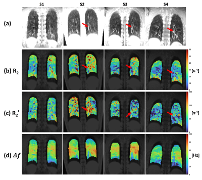 |
9 | Estimation of Lung Parenchyma Transverse Relaxation Rates at 0.55 Tesla
Bochao Li1, Nam Gyun Lee1, and Krishna Shrinivas Nayak1,2
1Department of Biomedical Engineering, University of Southern California, Los Angeles, CA, United States, 2Ming Hsieh Department of Electrical and Computer Engineering, University of Southern California, Los Angeles, CA, United States
Lung MRI with excellent image quality has been demonstrated at 0.55T, largely due to reduced susceptibility effects. Here, we present joint estimation of lung parenchyma transverse relaxation rates $$$R_{2}$$$, $$$R_{2}^{\prime}$$$, and off-resonance $$$\Delta f$$$. A custom echo-shifted turbo spin echo (TSE) pulse sequence with ECG triggering and breath hold was performed in 4 healthy volunteers, resulting in the mean $$$R_{2}$$$ of 23.4s-1, $$$R_{2}^{\prime}$$$ of 53.9s-1, and $$$\Delta f$$$ of 45.7Hz. This work demonstrates the feasibility of echo-shifted TSE for the estimation of three parameters of interest in lung MRI, and confirms prior measurements made with separate pulse sequences.
|
||
3373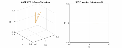 |
10 | High-Resolution Half-Spoke and Full-Spoke 4D Lung MRI with Golden-Angle 3D Radial Acquisition and Motion-Resolved Sparse Reconstruction
Can Wu1, Guruprasad Krishnamoorthy2, Ergys Subashi1, Victoria Yu1, and Ricardo Otazo1,3
1Department of Medical Physics, Memorial Sloan Kettering Cancer Center, New York, NY, United States, 2Philips Healthcare, MR R&D, Rochester, MN, United States, 3Department of Radiology, Memorial Sloan Kettering Cancer Center, New York, NY, United States
This work aims to develop high-resolution 4D lung MRI with golden-angle 3D radial (kooshball) acquisition and motion-resolved sparse reconstruction. The golden-angle radial k-space lines were continuously acquired during free-breathing and retrospectively binned to multiple respiratory phases with respect to the respiratory signal obtained from a built-in motion-sensing camera. The sparsity along the respiratory motion dimension was exploited using compressed sensing reconstruction to suppress artifacts and improve SNR. The feasibility of half-spoke ultrashort echo time (UTE) and full-spoke 4D lung MRI was demonstrated on healthy volunteers on a clinical 3T MRI scanner.
|
||
3374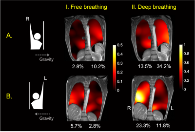 |
11 | Regional Lung Ventilation Mapping at 0.55T based on Feature Tracking
Yijing Yang1, Ziwei Zhao1, Ye Tian1, Roberta M. Kato2, Sophia X. Cui3, C.-C. Jay Kuo1, and Krishna S. Nayak1
1Ming Hsieh Department of Electrical and Computer Engineering, University of Southern California, Los Angeles, CA, United States, 2Children's Hospital of Los Angeles, Los Angeles, CA, United States, 3Siemens Medical Solutions USA, Inc., Los Angeles, CA, United States
Assessment of regional lung ventilation has significant clinical value for the diagnosis and follow-up of pulmonary diseases. High-performance 0.55T systems have provided new opportunities for pulmonary imaging due to prolonged T2* and reduced susceptibility. Here, we describe an image-based regional lung ventilation assessment method based on real-time bSSFP imaging with 0.3s temporal and 1.64x1.64 mm2 spatial resolution, and feature tracking. In healthy adult volunteers, we demonstrate the ability to detect posture-related left-right differences in ventilation, and test-retest repeatability. We detected -5% to 64% regional volume changes throughout the respiratory cycle from end of exhalation to total lung capacity.
|
||
3375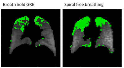 |
12 | Feasibility of VDP Calculation using 19F Free Breathing Spiral Acquisition with Post-Acquisition Denoising
Sang Hun Chung1, Khoi Minh Huynh1, Yong Chen2, Pew-Thian Yap3, Jennifer L. Goralski4,5,6, Scott H. Donaldson4,5, and Yueh Z. Lee3,4
1Biomedical Engineering, University of North Carolina, Chapel hill, NC, United States, 2Department of Radiology, Case Western Reserve University, Cleveland, OH, United States, 3Department of Radiology and Biomedical Research Imaging Center, University of North Carolina, Chapel hill, NC, United States, 4Division of Pulmonary and Critical Care Medicine, University of North Carolina, Chapel hill, NC, United States, 5Marsico Lung Institute/UNC Cystic Fibrosis Center, University of North Carolina, Chapel hill, NC, United States, 6Division of Pediatric Pulmonology, University of North Carolina, Chapel hill, NC, United States
19F lung ventilation MRI is usually acquired with extended breath holds that limit the acquisition to relatively healthy patients. New denoising methods have the potential to decrease the scan time significantly while achieving comparable SNRs. We compare 19F breath held scans to spiral acquisition based scans performed while free breathing combined with post-acquisition denoising in human subjects at 3T. VDP and SNR were evaluated. The denoised spiral scans yield higher SNRs (29.3 average increase) but overestimated VDPs (8.5% average increase). The differences in VDP were correlated (R-squared 0.74). The approach shows the potential for free-breathing 19F MRI ventilation imaging.
|
||
3376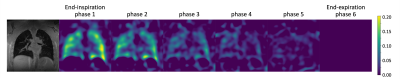 |
13 | Motion-Compensated Low-Rank Reconstruction with Iterative Registration for Ventilation Analysis on Ultrashort Echo Time (UTE) Lung MRI
Fei Tan1, Xucheng Zhu2, and Peder Larson1
1Radiology and Biomedical Imaging, University of California, San Francisco, San Francisco, CA, United States, 2GE Healthcare, Menlo Park, CA, United States
In this abstract, we introduce motion-compensated low-rank constrained reconstruction with iterative registration. This method jointly improves the motion field estimation and the reconstruction, providing both functional and structural images. In addition, we test the effect of the regularization parameter on structural images and ventilation maps. We conclude that the motion-compensated low-rank constrained with iterative registration method is suitable for regional ventilation quantification.
|
||
3377 |
14 | Feasibility of Biliary Imaging at 0.55T: A Comparison to 1.5T
Anupama Ramachandran1, Kathleen Ropella-Panagis1, Nancy Dudek1, Joel Morehouse1, Vikas Gulani1, Mishal Mendiratta-Lala1, and Nicole Seiberlich1
1Department of Radiology, University of Michigan, Ann Arbor, MI, United States MRCP exams were performed in three volunteers on 0.55T and 1.5T MRI systems. Images were rated on overall image quality and visualization of ducts by two board certified abdominal radiologists. All images on both systems were rated as excellent diagnostic quality. A paired sample t-test did not show significant differences in acquisition time between the two field strengths. The advantages of performing an MRCP exam on a 0.55T MRI system may include reduced susceptibility artifacts, reduced artifacts from cholecystectomy clips, improved patient comfort due to larger bore sizes, and potentially lower overhead costs. |
||
3378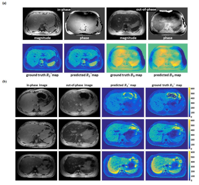 |
15 | Derivation of liver R2* and B0 maps from dual-echo MR images via deep learning
Yan Wu1, Yongwook Kee1, Marc Alley1, John Pauly2, Lei Xing1, and Shreyas Vasanawala1
1Stanford University, Stanford, CA, United States, 2Stanford University, Stanford University, CA, United States
Quantitative R2* map is an important liver disease indicator. However, the availability of R2* map is limited by the long scan time. In this study, we present a new paradigm to predict R2* and B0 maps from dual echo images. A self-attention deep convolutional neural network is trained and validated, where promising accuracy has been obtained. The proposed quantitative parametric mapping approach has a potential to eliminate the necessity for additional data acquisition other than clinical routine.
|
||
3379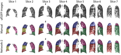 |
16 | MRI lung lobe segmentation of pediatric cystic fibrosis patients using a neural network trained with publicly accessible CT datasets
Orso Pusterla1,2,3, Rahel Heule4,5, Francesco Santini1,3,6, Thomas Weikert6, Corin Willers2, Simon Andermatt3, Robin Sandkühler3, Sylvia Nyilas7, Philipp Latzin2, Oliver Bieri1,3, and Grzegorz Bauman1,3
1Division of Radiological Physics, Department of Radiology, University Hospital Basel, University of Basel, Basel, Switzerland, 2Division of Pediatric Respiratory Medicine and Allergology, Department of Pediatrics, Inselspital, Bern University Hospital, University of Bern, Bern, Switzerland, 3Department of Biomedical Engineering, University of Basel, Basel, Switzerland, 4High Field Magnetic Resonance, Max Planck Institute for Biological Cybernetics, Tübinge, Germany, 5Department of Biomedical Magnetic Resonance, University of Tübingen, Tübingen, Germany, 6Department of Radiology, University Hospital Basel, University of Basel, Basel, Switzerland, 7Department of Diagnostic, Interventional and Pediatric Radiology, Inselspital, Bern University Hospital, University of Bern, Bern, Switzerland
Pulmonary biomarkers quantifications on a lobar level provide improved specificity against whole-lung analyses. However, lobar quantifications of pulmonary MR data are hardly accessible due to the complex work required for the manual segmentations. Supervised neural networks have shown the premise for automatic segmentation, but it is challenging to gather labelled data for the training. To overcome these limitations, in this work, we “translate” publicly accessible chest CT datasets and lobe segmentations to pseudo-MR data, and we then train a network able to segment consistently lung lobes of acquired MRI data. The cross-modality approach has excellent prospects to automatize MRI analyses.
|
||
 Back to Meeting Home
 Back to the Program-at-a-Glance
The International Society for Magnetic Resonance in Medicine is accredited by the Accreditation Council for Continuing Medical Education to provide continuing medical education for physicians.
 Back to Meeting Home
 Back to the Program-at-a-Glance
 View the Presentation