Online Gather.town Pitches
Body: GU & Breast III
Joint Annual Meeting ISMRM-ESMRMB & ISMRT 31st Annual Meeting • 07-12 May 2022 • London, UK

Online Gather.town Pitches
Body: GU & Breast III
| Booth # | ||||
|---|---|---|---|---|
3994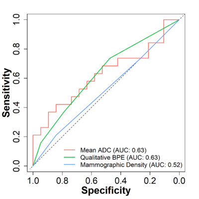 |
1 | Association of Fibroglandular Tissue ADC features on Diffusion-weighted MRI with Breast Cancer Risk
Wesley Surento1, Anum S. Kazerouni2, Janis Yee2, Debosmita Biswas2, Daniel S. Hippe3, Habib Rahbar2, and Savannah C. Partridge2
1Department of Biomedical Informatics and Medical Education, University of Washington, Seattle, WA, United States, 2Department of Radiology, University of Washington, Seattle, WA, United States, 3Clinical Research Division, Fred Hutchinson Cancer Research Center, Seattle, WA, United States
In this study, we investigated the association of fibroglandular tissue (FGT) apparent diffusion coefficient (ADC) measures with risk of breast cancer. Whole breast FGT regions were segmented on diffusion-weighted MRI (DWI) using a semi-automated fuzzy c-means-based algorithm. In high-risk screening cohort, ADC measures of FGT were compared in subjects with subsequent cancer diagnosis versus matched negative controls. Our findings indicate a modest negative association between FGT ADC and subsequent cancer diagnosis, with greater predictive value than mammographic density in the same cohort, pointing to the possible utility of DWI in the assessment of breast cancer risk.
|
||
3995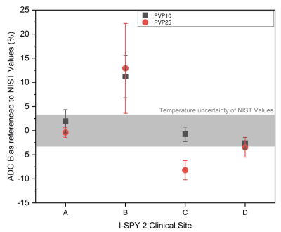 |
2 | Multi-site, longitudinal assessment of quantitative breast MRI variability using the CaliberMRI phantom
Jessica Gibbs1, Nu Le1, Todor Karaulanov2, Lisa Wilmes1, David Newitt1, Kathryn Keenan3, Bonnie Joe1, and Nola Hylton1
1University of California, San Francisco, San Francisco, CA, United States, 2CaliberMRI, Boulder, CO, United States, 3National Institute of Standards and Technology, Boulder, CO, United States Quantitative breast MRI data was acquired at seven clinical sites using the CaliberMRI phantom. T1 and DWI data were acquired, and the qCal software was used for analysis. Protocol adherence was assessed, quantitative measures were automatically derived, and variability between measurements was evaluated. Going forward, the phantom will be used for calibration as part of a program to assess quantitative accuracy in a large multi-site clinical trial. The phantom program will be expanded to other sites in the trial, with standardized reporting used to understand variability across sites. |
||
3996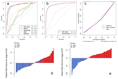 |
3 | Whole-lesion ADC histogram and texture analysis of preoperatively differentiating benign endometrial lesions from stage IA endometrial cancer Video Not Available
Jieying Zhang1, Xiaoduo Yu1, Shuang Chen1, Lizhi Xie2, Yan Chen1, and Han Ouyang1
1Diagnostic Radiology, National Cancer Center/National Clinical Research Center for Cancer/Cancer Hospital, Chinese Academy of Medical Sciences and Peking Union Medical College, Beijing, China, 2MR Research, GE Healthcare, Beijing, China Making an accurate preoperative diagnosis of endometrial lesions is critical to avoid unnecessary surgical procedures and protect the patients’ fertility. Noninvasive pre-surgical evaluation of abnormalities in the uterine cavity remains to be challenging. The volumetric ADC histogram analysis provides a mechanism for quantifying the overall heterogeneity and comprehensive assessment of a given abnormality. We demonstrated that whole-lesion ADC histogram-based texture analysis could serve as a quantitative technique of differentiating benign endometrial lesions (BELs) from stage IA endometrial cancer (EC) preoperatively. The combined ADC histogram model showed high classification ability for both premenopausal and postmenopausal patients. |
||
3997 |
4 | Preliminary study of extracellular volume fraction in identifying pathological risk factors of early cervical cancer
Wei Wang1, Yunxi Li1, Mengchao Zhang 1, and Yueluan Jiang2
1Department of Radiology, China Japan Union Hospital, Changchun, China, 2MR Scientific Marketing, Siemens Healthineers, Beijing, China
This study explored the feasibility of ECV based on T1 mapping evaluate early cervical cancer pathological characters, such as deep stromal invasion (DSI) and lymphovascular space invasion (LVSI). The ECV in the group with exceeding deep 1/2 of stromal invasion was significantly higher than that of the non-exceeding 1/2 group. DSI group was significantly higher than that in the non-DSI group. Our research shows that ECV based on T1 mapping could represent an emerging preoperative imaging biomarker for the classification of DSI of early cervical cancer.
|
||
3998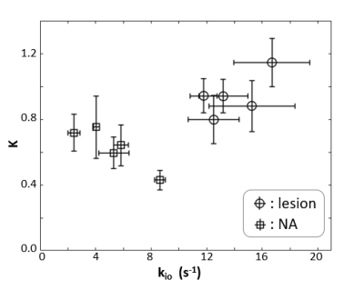 |
5 | Transcytolemmal water exchange in prostate manifest via DCE-MRI and DWI kurtosis imaging
Xin Li1, Ryan P Kopp2,3, Eric M Baker1, Brendan Moloney1, William D. Rooney1, Charles S Springer1, Fergus V Coakley4, and Mark G. Gartotto2,3
1Advanced Imaging Research Center, Oregon Health & Science University, Portland, OR, United States, 2Portland VA Medical Center, Portland, OR, United States, 3Urology, Oregon Health & Science University, Portland, OR, United States, 4Diagnostic Radiology, Oregon Health & Science University, Portland, OR, United States Transcytolemmal water exchange effects are quantified with SSM-DCE MRI and DWI kurtosis imaging with b-values up to 2000 s/mm2. The two-dimensional scatter plot of the unidirectional cellular water efflux rate constant (kio) and the diffusion kurtosis (K) shows good separation between lesion and normal appearing prostate tissue. |
||
3999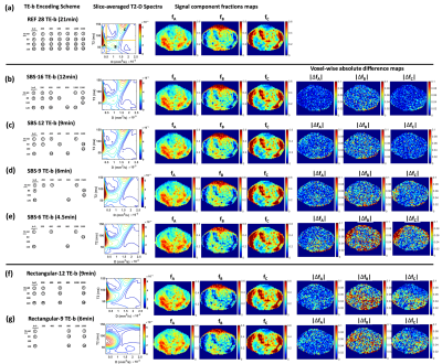 |
6 | A Data-Driven Sequential Backward Selection Framework to Accelerate Diffusion-Relaxation Prostate Microstructure Mapping
Zhaohuan Zhang1, Sohrab Afshari Mirak1, Melina Hosseiny1, Afshin Azadikhah1, Amir Bajgiran1, Alan Priester2, Kyunghyun Sung1, Anthony E Sisk3, Robert E Reiter2, Steven Raman1, Dieter R Enzmann1, and Holden H Wu1
1Department of Radiology, UCLA, Los Angeles, CA, United States, 2Department of Urology, UCLA, Los Angeles, CA, United States, 3Department of Pathology, UCLA, Los Angeles, CA, United States
Diffusion-Relaxation Correlation Spectrum Imaging (DR-CSI) can provide unique microstructural information for prostate cancer characterization, but requires longer scan times for two-dimensional encoding of TE and b-values. This study developed a data-driven sequential backward selection framework that determined subsampled encoding schemes for DR-CSI, achieving 70% reduction of scan time to 6min while maintaining accurate ex vivo prostate microstructure mapping.
|
||
4000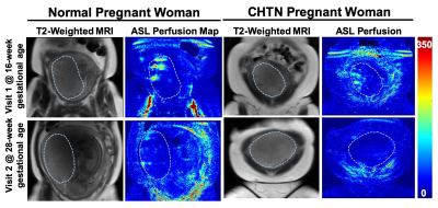 |
7 | Assessment of Placental Perfusion in Normal and Hypertensive Pregnancies using pCASL at 3T: Preliminary Findings
Yiming Wang1, Christina L. Herrera2, David M. Owen2, Quyen N. Do1, Yin Xi1,3, Matthew A. Lewis1, Baowei Fei1,4,5, Catherine Y. Spong2, Diane M. Twickler1,2, and Ananth J. Madhuranthakam1,4
1Department of Radiology, UT Southwestern Medical Center, Dallas, TX, United States, 2Department of Obstetrics and Gynecology, UT Southwestern Medical Center, Dallas, TX, United States, 3Department of Population and Data Sciences, UT Southwestern Medical Center, Dallas, TX, United States, 4Advanced Imaging Research Center, UT Southwestern Medical Center, Dallas, TX, United States, 5Department of Bioengineering, University of Texas at Dallas, Richardson, TX, United States
Measuring placental perfusion can provide important information about its function. Arterial Spin Labeled (ASL) MRI is a non-contrast perfusion imaging method particularly suitable in pregnancy as it does not require an exogenous contrast. We performed a preliminary assessment of placental perfusion in pregnant subjects, both normal and with CHTN, at 16-20 week and 24-28 week gestational ages. Perfusion reduction was observed in all 5 normal subjects and in 75% (12 of 16) of the subjects with CHTN. This could provide important information about perfusion changes during pregnancy and can serve as a precursor for a larger-scale longitudinal study in CHTN.
|
||
4001 |
8 | Standardization of breast dynamic contrast-enhanced MRI signal for assessment of background parenchymal enhancement rate
Milica Medved1, Keiko Tsuchiya2, Xiaobing Fan1, Gregory S Karczmar1, and Hiroyuki Abe1
1Department of Radiology, The University of Chicago, Chicago, IL, United States, 2Department of Radiology, Shiga University of Medical Science, Otsu, Japan
Breast DCEMRI signal is standardized for variable imaging protocols and the linear rate of background parenchymal enhancement (BPE) is calculated, to quantitatively and objectively describe changes in BPE rates following preventative tamoxifen treatment. This is in contrast with the current practice of using 4 subjective categories to describe BPE. Decreased BPE rates post-treatment agree with earlier results showing that BPE, like breast density, correlates with breast cancer risk. Standardization for imaging parameters and contrast agent relaxivity increases the observed effect size, pointing to increased sensitivity to treatment-induced changes and the potential as a tool for individual breast cancer risk management.
|
||
4002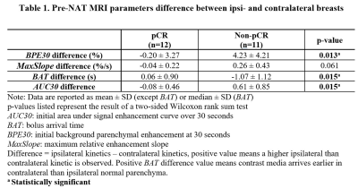 |
9 | Bilateral asymmetry of early parenchymal kinetics predicts response of breast cancer to neoadjuvant therapy
Zhen Ren1, Federico D. Pineda1, Frederick M. Howard2, Elle Hill1, Teodora Szasz3, Rabia Safi1, Milica Medved1, Rita Nanda2, Thomas E. Yankeelov4,5,6,7,8,9, Hiroyuki Abe1, and Gregory S. Karczmar1
1Radiology, The University of Chicago, Chicago, IL, United States, 2Medicine, The University of Chicago, Chicago, IL, United States, 3Research Computing Center, The University of Chicago, Chicago, IL, United States, 4Biomedical Engineering, The University of Texas at Austin,, Austin, TX, United States, 5Diagnostic Medicine, The University of Texas at Austin, Austin, TX, United States, 6Oncology, The University of Texas at Austin, Austin, TX, United States, 7Institute for Computational and Engineering Sciences, The University of Texas at Austin, Austin, TX, United States, 8Livestrong Cancer Institutes, The University of Texas at Austin, Austin, TX, United States, 9Imaging Physics, MD Anderson Cancer Center, Houston, TX, United States
We retrospectively reviewed data from 23 patients who received neoadjuvant therapy (NAT) and were scanned with a protocol that included ultrafast DCE-MRI (temporal resolution = 3-7 seconds) for the first minute after contrast injection prior to NAT. We measured parenchymal kinetics from ipsi- and contra-lateral normal parenchyma separately, so that new parameters related to bilateral parenchymal enhancement asymmetry could be calculated. The results showed that patients with similar pre-NAT parenchymal enhancement kinetics in ipsi- and contralateral normal parenchyma were more likely to achieve pCR post NAT ($$$p < 0.02$$$).
|
||
4003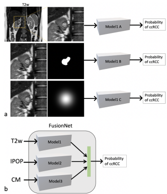 |
10 | Deep learning identification of clear cell renal carcinoma cancer using MR imaging
Junyu Guo1, Keith Husley1, Yin Xi1, and Ivan Pedrosa1
1Radiology, UT southwestern medical center, Dallas, TX, United States Clear cell renal carcinoma cancer (ccRCC) is the most common subtype among renal masses. ccRCC identification helps in decision making between active surveillance and definitive intervention. A clear cell likelihood score (ccLS) using subjective interpretation of multiparametric MRI by radiologists was proposed recently. In this study, we investigate whether deep learning (DL) using the three main MR sequences for ccLS can facilitate the diagnosis of ccRCC. We compared the results of twelve trained DL models with the reported ccLS performance. Our results demonstrate that DL may achieve a performance comparable to radiologists and provide useful information for identification of ccRCC. |
||
4004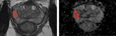 |
11 | Development of a MRI radiomic-based ML model to predict aggressiveness of prostate cancer
Ignacio Dominguez1, Paola Caprile2, Odette Rios2, Ignacio San-Francisco3, and Cecilia Besa 1
1Radiology, Pontificia Universidad Catolica de Chile, Santiago, Chile, 2Physics, Pontificia Universidad Catolica de Chile, Santiago, Chile, 3Urology, Pontificia Universidad Catolica de Chile, Santiago, Chile
We developed a non-invasive tool to predict the GS classification of PCa based on mpMRI information using ML. This retrospective study included 86 male patients with positive PCa fusion (mpMRI-ultrasound) biopsy. A radiomic analysis was performed considering first order, textural, shape, and clinical information. The best model found included image (T2w - ADC) and clinical information. The mean AUC was 0.91 [0.75−0.99] (p <0.05), with a validation AUC of 0.91 for a classification of high-lower aggressiveness (GS≥7 vs GS=6). Combining MRI-based radiomic and clinical information can significantly improve the model performance to classify PCa aggressiveness.
|
||
4005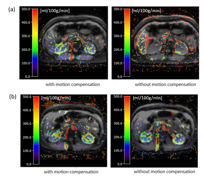 |
12 | Navigator-based slice tracking for kidney pCASL using EPI acquisition
Ke Zhang1,2, Simon M.F. Triphan1, Felix T. Kurz2, Chrstian H. Ziener2, Hans-Ulrich Kauczor1, Heinz-Peter Schlemmer2, and Oliver Sedlaczek1,2
1Department of Diagnostic and Interventional Radiology, Heidelberg University Hospital, Heidelberg, Germany, 2Department of Radiology, German Cancer Research Center, Heidelberg, Germany
Renal perfusion is an important physiological parameter in health and disease (1). To measure kidney perfusion using arterial spin labelling (ASL) the respiratory motion is a major problem. In this study, respiratory motion information is acquired from a projection signal and used to adjust the position of the excited slice in real time. The feasibility of free-breathing multi-slice kidney perfusion imaging using EPI based pseudocontinuous ASL (pCASL) with navigator-based slice tracking method is investigated.
|
||
4006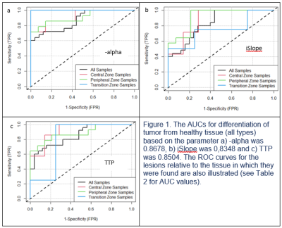 |
13 | Observations on Rapid Dynamic Contrast Enhanced MRI in Prostate Cancer
Armando Tartaro1,2, Roberto Renzetti3, Michele De Cristofaro Aulisa4, Federica Morrone5, Andrea De Nicola6,7, Ekaterina Bliakharskaia7, and Paul E Summers7
1Department of Clinical, Oral and Biotechnology Sciences, University "G. d'Annunzio" Chieti-Pescara, Chieti-Pescara, Italy, 2Magnetic Resonance Service, Ospedale di Popoli, AUSL, Pescara, Italy, 3UOC of Urology, Ospedale di Pescara, AUSL, Pescara, Italy, 4Faculty of Medicine, University "G. d'Annunzio“, Chieti-Pescara, Chieti-Pescara, Italy, 5Radiology Department, Centro Morrone, Caserta, Italy, 6Ospedale di Chieti, Chieti, Italy, 7QMRI Tech, Pescara, Italy
Prostate dynamic contrast-enhanced (DCE) MRI is often criticized for its weaknesses in terms of discriminating transition zone lesions and tumors in general. Using high-temporal resolution DCE MRI in patients who went on to prostatectomy, we evaluated time to peak (TTP), as well as the enhancement rate factor, and initial enhancement slope of a simple empirical mathematical model of early contrast uptake. Differences between tumors and surrounding healthy tissues were seen for the mean values of all three parameters. The AUCs were good (>0.8 in all cases except for TTP between tumor and transition zone) but case numbers limited.
|
||
 Back to Meeting Home
 Back to the Program-at-a-Glance
The International Society for Magnetic Resonance in Medicine is accredited by the Accreditation Council for Continuing Medical Education to provide continuing medical education for physicians.
 Back to Meeting Home
 Back to the Program-at-a-Glance
 View the Presentation