Online Gather.town Pitches
Body: GU & Breast II
Joint Annual Meeting ISMRM-ESMRMB & ISMRT 31st Annual Meeting • 07-12 May 2022 • London, UK

Online Gather.town Pitches
Body: GU & Breast II
| Booth # | ||||
|---|---|---|---|---|
3671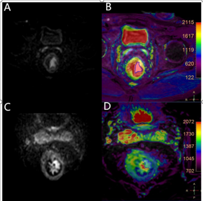 |
1 | Diagnosis value of T2 mapping on the of vascular invasion in rectal Cancer
Xiwei Li1, Anliang Chen1, Ailian Liu1, and Zhigang Wu2
1the First Affiliated Hospital of Dalian Medical University, Dalian, China, 2Philips Healthcare, Beijing, China
Rectal cancer is a malignant tumor with extremely high morbidity and mortality. It will change the physical microenvironments, but it is still a challenge to differentiate the rectal cancer with and without vascular invasion. T2 mapping imaging is a quantitative biomarker with good repeatability and stability. T2 mapping can non-invasively visualize and quantify tissue components (such as edema, fibrosis etc )without contrast agent. This study aims to assess the performance of T2 mapping on differentiating rectal cancer with and without vascular invasion, which may yield higher diagnostic confidence.
|
||
3672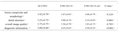 |
2 | Isotropic 3D high-resolution T2-weighted breast MRI with a deep learning constrained Compressed SENSE reconstruction: a pilot study Video Not Available
Yang Fan1, Yang Jieyin1, Sun Jiayu1, Zhang Xiaoyong2, and Ling Chuntang 3
1Department of Radiology, West China hospital of Sichuan University, Chengdu, China, 2Department of Clinical Science, Philips Healthcare, Chengdu, China, 3Department of Application, Philips Healthcare, Chongqing, China 2D T2-weighted (T2WI) breast MRI is prominently used for the identification of the colliquative necrosis and cysts, and it can also contribute to the characterization of lesions as benign or malignant. However, 2D imaging is routinely used in clinical practice, which has lower resolution, slice gaps, and may suffer distortion to delineate the breast lesions. In this study, we applied the Compressed-sensing Artificial Intelligence (CS-AI) framework to further increase the spatial resolution and reduce the scan time. The results of this study demonstrated that the high-resolution T2-weighted 3D CS-AI can provide further benefits to improve the depiction of diagnostic findings of breast lesions. |
||
3673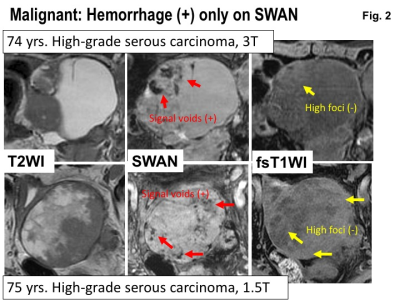 |
3 | Susceptibility-weighted MR sequence for the evaluation of intra-tumoral hemorrhage: Differentiation of benign and malignant ovarian tumors
Mayumi Takeuchi1, Kenji Matsuzaki2, and Masafumi Harada1
1Department of Radiology, Tokushima University, Tokushima, Japan, 2Department of Radiological Technology, Tokushima Bunri University, Sanuki-city, Japan
Intra-tumoral hemorrhage is one of the suggestive pathological findings of malignancy. Surgically proven 16 benign and 64 malignant solid or complex ovarian tumors were retrospectively evaluated. High intensity hemorrhagic foci on T1WI were detected in 16 of 64 malignant lesions (25%), whereas signal voids due to hemorrhage on susceptibility-weighted sequence (SWS) were detected in 41 of 64 lesions (64%). Neither high intensity foci on T1WI nor signal voids on SWS was detected in all 16 benign tumors. We conclude that the demonstration of intra-tumoral hemorrhage in patients with ovarian tumors by SWS may provide valuable diagnostic findings.
|
||
3674 |
4 | The value of APTw imaging combined with ALP to identify Prostate Cancerwith or withoutBone Metastasis Video Permission Withheld
Chen Lihua1, Song Qingwei1, Gao Mingli1, Wang Jiazheng2, Lin Liangjie2, Wu Zhigang2, and Liu Ailian1
1The First Affiliated Hospital of Dalian Medical University, DaLian, China, 2Philips Healthcare, Beijing, China
Amide proton transfer-weighted (APTw) imaging is a novel MRI imaging tool for detection of amide protons in mobile cellular proteins and peptides. This study aims to assess and differentiate prostate cancers with and without bone metastasis using APTw MRI and alkaline phosphatase (ALP).Results show that prostate cancers with bone metastasis were associated with significantly higher APTw and ALP values than those without bone metastasis. The diagnostic efficiency is the highest using APT combined with ALP. Therefore, APTw imaging together with ALP may provide an more effective tool to predict the aggressiveness of prostate cancer.
|
||
3675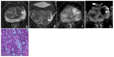 |
5 | Diagnostic performance of transition zone prostate cancer: a comparison between PI-RADS v2.1 and PI-RADS v2
Dan Zhang1, Zhi Chen1, Na Song1, Zhuo Wang1, Shao Zhang1, Xiao Chen1, Xiao Wei2, and Bing Chen1
1The Department of Radiology, The General Hospital of Ningxia Medical University, Yinchuan, China, 2GE Healthcare,MR Research, Beijing, China
In this study, we aim to investigate the diagnostic value of PI-RADS v2.1 and PI-RADS v2 (prostate imaging reporting and data system version 2.1 and version 2) in diagnosing transition zone prostate cancer. It was concluded that the diagnostic value of PI-RADS v2.1was not lower than that of PI-RADS v2.
|
||
3676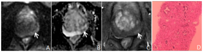 |
6 | Diagnostic performance of peripheral zone prostate cancer: a comparison between PI-RADS v2.1 and PI-RADS v2
Dan Zhang1, Zhi Chen1, Na Song1, Zhuo Wang1, Shao Zhang1, Xiao Chen1, Xiao Wei2, and Bing Chen1
1The Department of Radiology, The General Hospital of Ningxia Medical University, Yinchuan, China, 2GE Healthcare,MR Research, Beijing, China
In this study, we aim to investigate the diagnostic value of PI-RADS v2.1 and PI-RADS v2 (prostate imaging reporting and data system version 2.1 and version 2) for peripheral zone prostate cancer. It was concluded that the diagnostic value of PI-RADS v2.1 was higher.
|
||
3677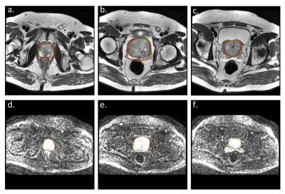 |
7 | Automatic segmentation of prostate gland on multiparametric MR images using deep learning convolutional neural network: a multi-center study Video Not Available
Lili Xu1, Gumuyang Zhang1, Li Mao2, Xiuli Li2, Hao Sun1, and Zhengyu Jin1
1Peking Union Medical College Hospital, Beijing, China, 2Deepwise AI Lab, Beijing, China
Accurate prostate segmentation on MR images plays an important role in the management of prostate diseases. Recently proposed deep learning architecture has been successfully applied for medical image segmentation to overcome the shortcomings of manual segmentation. Our study proposed a 3D UNet model for automatic and accurate prostate gland segmentation on both DWI and T2WI images. This model was tested in 3 different external cohorts and showed satisfactory results on T2WI images. The segmentation performance on DWI images was inferior but still inspiring in the external testing group. This study might benefit the management of prostate diseases in the future.
|
||
3678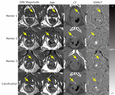 |
8 | Quantitative Susceptibility Mapping as an Alternative to CT for Localizing Gold Intraprostatic Fiducial Markers
Ashley Wilton Stewart1,2, Jonathan Goodwin3,4, Simon Daniel Robinson2,5,6, Kieran O’Brien1,2,7, Jin Jin1,2,7, Markus Barth1,2,8, and Steffen Bollmann1,2,8
1ARC Training Centre for Innovation in Biomedical Imaging Technology, The University of Queensland, St Lucia, Australia, 2Centre for Advanced Imaging, The University of Queensland, St Lucia, Australia, 3Department of Radiation Oncology, Calvary Mater Hospital, Newcastle, Australia, 4School of Information and Physical Sciences, University of Newcastle, Newcastle, Australia, 5High Field MR Center, Department of Biomedical Imaging and Image-Guided Therapy, Medical University of Vienna, Vienna, Austria, 6Department of Neurology, Medical University of Graz, Graz, Austria, 7Siemens Healthcare Pty Ltd, Brisbane, Australia, 8School of Information Technology and Electrical Engineering, The University of Queensland, St Lucia, Australia
Gold fiducial markers (FMs) for prostate radiotherapy are commonly localized using Computed Tomography (CT), though interest in MR-only workflows is growing. One current limitation with magnitude-based MR-only workflows is the ability to distinguish FMs from blood products and calcifications because all appear as signal voids. Quantitative Susceptibility Mapping (QSM) separates sources based on their susceptibility, and may be a potential solution. We apply the QSMxT pipeline to gradient-echo images of a prostate cancer patient with FMs and a calcification. We found that QSM provided a contrast that could differentiate the FMs from calcifications to a degree comparable to CT.
|
||
3679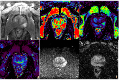 |
9 | Prostate cancer Diagnosis and risk stratification with combination of DISCO and MUSE: a feasibility study.
Dan Zhang1, Zhi Chen1, Na Song1, Zhuo Wang1, Shao Zhang1, Xiao Chen1, Xiao Wei2, and Bing Chen1
1The Department of Radiology, The General Hospital of Ningxia Medical University, Yinchuan, China, 2GE Healthcare,MR Research, Beijing, China
In this study, we aim to investigate the value of DISCO quantitative parameters combined with MUSE in distinguishing benign and malignant prostate lesions. It was concluded that Ktrans, Kep, Ve and ADC can be used as imaging biomarkers to distinguish benign and malignant prostate lesions; Ktrans and ADC are helpful to predict aggressiveness of the tumor,and can be used as independent predictors of prostate cancer.
|
||
3680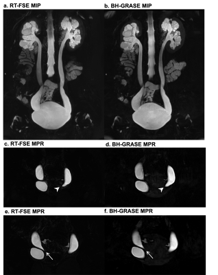 |
10 | Static-fluid MR Urography with three-dimensional Gradient and Spin-echo and Respiratory-trigger Fast Spin Echo sequences: A Comparison Study Video Not Available
Wei Wang1, Jing Liu1, Wei Li1, Ke Xue2, Yongming Dai2, and Jianxing Qiu1
1Peking University First Hospital, Beijing, China, 2United Imaging Healthcare, Shanghai, China This study is targeted to compare the performance of three-dimensional (3D) breath-hold gradient and spin-echo sequence (GRASE) and conventional respiratory-trigger fast spin-echo sequence (FSE) for magnetic resonance urography (MRU). Image quality and diagnostic performance of urinary tract dilation from two data sets were evaluated by three radiologists. The results showed that 3D GRASE MRU had a better performance compared with the FSE MRU, indicating 3D GRASE technique could be a feasible option for 3D MRU. |
||
3681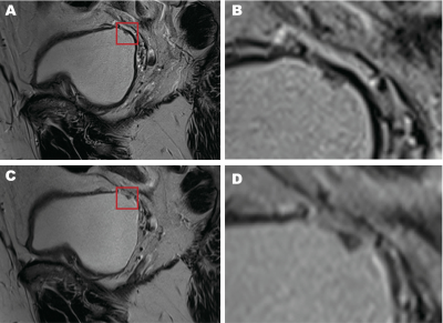 |
11 | Added value of deep learning-accelerated T2-weighted imaging of the bladder on image quality and lesion evaluation Video Not Available
Gumuyang Zhang1, Li Chen1, Hailong Zhou1, Yunna Wang1, Jinxia Zhu1, Marcel Dominik Nickel2, Elisabeth Weiland3, Hao Sun1, and Zhengyu Jin1
1Peking Union Medical College Hospital, Beijing, China, 2Siemens Healthineers Ltd., Beijing, China, 3MR Application Predevelopment, Siemens Healthcare GmbH, Erlangen, Germany
This study evaluated deep learning accelerated T2w imaging (T2DL) of the bladder in terms of acquisition time (TA), overall image quality, presence of artifacts, diagnostic confidence, sharpness of lesions, and VI-RADS T2 score compared to a standard T2w (T2S) sequence in twenty-five patients. Two radiologists evaluated the images independently. TA of T2DL was reduced by nearly 50% compared to T2S. Overall image quality and sharpness of lesions were superior in T2DL, while artifacts, diagnostic confidence and T2 score were similar between T2S and T2DL. T2DL has the potential to replace T2S in bladder MR.
|
||
3682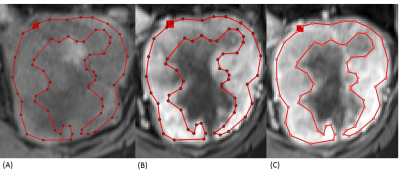 |
12 | The Comparison of Enhancing-part and Whole-lesion of ROI Measurements in MR Histogram Analysis of Clear Cell RCC and None Clear Cell RCC
Ming-Cheng Liu1,2, Yi-Jui Liu2,3, Pin-Sian Lyu3, Guan-Xin Pan3, Si-Wa Chan4,5,6, Yen-Ting Lin1,7, Siu-Wan Hung1,8,9, Jyh-Wen Chai1, and Kun-Yuan Chiu10,11
1Radiology, Department of Radiology, Taichung Veterans General Hospital, Taiwan, Taichung, Taiwan, 2Ph.D. Program of Electrical and Communications Engineering, Feng Chia University, Taichung, Taiwan, Taichung, Taiwan, Taiwan, 3Department of Automatic Control Engineering, Feng Chia University, Taichung, Taiwan, Taichung, Taiwan, 4Department of Medical Imaging, Taichung Tzu Chi Hospital, Buddhist Tzu Chi Medical Foundation, Taichung, Taiwan, Taichung, Taiwan, 5School of Medicine, Tzu Chi University, Hualien, Taiwan, Taichung, Taiwan, 6Department of Medical Imaging Radiological Science, Central Taiwan University of science and Technology, Taichung, Taiwan, Taichung, Taiwan, 7Institute of Clinical Medicine, National Yang-Ming University, Taiwan, Taichung, Taiwan, 8School of Medical Imaging and Radiological Sciences, Chung Shan Medical University, Taiwan, Taichung, Taiwan, 9Department of Veterinary Medicine, National Chung Hsing University, Taiwan, Taichung, Taiwan, 10Division of Urology, Department of Surgery, Taichung Veterans General Hospital, Taiwan, Taichung, Taiwan, 11Department of Applied Chemistry, National Chi Nan University, Taiwan, Taichung, Taiwan
Tumor necrosis, hemorrhage, and cystic change may influence the result of MR histogram analysis between ccRCC and ncRCC. We conducted EP ROI and WL ROI measurements of these two tumors. Five most important metrics were found in differentiation, including three EP ROI metrics on the T1 A and V phase, and the T2W images, and two WL ROI metrics on the DWI b800 images and the T2W images. Both EP ROI and WL ROI played a role in the diagnosis ccRCC and ncRCC. The addition of these two ROI measurements of histogram analysis to routine MRI evaluation may benefit diagnosis.
|
||
3683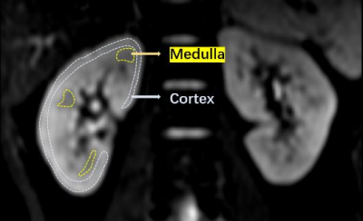 |
13 | Application of DKI in the assessment of early renal injury in patients with DM Video Not Available
You-Zhen Feng1, Zhong-Yuan Cheng1, Long Qian2, and Xiang-Ran Cai1
1Medical Imaging Center, the First Affiliated Hospital of Jinan University, Guangzhou, China, 2MR Research,GE Healthcare, Beijing, China Apply DKI technology to the assessment of early renal function changes in diabetic patients. These results indicate that the microstructure of kidney tissue may have changed in diabetic patients without clinically impaired renal function. In addition, the results of correlation analysis between DKI parameters and clinical laboratory indicators of renal function (HBA1, Uscr, and eGFR) show that there is a linear correlation between most of the parameters, which objectively confirms the feasibility of DKI parameters to reflect changes in renal function. |
||
3684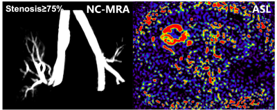 |
14 | The feasibility of ASL combined with Q-flow MRI in evaluating hemodynamic changes of transplant renal artery stenosis Video Permission Withheld
Liang Pan1, Jie Chen1, Wei Xing1, and Jilei Zhang2
1Third Affiliated Hospital of Soochow University, Changzhou, China, 2Philips Healthcare, Shanghai, China
Transplant renal artery stenosis (TRAS) is a less common but important etiology of renal dysfunction following transplantation. It is of great importance to include TRAS in the differential diagnosis for allograft dysfunction early after transplantation. We used ASL combined with 2D-PC Q-flow MRI to assess hemodynamic changes of TRAS. The results reflected that there were differences in the mean flux and velocity of the stenotic segment of transplant renal artery and renal allograft perfusion between different degrees of TRAS, and the mean flux and velocity were more closely related to the degree of TRAS compared with renal allograft perfusion.
|
||
3685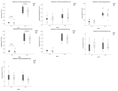 |
15 | A time-saving and optimized IVIM protocol for diagnosis of prostate cancer
qiqi zhou1, Weiyin Vivian Liu2, Qian Tang3, Ling Song1, Chao Liu1, Hu Chen1, and Lin Xu1
1Department of Radiology,Taihe hospital, Hubei University of Medicine, Hubei, China, 2GE Healthcare, Beijing, China, 3Biomedical Engineering College, Hubei University of Medicine, Hubei, China
It is of importance to acquire diffusion-weighted images with high image quality in a short time whereas higher b value and a greater number of b values reduce image quality and prolong scan time. In this study, we aimed to build an optimized protocol for IVIM in diagnosis of prostate cancer.We observed mean of all ADC, D, D* and f derived from IVIM with 5 b-value scheme showed almost the same diagnosis efficiency to IVIM with 13 b-value scheme, suggesting sensitivity and specificity were not different between different b-value scheme.
|
||
 Back to Meeting Home
 Back to the Program-at-a-Glance
The International Society for Magnetic Resonance in Medicine is accredited by the Accreditation Council for Continuing Medical Education to provide continuing medical education for physicians.
 Back to Meeting Home
 Back to the Program-at-a-Glance
 View the Presentation