Online Gather.town Pitches
Body: Liver/GI/Chest III
Joint Annual Meeting ISMRM-ESMRMB & ISMRT 31st Annual Meeting • 07-12 May 2022 • London, UK

Online Gather.town Pitches
Body: Liver/GI/Chest III
| Booth # | ||||
|---|---|---|---|---|
4199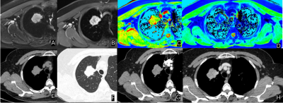 |
1 | The Diagnostic Value of Extracellular Volume Fraction in Different Pathological Types of Lung Cancer – a preliminary study
Peng Zhao1, Yongqing Yang2, Yu Wang2, Wenjing Ma1, Xiangtao Lin2, Liu Mengxiao3, Lu Zu2, Tao Yang2, Letian Yuan1, Yani Cheng2, Shanshan Sun1, Shuo Cai1, and Wei Zhang2,4
1Shandong Provincial Hospital Affiliated to Shandong First Medical University, Jinan, China, 2Shandong Provincial Hospital Affiliated to Shandong University, Jinan, China, 3MR Scientific Marketing,Diagnostic Imaging, Siemens Healthineers Ltd., Shanghai, China, 4The People's Hospital of LaoLing, LaoLing, China
Chest scans were performed on 42 lung cancer patients using MRI and CT scanners to calculate ECV. Using one-way analysis of variance, the ECV calculated based on CT is statistically different between SCLC and NSCLC, there is no statistical difference between squamous cell carcinoma and adenocarcinoma, and the ECV calculated based on MRI is statistically different among the three.It can be seen that ECV can be accurate and non-invasive assessment of the pathological type of lung cancer, and MRI-ECV has a higher diagnostic efficiency than CT-ECV, can provide some help for the choice of clinical treatment options.
|
||
4200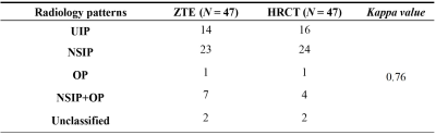 |
2 | Evaluation of interstitial lung diseases using free-breathing 3D isotropic ZTE-MRI in comparison with High-resolution computed tomography
Qiuxi Lin1, Yu Deng1, Xinchun Li1, Weiyin Vivian Liu2, Lei Zhang2, Ziyi Zhang3, Qun Luo3, Qi Wan1, and Chongpeng Sun1
1Department of Radiology, The first affiliated hospital of Guangzhou Medical University, Guangzhou, China, 2GE Healthcare, Beijing, China, 3Department of Respiratory and Critical Care Medicine, The first affiliated hospital of Guangzhou Medical University, Guangzhou, China
Pulmonary MRI provides qualitative structural and quantitative functional images of the lungs without ionizing radiation. In this study, free-breathing 3D isotropic high-resolution pulmonary zero echo time (ZTE) imaging was applied to detect pulmonary abnormalities and make diagnosis of interstitial lung diseases (ILDs). The pulmonary interstitial abnormalities including ground-glass opacity, reticular opacity, honeycombing, traction bronchiectasis and consolidation can be well depicted. The diagnosis performance of ZTE in ILDs is comparable with HRCT. ZTE might assist long-term follow-ups in ILDs evaluation without radiation exposure.
|
||
4201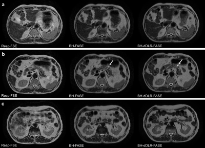 |
3 | Feasibility of an abdominal thin-slice breath-hold single-shot FSE sequence processed using a deep learning-based noise-reduction approach
Taku Tajima1,2, Hiroyuki Akai3, Koichiro Yasaka4, Akira Kunimatsu1, Masaaki Akahane2, Naoki Yoshioka2, Osamu Abe4, Kuni Ohtomo5, and Shigeru Kiryu2
1Department of Radiology, International University of Health and Welfare Mita Hospital, Tokyo, Japan, 2Department of Radiology, International University of Health and Welfare Narita Hospital, Chiba, Japan, 3Department of Radiology, The Institute of Medical Science, The University of Tokyo, Tokyo, Japan, 4Department of Radiology, Graduate School of Medicine, The University of Tokyo, Tokyo, Japan, 5International University of Health and Welfare, Tochigi, Japan
Single-shot fast spin echo (single-shot FSE) sequence is an accelerated T2-weighted imaging (T2WI) in pancreatic MRI. Fast advanced spin echo (FASE) is one of similar modalities. However, single-shot FSE suffers from image blurring and relatively low tissue contrast. We hypothesized that denoising approach with deep learning-based reconstruction (dDLR) would facilitate accelerated breath-hold thin-slice single-shot FSE MRI. We assessed the image quality of respiratory-triggered FSE T2WI (Resp-FSE) and breath-hold FASE with and without dDLR (BH-dDLR-FASE and BH-FASE, respectively) at 1.5 T. The image quality of BH-dDLR-FASE was superior to BH-FASE and Resp-FSE, and BH-dDLR-FASE had a shorter acquisition time than Resp-FSE.
|
||
4202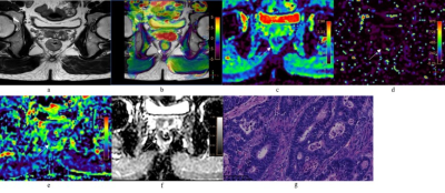 |
4 | Amide proton transfer weighted and intravoxel incoherent motion imaging in evaluation of prognostic factors for rectal adenocarcinoma
Juan Li1, cheng jingliang1, and Lin Liangjie2
1Department of MRI, The First Affiliated Hospital of Zhengzhou University, Zhengzhou, China, 2Philips Healthcare, Beijing, China
This study explored the application of APT and IVIM in evaluation of prognostic factors for rectal adenocarcinoma. Parameters of APT signal intensity (APT SI), pure diffusion coefficient (D), pseudo-diffusion coefficient (D*), perfusion fraction (f), and apparent diffusion coefficient (ADC) were measured. The results showed that APT SI was significantly lower in low- than high-grade adenocarcinoma. The D value was significantly higher in low- than high-grade adenocarcinoma. The D value was significant lower in positive than in negative extramural vascular invasion (EMVI). This suggests that APT and IVIM were helpful to assess rectal adenocarcinoma, including histopathological grade and the EMVI status.
|
||
4203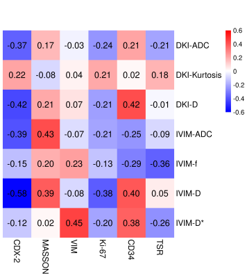 |
5 | DKI and IVIM for Assessment of Rectal Cancer: Correlations between Imaging Parameters and Tumor Tissue Composition
Jie Yuan1, Mengxiao Liu2, Songhua Zhan1, Zhigang Gong1, and Qiang Shen1
1Shuguang Hospital Affiliated to Shanghai University of Traditional Chinese Medicine, Shanghai, China, 2MR scientific Marketing, Diagnostic Imaging, Siemens Healthineers Ltd, Shanghai, China
It is important to combine histopathological and non-invasive imaging measurements to assess tumor components and predict tumor invasiveness to define disease treatment and prognostic assessment. In this study, we assessed the relationships between DKI and IVIM parameter measurements and the corresponding tumor tissue composition, regardless of the status of the malignant tumor. To this end, we selected a range of tissue parameters informative for important biological properties of the tumor, such as Ki-67, the tumor: stroma ratio, CD34 etc.
|
||
4204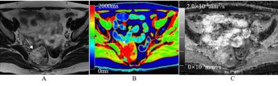 |
6 | Application of native T1 mapping for distinguishing histopathologic subtypes, grades, and stages of rectal adenocarcinoma
Juan Li1, Marcel Dominik Nickel2, cheng jingliang1, and Zhu Jinxia 3
1Department of MRI, The First Affiliated Hospital of Zhengzhou University, Zhengzhou, China, 2MR Application Predevelopment, Siemens Healthcare GmbH, Erlangen, Germany, 3MR collaboration, Siemens Healthcare Ltd., Beijing, China
This study evaluated the clinical significance of using native T1 mapping for distinguishing histopathologic subtypes, grades, and stage of rectal adenocarcinoma by measuring parameters such as T1 relaxation time and apparent diffusion coefficient (ADC). The results showed that T1 and ADC values were significantly higher in rectal mucinous adenocarcinoma (MC) compared to non-mucinous rectal adenocarcinoma (AC). In the AC group, T1 values were significantly lower in the low-grade AC than in the high-grade AC. Our findings suggest that native T1 mapping can distinguish histopathologic subtypes of renal adenocarcinoma. Moreover, T1 values distinguish different grades of AC more accurately than ADC values.
|
||
 |
4205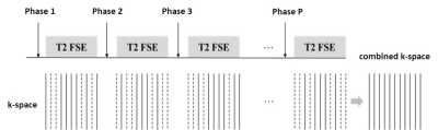 |
7 | T2-weighted dynamic approach for rectal MR imaging
Jing Cheng1, Jiansen Li2, Jun Yang2, Huimao Zhang3, Guobin Li2, Xin Liu1, and Dong Liang1
1Shenzhen Institutes of Advanced Technology, Chinese Academy of Sciences, Shenzhen, China, 2United Imaging Healthcare, Shanghai, China, 3Department of Radiology, the First Hospital of Jilin University, Changchun, China
T2-weighted MR imaging is the standardized protocol in clinical evaluation of rectal cancer and anatomic structures. Usually, spasmolytic agent injection is required before the examination to reduce the imaging artifacts caused by the rectum peristalsis. In this work, we propose a novel dynamic imaging approach that with a complementary k-space sampling strategy and deep learning-based image reconstruction method to illustrate the anatomical structures of the rectum without any agent injection. Experimental results demonstrate the superior performance of the proposed approach.
|
|
4206 |
8 | Comparative Study of histogram analysis of Intravoxel Incoherent Motion and Diffusion Kurtosis Imaging for the staging of liver fibrosis
Fengxian Fan1, Yanli Jiang1, Jing Zhang1, Jie Zou1, and Jialiang Ren2
1Department of Magnetic Resonance, Lanzhou University Second Hospital, Lanzhou, China, 2GE Healthcare, Shanghai, China
The purpose of this study is to compare the diagnostic accuracy of histogram analysis–derived parameters from intravoxel incoherent motion (IVIM) and diffusion kurtosis imaging (DKI) for staging of liver fibrosis (LF). The correlations between different histopathologic stages and histogram parameters were determined. The results showed that the histogram metrics of DKI maps demonstrate significant correlation with fibrosis stage, while there was no correlation between histogram metrics of IVIM and fibrosis stage. In conclusion, histogram analysis of DKI may serve as a valuable and robust tool for the staging of LF.
|
||
4207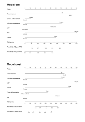 |
9 | Gadoxetic Acid–enhanced MRI for Predicting Postsurgical Recurrence in Hepatocellular Carcinoma:Compared with Four Clinical Staging Systems
Hong Wei1, Hanyu Jiang1, and Bin Song1
1Radiology, West China Hospital, Sichuan University, Chengdu, China
A total of 214 patients with pathologically confirmed Hepatocellular carcinoma (HCC) who underwent gadoxetic acid-enhanced magnetic resonance imaging (EOB-MRI) before curative resection between July 2015 and November 2020 were retrospectively included in this study. The preoperative model integrating EOB-MRI findings and serum AFP and AST levels achieved accurate recurrence prediction in HCC, with similar performance to that of the postoperative clinical-radiologic-pathologic model. Moreover, the preoperative model yielded superior predictive performance to four widely used clinical staging systems for HCC recurrence prediction. This model offered a potential noninvasive and reliable approach for individualized recurrence risk estimation before hepatectomy.
|
||
4208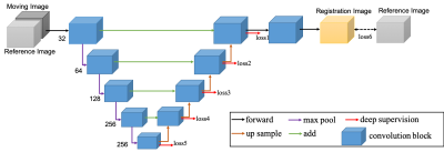 |
10 | Deep Learning Method Based Automatic Subtraction of Liver DCE-MR Images
Zhijun Geng1,2, Botong Wu3,4, Tiantian Liu3, Niange Yu3, Dandan Zheng3, Hua Guo4, and Chuanmiao Xie1,2
1The department of medical imaging, Sun Yat-sen University,Cancer Center, Guangzhou, China, 2States key laboratory in South China, Guangzhou, China, 3Shukun (Beijing) Technology Co., Ltd, Beijing, China, 4Center for Biomedical Imaging Research, Department of Biomedical Engineering,School of Medicine, Tsinghua University, Beijing, China
Subtraction images are an important part of routine multi-phase contrast-enhanced MRI for characterizing enhancement of lesions which are intrinsically hyperintense on T1-weighted imaging. Successful subtraction MRI is dependent on precise 3D co-registration of the pre- and contrast-enhanced source data. However, there still lack a robust, convenient, time efficient and labor free method for automatically image subtraction. This study developed a deep learning based nonrigid registration algorithm, measure the improvement in displacement after registration using anatomic landmarks and automatically generate the subtraction liver images.
|
||
4209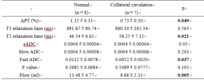 |
11 | Evaluation of hepatic microenvironment in cirrhotic patients with collateral circulation by multiparametric MRI
Wang Nan1, Jiazheng Wang2, Peng Sun2, Lihua Chen1, Qingwei Song1, and Ailian Liu1
1Department of Radiology, the First Affiliated Hospital of Dalian Medical University, Dalian, China, 2Philips Healthcare, Beijing, China
Cirrhosis of the liver not only increases the risk of liver cancer but also causes portal hypertension, leading to gastrointestinal bleeding, hepatic encephalopathy, ascites, and other complications. The opening of collateral circulation caused by portal hypertension plays an important role in the occurrence and progress of diseases. Therefore, precise diagnosis of collateral circulation (CC) in cirrhotic patients is important for clinical practice. Hereby, we compared quantitative parameters from multiparametric MRI (4D flow, APT, IVIM, T1/T2 mapping) between the CC and non-CC groups in cirrhotic patients, which showed significant differences.
|
||
4210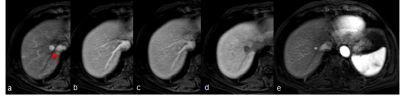 |
12 | Diagnostic Performance of Gd-EOB-DTPA for Hepatocellular Carcinoma Based on LI-RADS v2018, Compared With v2017 Video Not Available
Fei Xing1 and Xiance Zhao2
1the Third People’s Hospital of Nantong, Nantong, China, 2Philips Healthcare, Shanghai, China
The purpose of this study was to compare the diagnostic performance of the LI-RADS v2017 and v2018 for HCC in patients with cirrhosis using Gd-EOB-DTPA. We retrospectively analyzed Clinical data of 213 patients(246 observations)with cirrhosis who underwent Gd-EOB-DTPA from October 2015 to July 2020 were retrospectively collected. MRI major features were reviewed by two radiologists. The LI-RADS categories were assigned according to v2017 and v2018. Our study showed that Updated LR-5 criteria on Gd-EOB-DTPA, the diagnostic performance of LI-RADS v2018 for diagnosing HCC is superior to v2017, particularly in small HCC (10–19 mm), with a greater sensitivity and similar specificity.
|
||
4211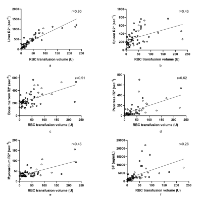 |
13 | Quantification of multi-organ iron overload using R2* mapping in patients with transfusion-dependent diseases
Xiaonan Wang1, Tongtong Sun1, Jinxia Zhu2, Stephan Kannengiesser3, and Hongyan Ni4
1Tianjin Medical University, Tianjin, China, 2Siemens Healthineers Ltd., Beijing, China, 3Siemens Healthcare GmbH, Erlangen, Germany, 4Tianjin First Central Hospital, Tianjin, China
We used R2* mapping to assess iron overload in multiple organs of patients with transfusion-dependent diseases and related the R2* values to transfusion volumes and serum ferritin (SF) levels. The organs assessed included the liver, spleen, pancreas, vertebral bone marrow, and myocardium. The results showed a correlation between the R2* values of multiple organs and between the R2* values of some organs and transfusion volumes. This study revealed differences in the distribution of iron overload in various organs and suggested that transfusion volumes could be used to predict the degree of liver iron overload in patients with transfusion-dependent diseases.
|
||
4212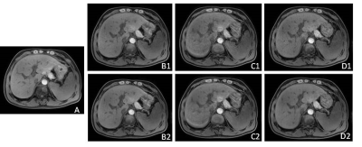 |
14 | 3D-isotropic T1WI in liver MRI with compressed sensing and artificial intelligence reconstruction Video Not Available
Shuang Zheng1, Lei Zhang1, Huimao Zhang1, Zhuo Wang1, Yuejiao Sun1, Yi Zhu2, and Ke Jiang2
1The First Hospital of Jilin University, Changchun, China, 2Philips Healthcare, Beijing, China
3D-isotropic volumetric data acquisition provides higher spatial resolution and signal-to-noise ratios, and facilitates multi-planar reformations. But the application was limited due to long scan time. Recently, compressed sensing (CS) has been proposed as a new method for reducing the number of k-space samples. However, one of the drawbacks of CS is a relatively lower SNR than for PI (parallel imaging). Artificial intelligence reconstruction has been introduced for improving imaging quality. The aim of this study is to investigate the feasibility of 3D-isotropic T1 weighted imaging (T1WI) with compressed sense AI reconstruction in the liver magnetic resonance imaging.
|
||
4213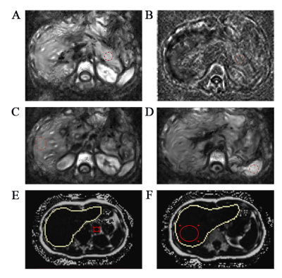 |
15 | Feasibility of Using Multi-frequency MR Elastography for the Assessment of Pancreatic Stiffness and Fluidity in Healthy Volunteers Video Permission Withheld
Siya Shi1, Yanji Luo1, and Liqin Wang1
1The First Affiliated Hospital, Sun Yat-Sen University, Guangzhou, China
Tomoelastography, a newly emerging imaging modality, is a multi-frequency magnetic resonance elastography technique using noise-robust data post-processing. The aim of our study was to investigate the assessment of stiffness and fluidity of pancreas in healthy volunteers with tomoelastography. Tomoelastography derived pancreatic stiffness and fluidity were near-perfect reproducible in healthy volunteers. There was no stiff or fluidic difference among different groups of sex, age, BMI or pancreatic anatomical region. Tomoelastography can provide stable and promising stiffness and fluidity measurements throughout the pancreas. Our results provide data that will enable pancreatic studies of tomoelastography as a potential clinical tool in future.
|
||
4214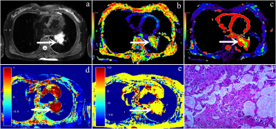 |
16 | Evaluation of amide proton transfer-weighted imaging and stretch index diffusion imaging for lung cancer staging and its correlation with Ki67
Pengyang Feng1, Nan Meng2, Zhun Huang1, Ting Fang2, Ziqiang Li3, Fangfang Fu4, Wei Wei4, Yan Bai4, Yang Yang5, Jianmin Yuan6, and Meiyun Wang*1
1Department of Radiology, Henan University People’s Hospital & Henan Provincial People’s Hospital, Zhengzhou, China, 2Department of Radiology, Zhengzhou University People’s Hospital & Henan Provincial People’s Hospital, Zhengzhou, China, 3Xinxiang Medical University & Henan Provincial People's Hospital, Xinxiang, China, 4Department of Radiology, Henan Provincial People’s Hospital, Zhengzhou, China, 5UIH Group, Central Research Institute, Beijing, China, 6UIH Group, Central Research Institute, Shanghai, China
Amide proton transfer-weighted imaging (APTWI) primarily reflects the protein content and acidity information of tissue structures. Stretch index diffusion imaging includes α, DDC values, these parameters reflect the heterogeneous characteristics of tumors. Ki67 is a good indicator to evaluate the proliferative activity of tumor cells. The results showed that APTWI and stretch index diffusion imaging had similar diagnostic performance in the diagnosis of lung cancer staging,and there was a certain correlation between the parameters and Ki67.
|
||
 Back to Meeting Home
 Back to the Program-at-a-Glance
The International Society for Magnetic Resonance in Medicine is accredited by the Accreditation Council for Continuing Medical Education to provide continuing medical education for physicians.
 Back to Meeting Home
 Back to the Program-at-a-Glance
 View the Presentation