Online Gather.town Pitches
Body: GI
Joint Annual Meeting ISMRM-ESMRMB & ISMRT 31st Annual Meeting • 07-12 May 2022 • London, UK

| Booth # | ||||
|---|---|---|---|---|
4215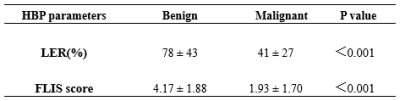 |
1 | Can Gd-EOB-DTPA enhanced MRI effectively in differentiate benign biliary obstruction from malignant obstruction?
Jinghui Hu1, Xiaoxiao Wang1, Weiqiang Dou2, Jing Ye1, and Xianfu Luo1
1Clinical Medical School of Yangzhou University, Northern Jiangsu People’s Hospital, Yangzhou, China, Yangzhou City, China, 2GE Healthcare, MR Research China, Beijing, P.R. China, Yangzhou City, China
This study aimed to investigate the feasibility of Gd-EOB-DTPA enhanced MRI in differentiating benign from malignant biliary obstruction at hepatobiliary phase (HBP) images. The liver enhancement ratio (LER) and the functional liver imaging (FLIS) score of benign and malignant biliary obstruction were significantly different. With these findings, Gd-EOB-DTPA enhanced MRI at HBP may be considered with an added clinical value in differentiating benign from malignant biliary obstruction.
|
||
4216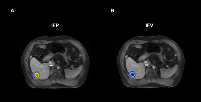 |
2 | Preoperative prediction of microvascular invasion in hepatocellular cancer using computation modeling of interstitial fluid pressure
Liyun Zheng1,2, Lifang Wu3, Chun Yang3, Ruofan Sheng3, Yongming Dai2, and Mengsu Zeng1,3
1Shanghai Institute of Medical Imaging, Shanghai, China, 2United Imaging Healthcare, Shanghai, China, 3Department of Radiology, Zhongshan Hospital, Fudan University, Shanghai, China Microvascular invasion (MVI) is a well-known major prognostic factor of hepatocellular carcinoma (HCC). Unlike macrovascular invasion, which can be easily identified via imaging before treatment, microvascular invasion (MVI) is visible only at microscopy. This study developed a new method for the noninvasive interstitial fluid pressure (IFP) measurement in HCC patients. Our results revealed that MVI status can be predicted by this IFP model preoperatively, |
||
4217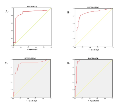 |
3 | Assessment of susceptibility-weighted Imaging in the staging of liver fibrosis Video Not Available
Xuan Jin1, Xinming Li1, Qiying Ke1, Tianyuan Zhang1, Jing Li1, Bingbing Bai1, Xianyue Quan1, and Chen Zhao2
1Department of Radiology, Zhujiang Hospital, Southern Medical University, Guangzhou, China, Guangzhou, China, 2Philips Healthcare, Guangzhou, China, Guangzhou, China
Diagnosing and staging liver fibrosis arouse worldwide concern, and the value of susceptibility-weighted Imaging (SWI) score combined with serum indexes in staging liver fibrosis is controversial. The objective of this study was to assess if SWI score combined with serum indexes could increase the accuracy of staging liver fibrosis. With the help of multivariate ordered logistic regression model, SWI score and serum indexes showed significant correlations with stages of liver fibrosis.
|
||
4218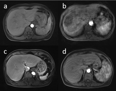 |
4 | Evaluation of the advantage of free-breathing liver DCE-MRI with golden-angle radial sparse parallel using different contrast agents
Lingjie Yang1, Xiaohui Duan1, Mengzhu Wang2, Wei Jiang1, Yu Wang1, Huijun Hu1, Weike Zeng1, and Jun Shen1
1Department of Radiology, Sun Yat-Sen Memorial Hospital, Sun Yat-Sen University, Guangzhou, China, 2MR Scientific Marketing, Siemens Healthineers, Guangzhou, China
To evaluate the image quality between two sequences (GRASP vs. CDT-VIBE), as well as two kinds of contrast agents (Gd-EOB-DTPA vs. gadobutrol) in liver DCE-MRI. The qualitative and quantitative evaluations regarding of the image quality, and the detection rates of lesions among four groups were analyzed and compared. The results showed that MR images with GRASP sequence had significantly better image quality and less artifacts than that with CDT-VIBE sequence especially in arterial phase, and MR images with injection of gadobutrol could improve the hepatic vessel clarity and reduce motion artifacts compared with that of Gd-EOB-DTPA in CDT-VIBE sequence.
|
||
4219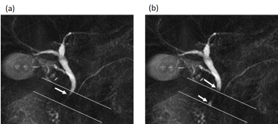 |
5 | Bile flow dynamics in patients with cholelithiasis: Evaluation with cine-dynamic MRCP using spatially selective inversion-recovery pulse Video Permission Withheld
Mayumi Higashi1, Masahiro Tanabe1, and Katsuyoshi Ito1
1Radiology, Yamaguchi University Graduate School of Medicine, Ube, Yamaguchi, Japan
We evaluated the changes in the bile flow dynamics in patients with cholelithiasis using cine-dynamic MRCP with a spatially selective IR pulse. The frequency and mean secretion grade of the antegrade bile flow were significantly increased in the gallstone group than in the non-gallstone group, while no significant differences in the frequency and mean secretion grade of the reverse bile flow were found between the two groups. Cine-dynamic MRCP with a spatially selective IR pulse can visualize the changes in the bile flow dynamics in patients with gallstones noninvasively.
|
||
4220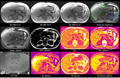 |
6 | An Automatic Abdomen MR-Quantification based on a Simultaneous Multi-Parameter Mapping within 3 Breath-holds and deep-learning Segmentation
Min-xiong Zhou1, Zheng Qu2, Yun Liu3, Haodong Zhong3, Yang Song4, Guang Yang3, Jianqi Li3, and Xu Yan4
1Shanghai University of Medicine & Health Sciences, Shanghai, China, 2West China School of Medicine/West China Hospital, Sichuan University, Chengdu, China, 3Shanghai Key Laboratory of Magnetic Resonance, East China Normal University, Shanghai, China, 4MR Scientific Marketing, Siemens Healthineers, Shanghai, China
For automatic abdomen quantification, a rapid and simultaneous multi-parameter mapping method was evaluated, which could acquire quantitative R1, R2* and PDFF indices within 3-4 breath-holds. By adopting automatic organ segmentation, it could directly generate MR quantitative information for liver, kidney and spleen, which could be potentially used for liver pathology evaluation. The result showed that the multi-parameter sequence could generate high-quality R1, R2* and PDFF maps, with well B1 field correction and fat signal separation.
|
||
4221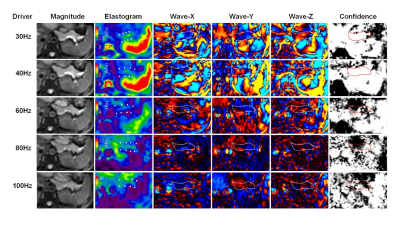 |
7 | Feasibility and Reproducibility of Multi-frequency MR elastography in healthy and diseased pancreas
Qike Song1 and Yu Shi1
1radiology, Shengjing Hospital of China Medical University, Shenyang,, China
To determine the feasibility of multi-frequency MRE for assessing pancreatic stiffness in healthy volunteers and patients with pancreatic adenocarcinoma (PDAC).A total of 40 healthy volunteers and 10 patients with PDAC were prospectively recruited between March 2020 and October 2021.Each volunteer and patient underwent 3.0-T pancreatic MRE at frequencies of 30Hz, 40Hz, 60Hz, 80Hz and 100Hz.Shear stiffness of healthy pancreas and PDAC, pancreatic width and volume, waist circumference, and wave distance were measured. Image quality assessment was performed according to image quality score (IQS: 1~4,≥3 considered qualified).
|
||
4222 |
8 | Differentiation between hepatocellular carcinoma (HCC) and hepatic metastasis (HM) by mDIXON quant and IVIM imaging Video Not Available
Xue Ren1, Jiazheng Wang2, Liangjie Lin2, Zhigang Wu2, Qingwei Song1, Renwang Pu1, Lihua Chen1, Ying Zhao1, Tao Lin1, Qihao Xu1, and Ailian Liu1
1Department of Radiology, the First Affiliated Hospital of Dalian Medical University, Dalian, China, 2Philips Healthcare, Beijing, China
Hepatocellular carcinoma (HCC) and hepatic metastasis (HM) are associated with distinct treatment and prognosis, while the differential diagnosis of HCC and HM still remains challenging. This study aims to explore the value of mDIXON quant and IVIM imaging in the differential diagnosis of HCC and HM. Results shown that the combination of R2*and IVIM measurements may offer a good biomarker for differential diagnosis of HCC and HM.
|
||
4223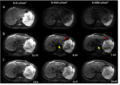 |
9 | High-Resolution Diffusion-Weighted Imaging of Hepatocellular Carcinoma (HCC)
Hui Zhang1, Chengyan Wang2, Peng Wu3, Huazheng Shi4, Weibo Chen3, and He Wang1
1Institute of Science and Technology for Brain-Inspired Intelligence, Fudan University, Shanghai, China, 2Human Phenome Institute, Fudan University, Shanghai, China, 3Philips Healthcare, Shanghai, China, 4Shanghai Universal cloud imaging dignostic center, Shanghai, China
Diffusion-weighted imaging (DWI) is a widely used tool for diagnosing hepatocellular carcinoma (HCC). However, clinical routine liver DWI suffers from poor spatial resolution, severe geometry distortion, and image blurring since single-shot (SSH) echo-planar imaging (EPI) is used. Here we proposed an efficient solution for clinical high-resolution whole-liver DWI. The efficacy and generalization capability of the method were validated on healthy volunteers and HCC patients.
|
||
4224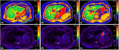 |
10 | Correlation between liver function indexes and T1/T2 relaxation time in patients with liver cirrhosis
Wang Nan1, Qingwei Song1, Jiazheng Wang2, Peng Sun2, Lihua Chen1, and Ailian Liu1
1Department of Radiology, the First Affiliated Hospital of Dalian Medical University, Dalian, China, 2Philips Healthcare, Beijing, China
Evaluation of liver reserve function is crucial for patients with cirrhosis and is helpful for the determination of the surgical plan formulation, the evaluation of treatment process and prognosis, and the operation of cirrhosis. However, there is still lacking a clinically useable non-invasive technique to measure the abnormality of liver function. The purpose of this study is to investigate the potential correlation between T1/T2 relaxation time and liver function indexes in patients with cirrhosis using MR T1/T2 mapping. We found that there was a moderate correlation between T1/T2 relaxation time and liver function assay indexes in liver cirrhosis.
|
||
4225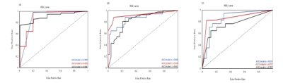 |
11 | Stage liver fibrosis and inflammatory activity using radiomic models based on easy access magnetic resonance images Video Permission Withheld
Huanhuan Wei1, Fangfang Fu2, Yaping Wu2, Yan Bai2, Nan Meng1, Wei Wei2, Kewei Liu3, Xianchang Zhang4, and Meiyun Wang2
1Academy of Medical Sciences, the People’s Hospital of Zhengzhou University, Zhengzhou, China, 2Department of Medical Imaging, Henan Provincial People's Hospital & the People’s Hospital of Zhengzhou University, Zhengzhou, China, 3The First People's Hospital of Zhoukou, Zhoukou, China, 4MR Collaboration, Siemens Healthineers Ltd, Beijing, China
Invasive pathological puncture biopsy is the diagnostic standard for liver fibrosis (LF), but it has certain risks and low repeatability. A non-invasive and reliable method is essential for early detecting and staging of LF. To this end, this study explored the feasibility of using radiomics approach based on easy access T1-weighted and T2-weighted fat saturation magnetic resonance imaging data to stage the liver fibrosis and inflammatory activity. The receiver operating curves analysis demonstrated that the trained radiomic models could effectively stage the LF and inflammatory activity with a high efficacy.
|
||
4226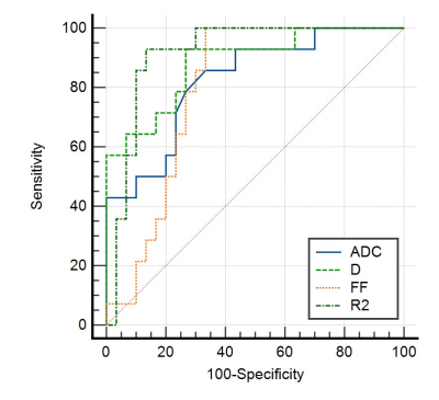 |
12 | The value of IVIM-DWI and IDEAL-IQ in evaluating pathological grading of Hepatocellular Carcinoma
Shaopeng Li1, Kexue Deng1, and Peng Wang2
1Department of Radiology, The First Affiliated Hospital of USTC, Southern District of Anhui Provincial Hospital, Hefei, China, 2Department of Radiology, The First Affiliated Hospital of USTC, Southern District of Anhui Provincial Hospital, hefei, China
During the evolution of hepatocellular carcinoma(HCC) from high differentiation to low differentiation, the number of tumor cells increased and arranged more closely, the limited diffusion of water molecules was more obvious. Meanwhile, the iron and fat content in the lesions decreased gradually. The ADC、D values derived from IVIM-DWI and FF、R2* values derived from IDEAL-IQ can accurately reflect the diffusion limitation and the changes of fat and iron content in HCC, so as to accurately evaluate the pathological grade of HCC.
|
||
4227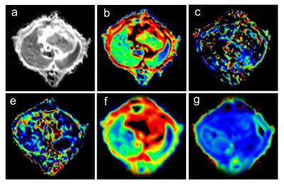 |
13 | The value of different diffusion models in evaluating liver regeneration after standard partial hepatectomy in rats
caixin Qiu1, shuangshuang xie1, yajie Sun2, yongquan Yu3, xuyang Wang2, jinxia Zhu4, Grimm Robert5, and wen Shen2
1Radiology Department, Tianjin First Center Hospital, TIANJIN, China, 2Tianjin First Center Hospital, TIANJIN, China, 3Weihai Central Hospital, SHAN DONG, China, 4MR Collaboration, Siemens Healthcare Ltd., BEIJING, China, 5MR Application Predevelopment, Siemens Healthcare GmbH, Erlangen, Germany
Different diffusion weighted imaging (DWI) models were used to evaluate microscopic changes of residual liver in rats after standard partial hepatectomy (70%PH). Multiple b-value DWI at multiple timepoints after 70%PH were acquired, and the data was analyzed using three diffusion models: conventional monoexponential DWI, intravoxel incoherent motion (IVIM), and diffusion kurtosis imaging (DKI) models. Compared with the monoexponential model of DWI, IVIM and DKI models can describe the changes of blood supply in the process of liver regeneration and provide added value in evaluating the microstructure of liver regeneration after standard partial hepatectomy.
|
||
4228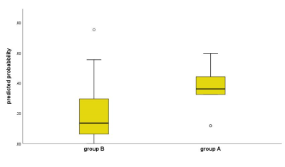 |
14 | The value of DCE-MRI quantitatively predicting vascular invasion of rectal cancer Video Not Available
Wan Dong1, Anliang Chen1, Yuhui Liu2, Qingwei Song2, Lizhi Xie3, and Ailian Liu2
1Radiology, the First Affiliated Hospital of Dalian Medical University, Dalian, China, 2the First Affiliated Hospital of Dalian Medical University, Dalian, China, 3GE Healthcare, MR Research, Beijing, China
Vascular invasion has been proved to be closely related to the poor prognosis of rectal cancer. The current gold standard for the diagnosis of rectal cancer vascular tumor thrombus is postoperative pathology. MRI is a common technology in evaluating malignant tumor. In this work, we explored the feasibility of DCE-MRI in quantitatively predicting vascular invasion of rectal cancer. Results showed that the Ktrans and Kep can differentiate the vascular invasion from normal status accurately. Therefore, DCE-MRI may serve as a feasible and non-invasive way in predicting vascular invasion of rectal cancer preoperatively, that is of great significance for clinical diagnosis.
|
||
4229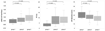 |
15 | Age-related changes in elasticity, fat degeneration, and morphology of the pancreas: evaluation using multiparametric MR imaging
Akihiko Kanki1, Kazuya Yasokawa1, Hidemitsu Sotozono1, Kiyoka Maeba1, Atsushi Higaki1, Akira Yamamoto1, and Tsutomu Tamada1
1Radiology, Kawasaki Medical School, Kurashiki, Japan
We evaluated relationships between age and shifted apparent diffusion coefficient, proton density fat fraction (PDFF), and pancreas size using 3-T MRI. PDFF increased with age, and anterior-posterior diameters decreased with age. Interestingly, elasticity of the pancreas decreased with age. Based on these results, decreases in the size of the pancreatic parenchyma with age appear attributable to not only fibrosis, but also fatty degeneration, resulting in decreased elasticity. Fat content should be evaluated at the same time as fibrosis of the pancreatic parenchyma.
|
||
4230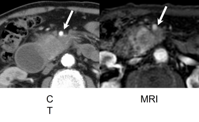 |
16 | Vascular Involvement and Resectability of Pancreatic Ductal Adenocarcinoma on Contrast-Enhanced MRI: Comparison with Pancreatic Protocol CT
Yoshifumi Noda1, Nobuyuki Kawai1, Avinash R. Kambadakone2, Tetsuro Kaga1, Takuma Ishihara3, Fuminori Hyodo4, Hiroki Kato1, and Masayuki Matsuo1
1Department of Radiology, Gifu University, Gifu, Japan, 2Department of Radiology, Massachusetts General Hospital, Boston, MA, United States, 3Innovative and Clinical Research Promotion Center, Gifu University Hospital, Gifu, Japan, 4Department of Radiology, Frontier Science for Imaging, Gifu University, Gifu, Japan
MRI is used as a problem-solving tool when contrast-enhanced CT is not available for assessing pancreatic ductal adenocarcinoma. In this study, we evaluated the diagnostic performance of vascular involvement and resectability on MRI and compared them with pancreatic protocol CT. Our results showed that no statistical difference in terms of the detection of vascular involvement and the resectability classification based on the NCCN guideline between CT and MRI.
|
||
4231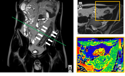 |
17 | Quantitative Evaluation of Crohn's Disease Using Native T1 Mapping: A Feasibility Study Video Permission Withheld
Daisuke Morimoto-Ishikawa1,2, Tomoko Hyodo3, Yoriaka Komeda4, Hiroyuki Fukushima1, Yu Ueda5, Keizou Miyagoshi1, Shigeyoshi Saito2, and Kazunari Ishii3
1Radiology Center, Kindai University Hospital, Osakasayama City, Japan, 2Department of Medical Physics and Engineering, Osaka University Graduate School of Medicine, Suita City, Japan, 3Department of Radiology, Kindai University, Osakasayama City, Japan, 4Department of Gastroenterology and Hepatology, Kindai University, Osakasayama City, Japan, 5Philips Japan, Tokyo, Japan We investigated the usefulness of native T1 mapping using a modified look-locker inversion recovery (MOLLI) sequence, which can be used to evaluate Crohn's disease (CD). The nativeT1 value was measured by setting an ROI on the intestinal wall of the nativeT1 map containing the short-axis image of the intestine. Simple endoscopic activity score in CD (SES-CD) was used as the reference standard for evaluating CD activity. A strong correlation was found between native T1 value and the SES-CD (r = 0.83; p = 0.001). |
||
4232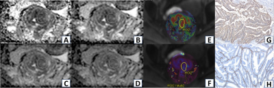 |
18 | Diffusion and perfusion MRI and radiomics signatures for preoperative evaluation of epithelial-mesenchymal transformation in rectal cancer
Yidi Chen1,2, Liling long3, Bin Song1, and Huiting Zhang4
1Radiology, West China Hospital, Sichuan University, Chengdu, China, 2Guangxi Medical University First Affiliated Hospital, Nanning, China, 3Radiology, Guangxi Medical University First Affiliated Hospital, Nanning, China, 4MR Scientific Marketing, Siemens Healthineers, Wuhan, China
This study demonstrated morphological characteristics of MRI can hardly diagnose the expression of E-cadherin and Vimentin in rectal cancer. ADC value (Ultra-high b-Value) was positively and negatively correlated with E-cadherin and Vimentin expression. Ktrans and Kep values were negatively and positively correlated with E-cadherin and Vimentin expression. ADC, Ktrans and Kep values had significant diagnostic efficiencies for low E-cadherin and high Vimentin expression. Radiomics signatures with machine learning have excellent diagnostic efficacy for E-cadherin and Vimentin expression, building the model which combined MRI quantitative parameters and radiomics features will improve the predictive performance for E-cadherin and Vimentin expression.
|
||
 Back to Meeting Home
 Back to the Program-at-a-Glance
The International Society for Magnetic Resonance in Medicine is accredited by the Accreditation Council for Continuing Medical Education to provide continuing medical education for physicians.
 Back to Meeting Home
 Back to the Program-at-a-Glance
 View the Presentation