Online Gather.town Pitches
Cardiovascular Anatomy, Function, Hemodynamics II
Joint Annual Meeting ISMRM-ESMRMB & ISMRT 31st Annual Meeting • 07-12 May 2022 • London, UK

Online Gather.town Pitches
Cardiovascular Anatomy, Function, Hemodynamics II
| Booth # | ||||
|---|---|---|---|---|
4542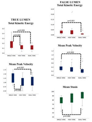 |
1 | Aortic hemodynamics with accelerated dual-venc 4D fow MRI in patients with type B aortic dissection Video Permission Withheld
Ozden Kilinc1, Stanley Chu1, Justin Baraboo1,2, Elizabeth K. Weiss1,2, Anthony Maroun1, Ning Jin3, Kelvin Chow1,4, Xiaoming Bi4, Rachel Davids4, Chris Mehta5, S. Chris Malaisrie5, Andrew Hoel6, Michael Markl1,2, and Bradley D. Allen1
1Department of Radiology, Feinberg School of Medicine, Northwestern University, Chicago, IL, United States, 2Department of Biomedical Engineering, Northwestern University, Chicago, IL, United States, 3Cardiovascular MR R&D, Siemens Medical Solutions USA, Inc., Cleveland, OH, United States, 4Cardiovascular MR R&D, Siemens Medical Solutions USA, Inc., Chicago, IL, United States, 5Division of Cardiac Surgery, Feinberg School of Medicine, Northwestern University, Chicago, IL, United States, 6Division of Vascular Surgery, Feinberg School of Medicine, Northwestern University, Chicago, IL, United States
4D flow MRI is a well-validated imaging technique for quantitative assessment of blood flow hemodynamics and has shown promise in evaluation of type B aortic dissection (TBAD). However, single velocity encoding (venc) 4D flow MRI is limited by its inability to fully capture wide range of velocities related to variable pathologic hemodynamic patterns such as true lumen and false lumen flow range of velocities in TBAD. We hypothesize that dual-venc acquisition improves velocity-to-noise ratio, better captures full dynamic range of velocities in TBAD and provides improved characterization of flow hemodynamics relative to single-venc acquisitions.
|
||
4543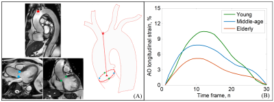 |
2 | Reference value of ascending aorta global longitudinal strain by cardiovascular magnetic resonance feature tracking
HongZhou Zhang1,2, Shuang Leng1,3, Ru-San Tan1,3, Ping Chai4,5, Jennifer Bryant1,3, Lynette Teo5,6, Ching Ching Ong4,5, Angela S. Koh1,3, Wen Ruan1, James W. Yip4,5, Ju Le Tan1,3, and Liang Zhong1,3
1National Heart Centre Singapore, Singapore, Singapore, 2Department of Cardiovascular Medicine, The Second Affiliated Hospital of Nanchang University, Nanchang, China, 3Duke-NUS Medical School, Singapore, Singapore, 4National University Heart Centre, Singapore, Singapore, 5Yong Loo Lin School of Medicine, National University of Singapore, Singapore, Singapore, 6National University Hospital, Singapore, Singapore
This study aimed to introduce a novel parameter—aorta (AO) global longitudinal strain (GLS)—for AO stiffness assessment by semi-automated feature tracking approach with standard cine cardiovascular magnetic resonance (CMR). The data demonstrated excellent intra and inter-observer variability of AO GLS of 4.4% and 5.0%, respectively. AO GLS was significantly negatively associated with age. The mean AO GLS was greater in females (10.1±3.5% versus 8.1±2.7%, P<0.001). The AO GLS decreased by 1.68% in females and 1.32% in males per decade respectively. Age was an independent predictor of the AO GLS in both males and females.
|
||
4544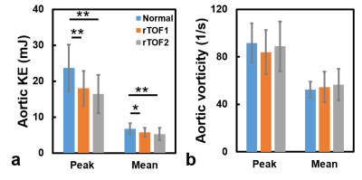 |
3 | Aortic kinetic energy in repaired tetralogy of Fallot patients with or without aortic root dilatation
Yu-Ru Yang1, Meng-Chu Chang1, Ming-Ting Wu2, Ken-Pen Weng3, and Hsu-Hsia Peng1
1Department of Biomedical Engineering and Environmental Sciences, National Tsing Hua University, Hsinchu, Taiwan, 2Department of Radiology, Kaohsiung Veterans General Hospital, Kaohsiung, Taiwan, 3Department of Pediatrics, Kaohsiung Veterans General Hospital, Kaohsiung, Taiwan
The aortic hemodynamic flow pattern and aortic kinetic energy (KE) is less discussed for repaired tetralogy of Fallot (rTOF) patients without aortic root dilatation. We aimed to evaluate aortic KE in rTOF patients with or without aortic root dilatation. The rTOF1 patients (indexed aortic root diameter<16 mm/m2) demonstrated aortic regurgitation. Both rTOF1 and rTOF2 (indexed aortic root diameter≥16 mm/m2) patients exhibited normal aortic vorticity and decreased aortic KE. In conclusion, in rTOF patients with preserved left ventricular ejection fraction, the altered velocity-derived KE and RF can provide an indication of aortapathy progress earlier than morphological aortic dilatation and aortic vorticity.
|
||
4545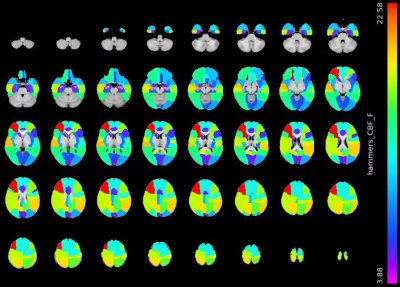 |
4 | Severe Cerebral Small Vessel Disease Burden is Associated with Brain Hypo-perfusion, and higher Framingham Risk score: An 8 year follow-up study
Xiaoqian Zhang1, Sirui Liu1, Feng Feng1, and Zhentao Zuo2,3
1Department of Radiology, Peking Union Medical College Hospital, Chinese Academy of Medical Sciences and Peking Union Medical College, Beijing, China, 2State Key Laboratory of Brain and Cognitive Science, Institute of Biophysics, Chinese Academy of Sciences, Beijing, China, 3Department of Human Anatomy, Histology and Embryology, School of Basic Medicine, Peking Union Medical College, Beijing, China
The study investigated relationship between cerebral small vessel disease (CSVD) burden, cerebral blood flow (CBF) and cardiovascular risk in the aging brains with normal cognitive function at baseline and conducted telephone cognitive follow-up for nearly 8 years. The results showed that severe CSVD burden was significantly associated with higher cardiovascular risk scores and decreased CBF in multiple areas, with involvement of nearly all cortical brain areas. Subjects with severe CSVD burden were more likely to suffer from long-term cognitive decline. This suggests the capability of CSVD burden serving as an imaging marker of predicting cognitive decline.
|
||
4546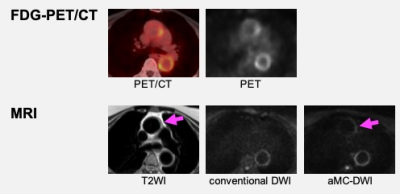 |
5 | Acceleration Motion Compensation Diffusion-weighted Imaging for Large Vessel Vasculitis: Phantom Model and Initial Clinical Experience Video Permission Withheld
Tomoko Hyodo1, Daisuke Morimoto-Ishikawa2,3, Hayato Kaida1, Yu Ueda4, Daisuke Tomita5, Atsuhiro Yamamoto5, Makoto Itoh2, Nao Yasuda2, Hiroyuki Fukushima2, Yuji Nozaki5, Itaru Matsumura5, and Kazunari Ishii1
1Radiology, Kindai University Faculty of Medicine, Osaka-Sayama, Japan, 2Radiology Center, Kindai University Hospital, Osaka-Sayama, Japan, 3Medical Physics and Engineering, Osaka University Graduate School of Medicine, Suita, Japan, 4Philips Japan, Tokyo, Japan, 5Hematology and Rheumatology, Kindai University Faculty of Medicine, Osaka-Sayama, Japan
The ascending aorta is poorly delineated on DWI due to the heartbeat, making it difficult to assess the activity of large vessel vasculitis using MRI. We investigated the utility of acceleration motion compensation diffusion-weighted imaging (aMC-DWI) compared with conventional DWI (cDWI) to detect active inflammation of the accenting aorta. The phantom experiment revealed that aMC-DWI showed a significantly higher median visual score than cDWI in both experimenters (P =.048 for both). The clinical study on 11 patients showed that aMC-DWI had higher sensitivity, specificity, and positive predictive value (with FDG-PET/CT as the reference method) than cDWI.
|
||
4547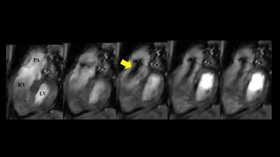 |
6 | High-resolution flow-sensitive CINE imaging visualizing valve, chamber, and regurgitant flow
Isao Shiina1, Michinobu Nagao2, Masami Yoneyama3, Yasuhiro Goto1, Yutaka Hamatani1, Kazuo Kodaira1, Takumi Ogawa1, Mana Kato1, and Shuji Sakai2
1Department of Radiological Services, Tokyo Woman's Medical University, Tokyo, Japan, 2Department of Diagnostic imaging & Nuclear Medicine, Tokyo Woman's Medical University, Tokyo, Japan, 3Philips Japan, Tokyo, Japan
Cardiac bSSFP CINE imaging is widely used in clinical practice for function and morphological evaluation, but we often experienced situations where it is difficult to visually evaluate blood flow and valves. TFEPI is a flow-sensitive imaging method and can be expected to evaluate blood flow and valves. In this study, we investigate the clinical usefulness of Flow sensitive CINE Imaging using TFEPI.
|
||
4548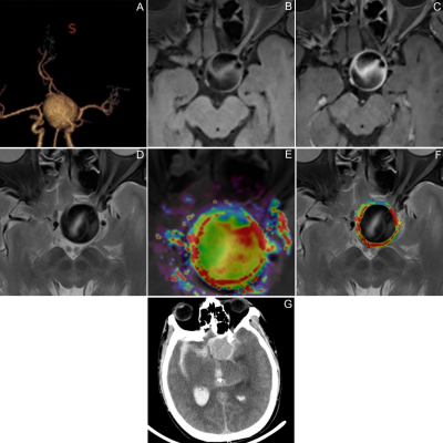 |
7 | Evaluation of the Instability of Intracranial Aneurysms Wall by Dynamic Contrast-enhanced Magnetic Resonance Imaging and Vessel Wall Imaging
Qichang Fu1, Yi Zhang1, Jingliang Cheng1, Sheng Guan2, and Chengcheng Zhu3
1Department of Magnetic Resonance, The First Affiliated Hospital of Zhengzhou University, Zhengzhou, China, 2Department of Interventional Neuroradiology, The First Affiliated Hospital of Zhengzhou University, Zhengzhou, China, 3Department of Radiology, University of Washington, Washington, WA, United States
This study used DCE-MRI and VWI to explore the value of Ktrans and aneurysm wall enhancement (AWE) in screening UIAs instability. Eighty-two patients were enrolled, including 51 patients with stable IAs and 31 patients with unstable IAs. All patients completed examinations based on a 3.0T Siemens Prisma magnetic resonance. Multivariate logistic regression revealed that AWEP (OR, 4.1; 95% CI, 2.06 to 8.16; P < 0.001) and Ktrans (OR, 2.77; 95% CI, 1.49 to 5.17; P = 0.01) were the only independent factors associated with unstable UIAs. Both Ktrans and AWEP were independent risk factors for the unstable state of UIAs.
|
||
4549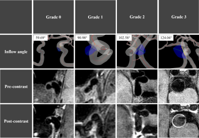 |
8 | Aneurysmal wall enhancement is positively correlated with inflow angle in sidewall aneurysms
Mingzhu Fu1, Fei Peng2, Wenwen Chen1, Aihua Aihua Liu2, and Rui Li1
1Department of Biomedical Engineering, Tsinghua University, Center for Biomedical Imaging Research, Beijing, China, 2Department of Interventional Neuroradiology, Beijing Tiantan Hospital, Beijing, China
In this study, we utilized HR-VWI and 3D TOF-MRA images to investigate the relationship between AWE grades and aneurysm inflow angle. Statistics indicated that the AWE grades were positively associated with the aneurysm inflow angle. This is probably because a sidewall aneurysm with high inflow angle is easier to grow and cause slower inflow velocity and lower wall shear stress which contribute to inflammation processes on aneurysmal wall and lead to a high grade of AWE. The results of this study showed that the AWE was positively associated with aneurysm inflow angle.
|
||
4550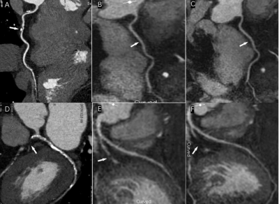 |
9 | Assessment of compressed sensing accelerated non-contrast-enhanced whole-heart coronary MR angiography at 3.0 T
Wenbo Zhang1, Jingliang zhang Cheng1, Liangjie Lin2, Yong zhang Zhang1, Keyan zhang Wang1, Huiyu zhang Huang1, Qi Ren1, and WenHua Zhang1
1MRI, The First Affiliated Hospital of Zhengzhou University, Zhengzhou, China, 2Philips Healthcare, Beijing, China
The feasibility of non-contrast-enhanced coronary MR angiography (CMRA) with acceleration by compressed sensing (CS) was assessed in patients with suspected coronary artery diseases on a 3.0 T scanner, compared with conventional coronary MRA. Results indicate that the whole-heart non-contrast CMRA accelerated by CS with a factor of 4 obtained relatively high sensitivity, positive predictive value, negative predictive value and accuracy in a significantly shortened acquisition time compared with conventional CMRA.
|
||
4551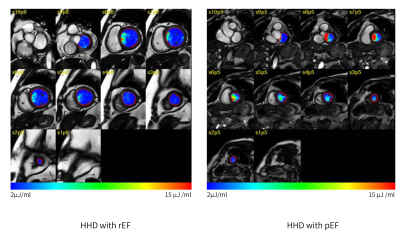 |
10 | Left ventricular blood flow kinetic energy in Hypertensive heart disease :A pilot study of 4D flow cardiovascular magnetic resonance
Kun Peng1, Yong Liu1, Ting Hua1, Xianling Zhang2, Xiance Zhao3, Jilei Zhang3, and Guangyu Tang1
1Department of Radiology, Shanghai Tenth People’s Hospital, School of Medicine,Tongji University, Shanghai, China, 2Department of Cardiology, Shanghai Tenth People’s Hospital, School of Medicine,Tongji University, Shanghai, China, 3Philips Healthcare, Shanghai, China
Four-dimensional flow magnetic resonance imaging (4D-flow MRI) technique has been developed to assess intra-cavity left ventricular (LV) blood flow. Hypertensive heart disease (HHD) is a common cardiovascular disease and manifested as left ventricular hypertrophy and diastolic/systolic dysfunction. The purpose of this study was to investigate whether 4D-flow CMR kinetic energy (KE) parameters are sensitive to distinguish HHD patients from age/gender-matched healthy controls, and to access if there are significant differences in different sub-groups of LV ejection fraction. We also aimed to investigate the association of LV KE to LV function parameters and LV mass.
|
||
4552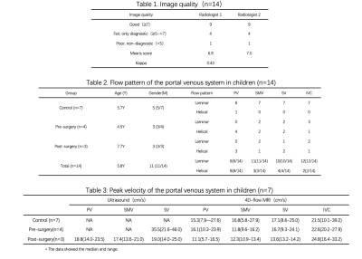 |
11 | The Value of Free-breathing 4D-Flow MRI in Children with Extrahepatic Portal Venous Obstruction: A Preliminary Study
Huiying Wu1, Ning Zhou1, Zhe Wen2, Lianwei Lu1, Mingjie Zhang1, Fuyu You2, Tao Liu2, Yunzhu Wu3, and Xiaochun Zhang*1
1Radiology, Guangzhou Women and Children’s Medical Center, Guangzhou, China, 2Pediatric Surgery, Guangzhou Women and Children’s Medical Center, Guangzhou, China, 3MR Scientific Marketing, Siemens Healthineers, Guangzhou, China
This preliminary study has investigated the feasibility of free-breathing 4D-flow MRI to evaluate portal hemodynamics in children with EHPVO. Free-breathing 4D-flow MRI is feasible and hopeful for the comprehensive 3D visualization and quantification of portal vein flow dynamics in 14 children and the peak velocity had similar diagnostic efficiency to ultrasound in our study. In the future, we intend to compare 4D-flow parameters with intraoperative manometry of the portal vein system and use the noninvasive radionics methodology to estimate portal vein hemodynamics in children.
|
||
4553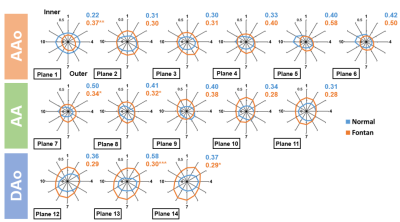 |
12 | Association of Aortic Wall Characteristics and Cardiac Function in Fontan Patient
Yu-Cheng Lin1, Ming-Ting Wu2, Ken-Pen Weng3, and Hsu-Hsia Peng1
1Department of Biomedical Engineering and Environmental Sciences, National Tsing Hua University, Hsinchu, Taiwan, 2Department of Radiology, Kaohsiung Veterans General Hospital, Kaohsiung, Taiwan, 3Department of Pediatrics, Kaohsiung Veterans General Hospital, Kaohsiung, Taiwan
The interaction of aortic wall characteristics and cardiac function in Fontan patients is unclear. 4D flow MRI was employed to quantify the wall shear stress (WSS) and oscillatory shear index (OSI) in the aorta. Fontan patients exhibited increased axial WSS, decreased axial OSI and non-roundness of WSS in descending aorta (DAo). We also found that the increased axial WSS in DAo correlated with decreased ejection fraction and increased indexed end-systolic volume. The cardiac dysfunction presented adverse interaction with aortic wall characteristics in patients with Fontan, especially in DAo, and might lead to higher risk of atherosclerosis and stenosis.
|
||
4554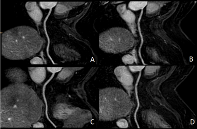 |
13 | Clinical application of 3.0T whole-heart non-contrast agent MRCA by using Compressed SENSE
XianKuo Hu1, Yang Zhang2, XiaoHu Li3, Yu Shan Yuan2, Bin Peng1, and YuanYuan Li2
1Fuyang People's Hospital, fuyang, China, 2Fuyang People's Hospital, FuYang, China, 3the First Affiliated Hospital of Anhui Medical University, hefei, China
MR Coronary angiography (MRCA) technology can observe the anatomical morphology of coronary arteries and detect lumen stenosis, dilation, and abnormal wall lesions [1]. MRCA technology is relatively complex with long scanning time [2]. Compressed SENSE (CS) technology can greatly shorten the scanning time and improve the rate of succussed examination. This study aim to explore the clinical feasibility of Compressed SENSE technology in non-contrast agent MRA and explore the best acceleration factor of CS.
|
||
4555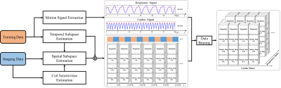 |
14 | Motion-resolved 4D Real-time Flow MRI
Aiqi Sun1, Bo Zhao2, Yichen Zheng3, Yuliang Long4, Peng Wu5, and He Wang1,6
1Institute of Science and Technology for Brain-Inspired Intelligence, Fudan University, Shanghai, China, 2University of Texas at Austin, Austin, TX, United States, 3Beijing PINS Medical Co., Ltd, Beijing, China, 4Department of Cardiology, Zhongshan hospital Fudan University, Shanghai, China, 5Philips Healthcare, Shanghai, China, 6Human Phenome Institute, Fudan, Shanghai, China
Conventional 4D flow MRI requires electrocardiogram gated cine acquisition and respiration control to measure flow dynamics associated with one synthetic cardiac cycle. This often leads to low acquisition efficiency and also cannot resolve beat-by-beat nor respiratory-related flow variations. This work presents a new 4D real-time flow MRI method which is able to simultaneously resolve respiratory and cardiac motion. Compared to conventional 4D flow MRI, the proposed method well captures the beat-by-beat flow variations and respiration-related flow dynamics. Its feasibility for aortic flow was demonstrated in several healthy subjects and also patients with atrial fibrillation.
|
||
4556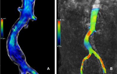 |
15 | Assessments of hemodynamic changes in abdominal aortic aneurysm in patients using 4D EPI flow MRI
Wen Zeng1, Chunchao Xia1, Xiaoyong Zhang2, and Zhenlin Li1
1West China Hospital, Sichuan University, Chengdu, China, 2Clinical Science, Philips Healthcare, Chengdu, China, Chengdu, China
Four-dimensional flow MRI (4D flow MRI) plays a significant role in cardiovascular imaging, which provides new insights into the pathophysiology of disease. In this study, 4D flow with echo-planar imaging was introduced to evaluate the hemodynamic parameters of patients with abdominal aortic aneurysm, including velocity, flow and wall shear stress. We propose that 4D EPI flow can find differences of the above parameters between abdominal aortic aneurysm and normal abdominal aorta and has potential to be used for hemodynamics assessment in AAA patients.
|
||
 Back to Meeting Home
 Back to the Program-at-a-Glance
The International Society for Magnetic Resonance in Medicine is accredited by the Accreditation Council for Continuing Medical Education to provide continuing medical education for physicians.
 Back to Meeting Home
 Back to the Program-at-a-Glance
 View the Presentation