Online Gather.town Pitches
Cardiovascular Anatomy, Function, Hemodynamics I
Joint Annual Meeting ISMRM-ESMRMB & ISMRT 31st Annual Meeting • 07-12 May 2022 • London, UK

Online Gather.town Pitches
Cardiovascular Anatomy, Function, Hemodynamics I
| Booth # | ||||
|---|---|---|---|---|
4130 |
1 | FLEXA 3DTOF: Fast 3D TOF MR Angiography using Thin-slab Two-Point Dixon Acquisition
Naoyuki Takei1, Shiori Amemiya2, Tsuyoshi Ueyama3, Keita Fujii3, Osamu Abe2, and Tetsuya Wakayama1
1GE Healthcare, Tokyo, Japan, 2Radiology, The University of Tokyo Graduate School of Medicine, Tokyo, Japan, 3Radiology, The University of Tokyo Hospital, Tokyo, Japan
Conventional 3DTOF MOTSA MRA technique without contrast agent is an established technique for carotid artery examination. The scan time is relatively long to have opportunity to improve. We have explored 3DTOF with two-point Dixon acquisition to achieve faster scan time by optimizing scan parameters and dealing with Water-Fat swap issue where B0 inhomogeneity and strong susceptibility exist at off-center scan in multi-slab acquisitions. The proposed Dixon MRA offers an alternative approach to conventional 3DTOF with about 4.2 times faster acquisition and better image contrast between artery and muscle for carotid MRA.
|
||
4131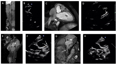 |
2 | Evaluation of Coronary Artery Damage in Kawasaki Disease with Non-Contrast Whole-Heart Coronary Magnetic Resonance Angiography
Yurong Ma1, Juan Liang1, Na Han1, Kai Ai2, and Jing Zhang1
1Department of Magnetic Resonance, Lanzhou University Second Hospital, Lanzhou, China, 2Philips healthcare, Xi'an, China
In this study, the non-contrast whole-heart coronary magnetic resonance angiography (NCE-CMRA) is used to evaluate the coronary artery damage in Kawasaki Disease (KD). The NCE-CMRA and ultrasound images of children with Kawasaki disease (KD) are retrospectively analyzed. Compared with echocardiography, the NCE-CMRA has its unique advantages in displaying coronary artery, such as high image resolution, good soft tissue contrast and strong signal to noise ratio. Furthermore, NCE-CMRA can clearly and directly show the damaged coronary artery. Therefore, it is a potential alternative to ultrasound for imaging of the coronary artery in children with KD.
|
||
4132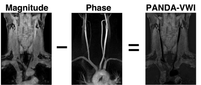 |
3 | PANDA: Simultaneous Phase-Contrast Bright and Dark-blood Angiography for Improvement of Carotid Plaque Assessment
Daichi Murayama1, Takayuki Sakai1, Masami Yoneyama2, Jihun Kwon2, and Shigehiro Ochi1
1Department of Radiology, Eastern Chiba Medical Center, Chiba, Japan, 2Philips Japan, Tokyo, Japan
TOF-MRA sometimes cannot delineate the vessel clearly depending on the vessel orientation. Additionally, insufficient fat suppression can occur in FS-T1-VISTA, it may obscure the plaque depiction. we have developed a new sequence, termed Phase-contrast ANgiography with Dark-blood Application (PANDA), which can simultaneously acquire bright-blood image (MRA) and vessel-wall images (VWI) using the phase contrast method. There was no difference in the visualization ability of carotid plaque between PANDA-VWI and FS T1 VISTA. PANDA-MRA better visualized around the aorta compared to TOF-MRA. PANDA is useful method for simultaneous acquisition of MRA and VWI images.
|
||
4133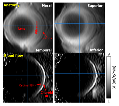 |
4 | High resolution retinal blood flow MRI using a new acquisition strategy for MRI of curved objects
Eric R. Muir1
1Radiology, Stony Brook University, Stony Brook, NY, United States
The retina has highly structured laminar organization, with two separate blood supplies, the retinal and the choroidal blood flow layers. Retinal MRI has generally used a single thick 2D slice. Extension to 3D retinal MRI is difficult due to the curvature of the eye and limited SNR at the needed high resolution. A new acquisition strategy for MRI of curved objects was developed, in which high-resolution is acquired perpendicular to the retina but with low-resolution tangent to the retina. This approach gave significant gains in SNR and provided laminar resolution of the retinal and choroidal blood flow in 3D.
|
||
4134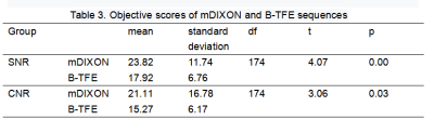 |
5 | Comparative analysis of the imaging quality in magnetic resonance coronary angiography with 3D mDIXON and B-TFE sequence on 3T Video Permission Withheld
Gang Zhang1, Wei Xing2, Tingting Li2, Yan Zheng1, Ying Huang2, Junjing He2, and Xiuzheng Yue3
1The First Affiliated Hospital of Henan University of CM, Zhengzhou, China, 2Department of Magnetic Resonance, The First Affiliated Hospital of Henan University of CM, Zhengzhou, China, 3Department of Magnetic Resonance, Philips Healthcare, Zhengzhou, China
Due to the narrow diameter, tortuous route of coronary arteries, and the interference of respiration and heartbeats during scanning, MR coronary angiography (MRCA) remains challenge. In 3.0T MR systems, an improved 3D Balanced Turbo Field Echo (B-TFE) sequence is often used for MRCA[1,2], however, 3D mDIXON sequences have been applied for clinical application recently.[3,4] In this study, the subjective imaging quality, objective imaging quality and image authenticity indexes of MRCA with two imaging sequences were compared to analysis. Results showed that B-TFE had better subjective evaluation than mDixon, but mDIXON had better objective quality evaluation for SNR and CNR.
|
||
4135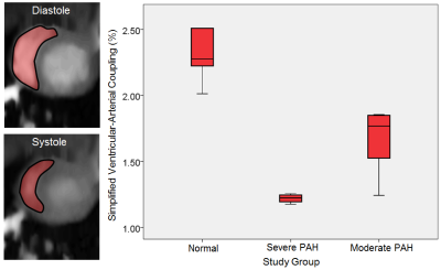 |
6 | Introduction of the Right Ventricular-Arterial Coupling Index in CMR Reflecting Lung Histological Changes in Experimental PAH
Ali Nahardani1,2, Katja Grün3, Martin Krämer2, Karl-Heinz Herrmann2, Andrea Schrepper4, Sara Moradi1, Jürgen R. Reichenbach2, Marcus Franz3, and Verena Hoerr1,2,5
1Heart Center Bonn, Department of Internal Medicine II, University Hospital Bonn, Bonn, Germany, 2Medical Physics Group, Institute of Diagnostic and Interventional Radiology, Jena University Hospital, Friedrich Schiller University Jena, Jena, Germany, 3Department of Internal Medicine I, Division of Cardiology, Angiology, Pneumology, and Intensive Medical Care, Jena University Hospital, Jena, Germany, 4Department of Cardiothoracic Surgery, Jena University Hospital, Jena, Germany, 5Translational Research Imaging Center (TRIC), Clinic for Radiology, University Hospital Muenster, Muenster, Germany
The current preclinical study aims to investigate the associativity between the ventricular-arterial coupling index (VAC) derived by CMR and microscopic vascular changes in pulmonary arterial hypertension (PAH). To this end, three stages of disease were incorporated in the study: healthy, moderate, and severe. All animals underwent CMR and echocardiography. The corresponding lung tissue was also assessed histologically. The right VAC could predict the presence of histopathological changes with r=-0.95 and had a strong correlation with tricuspid annular plane systolic excursion (r=0.90).
|
||
4136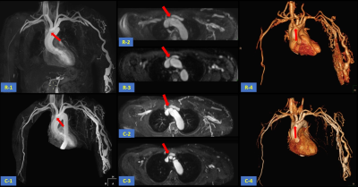 |
7 | The application of REACT for central veins in patients with end-stage renal disease Video Not Available
Fengming Tao1, Li Tao1, Wei Zhu1, Fajin Lv1, Yongmei Li1, Haitao YANG1, Zhiwei Zhang1, and Ke Jiang2
1The first affiliated hospital of Chongqing medical university, Chongqing, China, 2Philips Healthcare, Beijing, China
Relaxation-Enhanced Angiography without Contrast and Triggering (REACT) is a MR angiography technique without cardiac triggering, breath holding and contrast agent injection and is able to show robust blood-to-tissue contrast over multiple anatomies. This study aims to investigate the feasibility of REACT on central veins by comparing it with conventional CE-MRA and catheter angiography. Results showed that the image quality acquired from REACT was comparable to that from CE-MRA and there was no difference in central venous stenosis evaluated by REACT, CE-MRA and angiography.
|
||
4137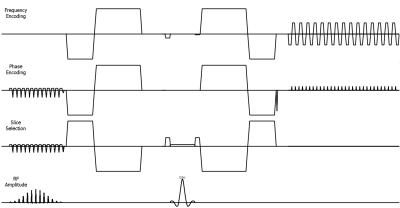 |
8 | iZoom with 2nd order flow compensated diffusion for Improving cardiac diffusion imaging: a preliminary study
Zhigang Wu1, Yajing Zhang2, Xiuquan Hu1, Jing Zhang1, Fei Zeng1, Xiaofang Xu1, Guangyu Jiang3, Yan Zhao3, Guillaume Gilbert4, and Jiazheng Wang1
1Philips Healthcare, Beijing, China, 2MR Clinical Science, Philips Healthcare (Suzhou), Suzhou, China, 3MR R&D, Philips Healthcare (Suzhou), Suzhou, China, 4Philips Healthcare, Precision Diagnosis, Hillmount, ON, Canada
Diffusion MRI could provide unique information non-invasively. However, it is still a technical challenge due to the intrinsic non-rigid motion during the cardiac cycle, displacement of the myocardium due to respiratory motion, field inhomogeneity, and short T1 and T2 values. Parallel imaging and Zoom imaging based on 2D RF (iZoom) could both reduce the distortion dramatically, second order flow compensated diffusion could be used to decrease the impact of motion. We propose a solution that combines iZoom, SENSE and the 2nd flow compensated diffusion to generate a robust cardiac diffusion MRI, its robustness was validated by a preliminary study.
|
||
4138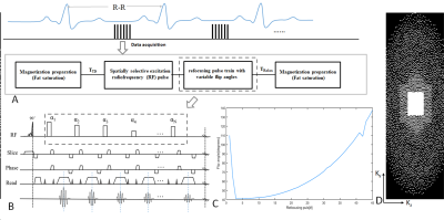 |
9 | High resolution Isotropic 3D Black Blood Thoracic Aorta Imaging With Low Rank Patch-Based Reconstruction
caiyun shi1,2, yuanyuan liu1, guanxu cheng3, yulong qi3, haifeng wang1, lei zhang1, xin liu1, hairong zheng1, dong liang1, and yanjie zhu1
1Shenzhen Institutes of Advanced Technology, Chinese Academy of Sciences, shenzhen, China, 2Shenzhen College of Advanced Technology, University of Chinese Academy of Sciences, shenzhen, China, 3Peking University Shenzhen Hospital, shenzhen, China
Three-dimensional (3D) black-blood MRI is a promising noninvasive imaging technique for assessing aortic atherosclerotic plaque. In this work, a low rank patch-based technique is proposed, and combined with a 3D modulated vFA-FSE sequence for high-resolution thoracic aorta imaging. The comparison was conducted on healthy volunteers and compared against a conventional GRAPPA acquisition to assess the feasibility of the proposed scheme. The results showed the reconstruction scheme was able to visualize the lumen areas clearly and improve the vessel sharpness and contrast ratio significantly.
|
||
4139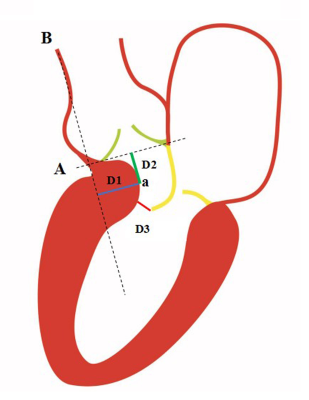 |
10 | Sub-aortic Complex is a Simple and Accurate Parameter to Predict Obstruction in Hypertrophic Cardiomyopathy: A CMR and Doppler Study
Zixian Chen1, Junqiang Lei1, Shunlin Guo2, Zheng Zhang3, Shihua Zhao4, and Minjie Lu4
1Department of Radiology, The First Hospital of Lanzhou University, Intelligent Imaging Medical Engineering Research Center of Gansu Province, Accurate Image Collaborative Innovation International Science and Technology Cooperation Base of Gansu Province, Radiological Clinical Medicine Research Center of Gansu Province, Lanzhou, China, 2Radiology, The First Hospital of Lanzhou University, Intelligent Imaging Medical Engineering Research Center of Gansu Province, Accurate Image Collaborative Innovation International Science and Technology Cooperation Base of Gansu Province, Radiological Clinical Medicine Research Center of Gansu Province, Lanzhou, China, 3Department of Cardiology, The first Hospital of Lanzhou University, Lanzhou, China, 4Department of Magnetic Resonance Imaging, Cardiovascular Imaging and Intervention Center, State Key Laboratory of Cardiovascular Disease,Fuwai Hospital, National Center for Cardiovascular Diseases,Chinese Academy of Medical Sciences and Peking Union Medical College, Beijing, China
To investigate the “sub-aortic complex (SAC)”, a new cardiac magnetic resonance (CMR) derived parameter, for the detection and grading of left ventricular outflow tract (LVOT) obstruction in hypertrophic cardiomyopathy (HCM) patients, compared with echocardiography and CMR 2D flow.
|
||
4140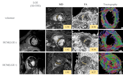 |
11 | Assessment of myocardial microstructural change using novel motion compensated technique in cardiac diffusion tensor imaging
Wataru Ueki1, Yoshiaki Morita1, Yu Ueda2, Masaru Shiotani1, Tatsuhiro Yamamoto1, Yasuhiro Nagai1, Yasutoshi Ohta1, Keizo Murakawa1, and Tetsuya Fukuda1
1National Cerebral and Cardiovascular Center, Suita, Japan, 2Philips Japan Ltd, Tokyo, Japan
The accelerated motion compensation (aMC) using higher order motion compensated gradients with asymmetric bipolar diffusion waveform can improve the image quality of cardiac DTI compared with conventional method and provide the relatively constant DTI marker. In LVH patients, aMC-DTI derived possible preliminary insights into detection of abnormal changes in myocardial microstructures in-vivo, even in segments without LGE.
|
||
 Back to Meeting Home
 Back to the Program-at-a-Glance
The International Society for Magnetic Resonance in Medicine is accredited by the Accreditation Council for Continuing Medical Education to provide continuing medical education for physicians.
 Back to Meeting Home
 Back to the Program-at-a-Glance
 View the Presentation