Online Gather.town Pitches
Vascular Imaging & Stroke I
Joint Annual Meeting ISMRM-ESMRMB & ISMRT 31st Annual Meeting • 07-12 May 2022 • London, UK

Online Gather.town Pitches
Vascular Imaging & Stroke I
| Booth # | ||||
|---|---|---|---|---|
4141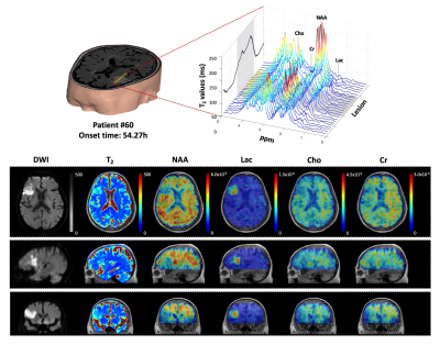 |
1 | Fast High-Resolution 3D MRSI and T2 Mapping for Clinical Outcome Prediction of Ischemic Stroke Patients
Ziyu Meng1, Tianyao Wang2, Bin Bo1, Rong Guo3,4, Yudu Li3,4, Yibo Zhao3,4, Xin Yu5, Zhi-Pei Liang3,4, and Yao Li1
1School of Biomedical Engineering, Shanghai Jiao Tong University, Shanghai, China, 2Radiology Department, The Fifth People's Hospital of Shanghai, Fudan University, Shanghai, China, 3Beckman Institute for Advanced Science and Technology, University of Illinois at Urbana-Champaign, Urbana, IL, United States, 4Department of Electrical and Computer Engineering, University of Illinois at Urbana-Champaign, Urbana, IL, United States, 5Department of Biomedical Engineering, Case Western Reserve University, Cleveland, OH, United States
Characterization of the underlying pathophysiological changes associated with edema progression is essential for treatment guidance in ischemic stroke. 1H-MRSI and quantitative T2 mapping can acquire image markers related to the physiological states of brain tissues. This study investigated the neurometabolite changes in relation to the development of vasogenic edema in ischemic stroke patients, using fast high-resolution 3D MRSI and T2 mapping techniques. Our results showed that neurometabolite concentrations were strongly correlated with T2 values within ischemic lesion of stroke patients and integrated T2 and neurometabolite biomarkers improved the prediction accuracy of stroke patients' clinical outcome.
|
||
4142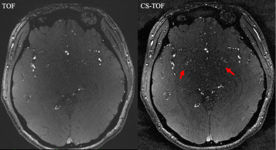 |
2 | 0.2 mm Isotropic Intracranial Perforating Arteries Imaging using Compressed Sensing TOF-MRA at 7T
Zhe Zhang1,2, Qingle Kong3, Jing Jing1,2, and Yongjun Wang1,2,4
1Tiantan Neuroimaging Center of Excellence, Beijing Tiantan Hospital, Capital Medical University, Beijing, China, 2China National Clinical Research Center for Neurological Diseases, Beijing, China, 3MR Collaboration, Siemens Healthineers Ltd., Beijing, China, 4Department of Neurology, Beijing Tiantan Hospital, Capital Medical University, Beijing, China
The impairment of microvessels can lead to neurologic diseases such as stroke and vascular dementia. The imaging of perforating arteries requires an extremely high resolution due to their small caliber size. In this study, we optimized the parameters of compressed sensing (CS) TOF-MRA to achieve isotropic 0.2 mm lenticulostriate artery (LSA) images within 10 minutes at 7T. More LSA stems and branches were delineated in CS TOF images compared with conventional TOF images. CS TOF-MRA can be a state-of-the-art method for detecting microvasculopathies of cerebral vascular diseases.
|
||
4143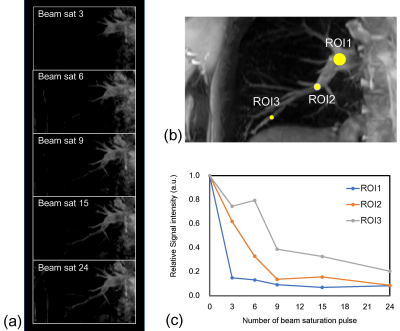 |
3 | Non-Contrast Time-Resolved MR Angiography with Consecutive Beam Pulse with Variable Saturation Flip Angle Video Permission Withheld
Hirohito Kan1,2, Kyosuke Mizuno3, Masahiro Takizawa4, Masashi Shimohira5, Tatsuya Kawai5, Tositaka Aoki6, Satoshi Tsubokura6, and Harumasa Kasai6
1Department of Integrated Health Scieneces, Nagoya University Graduate School of Medicine, Nagoya, Japan, 2Department of Radiology, Nagoya City University Graduate School of Medical Sciences, Nagoya, Japan, 3Department of Radiology, Nagoya City University Hospital, Nagoya, Japan, 4FUJIFILM Healthcare Corp., Tokyo, Japan, 5Department of Radiology, Nagoya City University Graduate School of Medical Scieneces, Nagoya, Japan, 6Nagoya City University Hospital, Nagoya, Japan
To develop and validate the non-contrast time-resolved MR angiography (NC TR-MRA) with consecutive beam pulse with variable saturation flip angle method, we performed a phantom study using steady flow and the volunteer study targeted to the pulmonary artery. The length of visualized flow signals was exhibited proportional to the number of beam saturation pulses in the phantom study. In human study, the visualization range of the pulmonary artery was also extended with increasing the number of beam pulses. The novel consecutive beam pulse with variable saturation flip angle method can depict blood vessels in each flow phase.
|
||
4144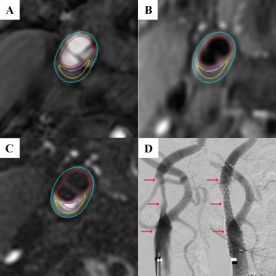 |
4 | Vulnerable plaque characteristics on MR vessel wall imaging predict hemodynamic instability during carotid artery stenting Video Permission Withheld
Qi Meng1, Chao Jiang2, Keqiang Zhao2, Huabin Zhang1, Hongliang Zhao3, Xihai Zhao4, Weiwei Wu2, and Zhuozhao Zheng3
1Department of Ultrasound, Beijing Tsinghua Changgung Hospital, School of Clinical Medicine, Tsinghua University, Beijing, China, 2Department of Vascular Surgery, Beijing Tsinghua Changgung Hospital, School of Clinical Medicine, Tsinghua University, Beijing, China, 3Department of Radiology, Beijing Tsinghua Changgung Hospital, School of Clinical Medicine, Tsinghua University, Beijing, China, 4Center for Biomedical Imaging Research, Department of Biomedical Engineering, Tsinghua University School of Medicine, Beijing, China
It has been shown that about 30%-70% of the patients underwent carotid artery stenting (CAS) suffered from perioperative hemodynamic instability (HI) characterized by transient hypotension and bradycardia. This study investigated the association between vulnerable plaque characteristics on MR vessel wall imaging and HI during CAS. We found that patients in HI group had significantly larger wall area and total vessel area, higher prevalence of intraplaque hemorrhage and vulnerable plaque, and larger volume of lipid-rich necrotic core (LRNC) of carotid plaque compared to those in non-HI group. The volume of LRNC and presence of vulnerable plaque could effectively predict HI.
|
||
4145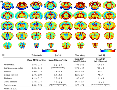 |
5 | Rapid high-resolution mapping of cerebral blood flow and volume by dynamic BOLD MRI with transient hypoxia in mice
Dongkyu Lee1, Thi Thuy Le1, Geun Ho Im1, and Seong-Gi Kim1
1Center for Neuroscience Imaging Research, Institute for Basic Science, Suwon, Korea, Republic of
Noninvasive perfusion mapping is important to determine perfusion changes due to molecular and neuropathological modifications as well as pharmaceutical interventions. Here, we adopted a dynamic BOLD-MRI method for quantifying whole brain perfusion, such as cerebral blood volume (CBV) and cerebral blood flow (CBF) without the use of exogenous contrast agents in mice. High-resolution perfusion maps allow us to determine regional perfusion values and cortical depth-dependency. The proposed technique is non-invasive and repeatable with every <1 min temporal resolution and can combine with evoked fMRI studies for determining a neural activity-induced quantitative change of perfusion parameters.
|
||
4146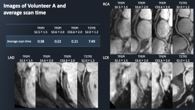 |
6 | Single-breath-hold whole heart coronary MRA using 3D turbo-field-echo-planar-imaging (TFEPI) with Compressed SENSE framework
Kazuo Kodaira1, Michinobu Nagao2, Masami Yoneyama3, Mana Kato1, Takumi Ogawa1, Yutaka Hamatani1, Isao Shiina1, Yasuhiro Goto1, and Shuji Sakai2
1Department of Radiological Services, Tokyo Woman's Medical University Hospital, Tokyo, Japan, 2Department of Diagnostic imaging & Nuclear Medicine, Tokyo Woman's Medical University Hospital, Tokyo, Japan, 3Philips Japan, Tokyo, Japan
Conventional whole-heart-coronary-magnetic-resonance-angiography (WHC-MRA) using NAV has a limitation of long scan time. Single-breath-hold-multi-shot-gradient-EPI can significantly reduce scan time of WHC-MRA, but it may be difficult depending on the gradient-spec of MRI-system. 3D-turbo-field-echo-planar-imaging (TFEPI) with Compressed-SENSE (C-SENSE) has the possibility to solve these problems. However, it is yet not applied for WHC-MRA. C-SENSE is suitable for subjects with a high sparse such as vessels, and can accelerate scan time of WHC-MRA while ensuring image quality. We propose a new combination of TFEPI with C-SENSE for single-breath-hold WHC-MRA, and examine image quality and scan time in comparison to the conventional methods.
|
||
4147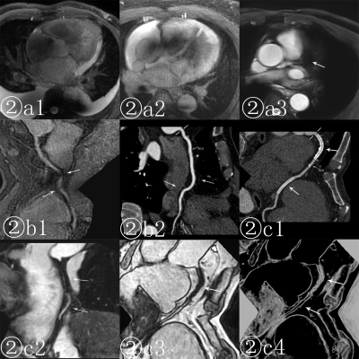 |
7 | The impact factors of the Quality of non-contrast agent Coronary angiography at 3.0T Magnetic Resonance Video Not Available
Junjing He1, Xiuzheng Yue2, Gang Zhang1, Wei Xing1, Tingting Li1, Yufu Hu1, and Yuhan Wang1
1Department of Magnetic Resonance, The First Affiliated Hospital of Henan University of CM, Zhengzhou, China, 2Philips Healthcare, Beijing, China
This study explores impact factors of the quality of 3.0T non-contrast agent magnetic resonance coronary angiography (MRCA), and uses an objective standard for assessing the image quality. A retrospective review was conducted on 72 patients who successfully completed 3.0T MRCA, and the correlation between their image quality and their clinical data, scan sequence, parameter settings, imaging technology and other factors were analyzed.The study found that BMI and respiratory motion were the key factors that affect the quality of non- contrast agent coronary MRA on 3.0T MRI. The mDixon-TFE imaging method can be used for MRCA as a routine scanning sequence.
|
||
4148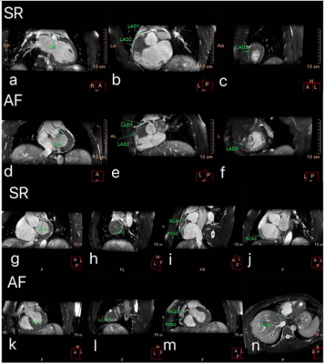 |
8 | The application of whole-heart coronary MRA with compressed sensing in patients with atrial fibrillation
FENG XIONG1, Yang Wu1, Longyan Zhang1, Xiaojing Ma1, Ke Yu1, Peng Sun2, and Haixia LI2
1Wuhan Asia General Hospital, WuHan, China, 2Philips Healthcare, Beijing, China
The whole-heart coronary MRA with compressed sensing (CS) technique is a novelly noninvasive, contrast agent-free, and radiation-free technique for evaluating coronary artery disease[2] [3].Patients with atrial fibrillation have irregular heartbeat, which brings uncertainty to the coronary MRA image quality. Results of this study indicated that there was no statistical difference in coronary artery image quality with CS acceleration factors of 2, 4, 6 between sinus rhythm and atrial fibrillation patients, which suggested that whole-heart coronary MRA with CS technique could also be applied inpatients with atrial fibrillation.
|
||
4149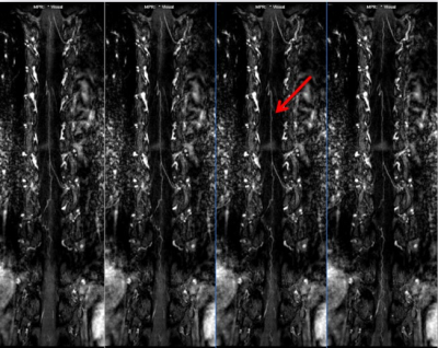 |
9 | Comparison of the Accuracy between High-resolution B-TFE and 3D T1-FFE MR Sequences on Human Spinal Artery Lesions
Yunjie Liao1 and Chen Thomas Zhao2
1Department of Radiology, the Third Xiangya Hospital, Central South University, Changsha, China, 2Philips Healthcare, Guangzhou, China
Spinal artery imaging has long been difficult in clinical detection and assessment of spinal vascular lesions, due to its anatomical complexity and very small diameter. Compared with DSA and CT, CE-MRA technology has the advantages of safety, noninvasive and radiation free. Traditional MR sequences, such as TOF and PC, perform poorly with lower SNR. Here, accuracy of B-TFE and 3D T1-FFE sequences were compared to provide radiologists a better choice in the spinal artery scenario. The results indicated that 3D T1-FFE would be preferred than B-TFE. And B-TFE should be used merely when patients are sensitive to contrast media
|
||
4150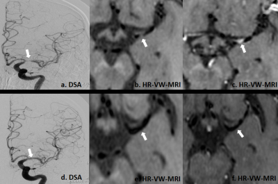 |
10 | Can HR-VW-MRI effectively evaluate the treatment effect on patients with MCA atherosclerotic stenosis after recanalization? Video Not Available
Kunjian Chen1, Weiqiang Dou2, Xinyi Wang1, Huimin Mao1, and Yu Guo1
1Department of Radiology, The First Affiliated Hospital of Shandong First Medical University, Jinan, China, 2MR Research, GE Healthcare, Beijing, China
This study mainly explored the feasibility of high-resolution vascular wall magnetic resonance imaging(HR-VW-MRI)in evaluating the treatment effect for patient with middle cerebral artery (MCA) recanalization. We included 21 patients with MCA stenosis diagnosed by DSA. All patients were treated with SeQuent Please drug-coated balloon recanalization and performed with HR-VW-MRI examination. Decreased responsible plaque area, length, enhancement index and lumen stenosis degree were found in MCA postoperatively. Additionally, the lumen stenosis degree evaluated by HR-VW-MRI was highly correlated with DSA. Therefore, HR-VW-MRI could be used as a valuable method to diagnose the effect of vascular recanalization.
|
||
4151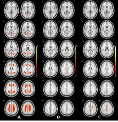 |
11 | Middle Cerebral Artery Morphologies Associated with White Matter Hyperintensities Video Permission Withheld
Boyu Zhang1, Zidong Yang1, Bei Wang1, Yajing Huo2, Huihui Lv2, Zhensen Chen1, Yan Han2, and He Wang1,3
1Institute of Science and Technology for Brain-Inspired Intelligence, Fudan University, Shanghai, China, 2Department of Neurology, Yueyang Hospital of Integrated Traditional Chinese and Western Medicine, Shanghai, China, 3Human Phenome Institute, Fudan University, Shanghai, China
The current report summarizes the morphological characteristics of middle cerebral artery quantified from MRA scans in a large sample of older Chinese population and shows that higher M1 radius is an independent predictor of white matter hyperintensities (WMH). Furthermore, as the pathological changes resulting in white matter lesions are heterogeneous, the current study suggest that the spatial distribution patterns of WMH could reflect the underpinning pathological mechanisms, though further studies are required.
|
||
 |
4152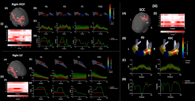 |
12 | Microstructural alterations in projection and association fibers in neonatal hypoxia-ischemia
Zuozhen Cao1, Xiaoxia Shen2, Fusheng Gao3, Tingting Liu1, Zhiyong Zhao1, Hongxi Zhang3, Lizhong Du2, Jiangyang Zhang4, Yi Zhang1, Can Lai3, Xiaolu Ma2, and Dan Wu1
1College of Biomedical Engineering & Instrument Science, Zhejiang University, HangZhou, China, 2Department of Neonatal Intensive Care Unit, Children's Hospital affiliated to Zhejiang University School of Medicine, HangZhou, China, 3Department of Radiology, Children's Hospital affiliated to Zhejiang University School of Medicine, HangZhou, China, 4Department of Radiology, New York University School of Medicine, New York, NY, United States
Diffusion MRI (dMRI) is known to be sensitive to hypoxic-ischemic encephalopathy (HIE) , however, the existing dMRI studies only used diffusion tensor metrics in a few selected brain regions and primarily focus on severe-to-moderate HIE. Here we investigated microstructural alterations using multi-shell dMRI across the whole-brain in severe, moderate, and mild HIE babies (n=5/13/13) in comparison with control neonates (n=11) with region-of-interest-based, tract-based, and fixel-based analysis. We found microstructural alternations in projection and association fibers, especially the inferior fronto-occipital fasciculus and inferior longitudinal fasciculus that are important for visual functions.
|
|
4153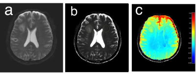 |
13 | Application of Electric Properties Tomography to Ischemic Stroke: Comparison of Conductivity between Infarct and Contralateral Brain
Onila Rasanjala1, Nguyen Trong Nguyen 2, Joohyun Kim3, Eunju Kim3, Ulrich Katscher4, Byung Hyun Baek5, and Ilwoo Park1,5
1Department of Artificial Intelligence Convergence, Chonnam National University, Gwangju, Korea, Republic of, 2Department of Biomedical Science, Chonnam National University, Gwangju, Korea, Republic of, 3Philips Korea, Seoul, Korea, Republic of, 4Philips Research Laboratories, Hamburg, Germany, 5Department of Radiology, Chonnam National University, Gwangju, Korea, Republic of
We demonstrate the feasibility of obtaining conductivity using EPT from patients with stroke. The ischemic lesion exhibited significantly higher conductivity values than the contralateral brain tissue in the cerebral brain, while the pons and cerebellum showed similar levels of conductivity values between the ischemic lesion and the contralateral brain tissue. The levels of conductivity value appeared to be specific to regions. The infratentorial normal brain exhibited a significantly higher level of conductivity than the supratentorial brain. In addition, the levels of conductivity were significantly different between the ischemic lesions of basal ganglia & thalamus and cerebral hemisphere.
|
||
4154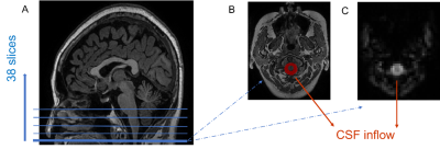 |
14 | Reduced coupling between global blood-oxygen-level-dependent signal and cerebrospinal fluid inflow is related to small vessel disease
Yao Zhang1, Ruiting Zhang1, Minming Zhang1, Yong Zhang2, and Peiyu Huang1
1The Second Affiliated Hospital, Zhejiang University School of Medicine, Hangzhou, China, 2GE Healthcare, Shanghai, China
The association between the glymphatic dysfunction and cerebral small vessel disease (SVD) is still unclear due a lack of in vivo measures. In this work, we employed a novel method based on the coupling between global blood-oxygen-level-dependent (gBOLD) signal and cerebrospinal fluid (CSF) inflow, which could detect alterations in CSF dynamics. We found that patients with severe SVD had significantly impaired glymphatic function. The dilation of peri-vascular space and the presence of diabetes were associated with worse glymphatic function. These results may provide useful knowledge for understanding the mechanism of SVD and improving clinical treatment.
|
||
4155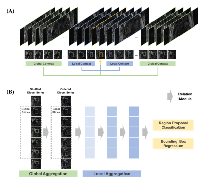 |
15 | Weakly-Supervised Carotid Vessel Wall Sub-pixel Segmentation Using Global-Local Context Aggregation and PolarMask
Jiaqi Dou1, Song Tian2, Yuze Li1, Ziming Xu1, Shuo Chen1, Yajie Wang1, and Huijun Chen1
1Center for Biomedical Imaging Research, Tsinghua University, Beijing, China, 2MSC Clinical & Technical Solutions, Philips Healthcare, Beijing, China
Carotid vessel wall segmentation on 3D black-blood MRI is a key step in plaque burden assessment and atherosclerotic lesions identification. In this study, a two-stage weakly-supervised carotid vessel wall segmentation approach was developed using limited manual delineations on 3D black-blood MR images. First, a global-local context aggregation strategy was used to identify bilateral carotid arteries robustly. Then, distances from artery center to boundaries were regressed in a polar coordinate by PolarMask to segment the vessel wall. The proposed segmentation approach outperformed Attention UNet (Quantitative score: 0.795±0.170 vs. 0.729±0.208) and showed great potential in quantitative atherosclerosis analysis.
|
||
 Back to Meeting Home
 Back to the Program-at-a-Glance
The International Society for Magnetic Resonance in Medicine is accredited by the Accreditation Council for Continuing Medical Education to provide continuing medical education for physicians.
 Back to Meeting Home
 Back to the Program-at-a-Glance
 View the Presentation