Online Gather.town Pitches
Heart III
Joint Annual Meeting ISMRM-ESMRMB & ISMRT 31st Annual Meeting • 07-12 May 2022 • London, UK

| Booth # | ||||
|---|---|---|---|---|
4770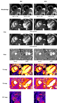 |
1 | Validation of Beat Sensor Cardiac Triggering against ECG Cardiac Triggering in both Volunteer and Patient Cohorts
Yue Pan1,2, Juliet Varghese1, Ning Jin3, Carmel Hayes4, Peter Speier4, Rizwan Ahmad2,5, and Orlando Simonetti1,2,6
1Davis Heart and Lung Research Institute, The Ohio State University Wexner Medical Center, Columbus, OH, United States, 2Department of Biomedical Engineering, The Ohio State University, Columbus, OH, United States, 3Siemens Medical Solutions USA, Malvern, PA, United States, 4Siemens Healthcare GmbH, Erlangen, Germany, 5Department of Electrical and Computer Engineering, The Ohio State University, Columbus, OH, United States, 6Department of Internal Medicine, The Ohio State University Wexner Medical Center, Columbus, OH, United States
In this study, the performance of Beat Sensor Cardiac (BSC) triggering was qualitatively and quantitatively compared with electrocardiogram (ECG) triggering. MR images typically acquired in a comprehensive exam including localizer, morphology, cine, 2D flow, parametric mapping and late gadolinium enhancement (LGE) were acquired using both BSC and ECG triggering in volunteers and patients. The overall image quality for BSC was equivalent to ECG. Quantitative measurements of function, flow, and parametric maps in healthy volunteers showed no significant differences except for peak aortic velocity.
|
||
| 4771 | 2 | CMR Risk Stratification for Dilated Cardiomyopathy with LVEF≥35%: Cohort Study with a Midterm Follow-up Video Permission Withheld
Shuang Li1, Wenjing Yang1, Di Zhou1, Yining Wang1, Xiaohan Fan1, Arlene Sirajuddin2, Andrew E. Arai2, Shihua Zhao1, and Minjie Lu1
1fuwai hospital, Beijing, China, 2National Heart, Lung and Blood Institute (NHLBI), National Institutes of Health (NIH), Bethesda, MD, United States
This study aims to identify the risk factors for adverse events in dilated cardiomyopathy (DCM) patients with LVEF≥35% and establish a scoring model to predict adverse event risk. 269 consecutive DCM patients with LVEF ≥35% who underwent gadolinium-enhanced cardiac magnetic resonance (CMR) imaging were enrolled in this study. The primary endpoint was a composite of SCD or aborted SCD. Secondary endpoints were all-cause mortality, heart transplantation and hospitalization for heart failure. The nomogram we created using these parameters provides a novel way to clinically assess and risk stratify this patient population.
|
||
4772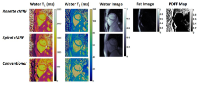 |
3 | Cardiac MRF Using Rosette Trajectories for Simultaneous Myocardial T1, T2, and Proton Density Fat Fraction Mapping
Yuchi Liu1, Jesse Hamilton1,2, Yun Jiang1,2, and Nicole Seiberlich1,2
1Department of Radiology, University of Michigan, Ann Arbor, MI, United States, 2Department of Biomedical Engineering, University of Michigan, Ann Arbor, MI, United States This work aims to extend rosette cardiac MRF to enable simultaneous myocardial T1, T2, and PDFF mapping from a single scan. The accuracy of rosette cMRF in T1, T2, and PDFF quantification is validated in the ISMRM/NIST system phantom and an in-house built fat fraction phantom, respectively. Studies in healthy subjects show that rosette cMRF potentially enables more accurate T1 and T2 measurements than spiral cMRF in the myocardium due to reduced fat contamination. |
||
4773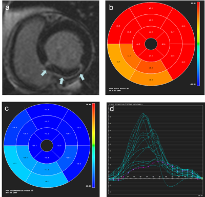 |
4 | Relationship between Microvascular Obstruction and Regional Myocardial Strain by CMR after ST-segment-elevation Myocardial Infarction
Yanan Zhao1, Jianing Cui1, Xueqian Liu2, Tao Li1, and Xiuzheng Yue3
1Department of Radiology, Chinese People’s Liberation Army General Hospital, Beijing, China, 2Qinhuangdao Workers' Hospital, Qinhuangdao, China, 3Philips Healthcare, Beijing, China
Effective risk assessment and stratification are essential for the clinical management of ST-segment elevation myocardial infarction (STEMI) patients. CMR imaging has become a beneficial imaging modality to assess myocardial morphology, function and infarct characteristics simultaneously. This study quantitatively evaluated relationship between microvascular obstruction and regional myocardial strain by Cardiac MRI after ST-segment-elevation myocardial infarction. Our data showed that regional strain parameters may be s new noninvasive imaging markers allowing comprehensive evaluation regional myocardium deformation.
|
||
4774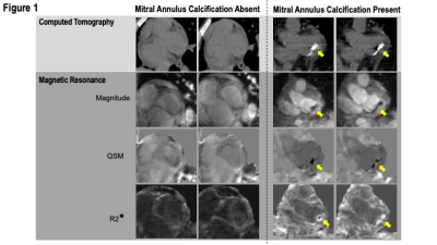 |
5 | Cardiac Quantitative Susceptibility Mapping for Mitral Annular Calcium: Validation using Quantitative Calcium Scoring on Computed Tomography
Pablo Villar-Calle1, Jiahao Li2, Alice Saffioti1, Justin Johannesen1, Katherine Tak1, Rachel Meier1, Jiwon Kim1, Quynh A Truong3, Yi Wang2, Pascal Spincemaille3, and Jonathan W Weinsaft1
1Medicine, Weill Cornell Medicine, New York NY, United States, New York, NY, United States, 2Meinig School of Biomedical Engineering, Cornell University, Ithaca NY, United States, New York, NY, United States, 3Radiology, Weill Cornell Medicine, New York NY, United States, New York, NY, United States
This study tested cardiac MRI cardiac quantitative susceptibility mapping (QSM) for measurement of mitral annular calcification (MAC) using the reference of computed tomography (CT). QSM entailed a free-breathing ECG-triggered multi-echo 3D GRE sequence with diaphragmatic navigation via ECG-gating (1D respiratory navigator, 4mm window, acquisition time ~7 minutes, navigator efficiency 36%, spatial resolution 1.5×1.5×5mm3). Among 24 patients undergoing MRI and CT (mean 3.9±3.4 months), annular susceptibility decreased stepwise (p<0.001) and R2* increased (p<0.05) among patients stratified by CT mitral annular calcium score. Both QSM (AUC=0.950, p=0.005) and R2* (AUC=0.938, p=0.007) yielded excellent diagnostic performance for advanced mitral annular calcification on CT.
|
||
4775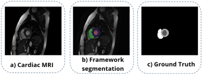 |
6 | A NOVEL OPEN-SOURCE CARDIAC SEGMENTATION FRAMEWORK FOR CINE MRI
Manuel A. León1, Rodrigo Salas1, Sergio Uribe2,3,4,5, and Julio Sotelo1,3,5
1School of Biomedical Engineering, Universidad de Valparaíso, Valparaíso, Chile, 2Department of Radiology, School of Medicine, Pontificia Universidad Católica de Chile, Santiago, Chile, 3Millennium Nucleus in Cardiovascular Magnetic Resonance, CardioMR, Santiago, Chile, 4Institute for Biological and Medical Engineering, Schools of Engineering, Medicine and Biological Sciences, Pontificia Universidad Católica de Chile, Santiago, Chile, 5Biomedical Imaging Center, Pontificia Universidad Católica de Chile, Santiago, Chile
Cardiac contour delineation currently presents several proposals for automatic segmentation in short-axis MRI. However, most of them have drawbacks such as low accuracy with images from other MRI scanners, lack of description of the algorithms, and difficult access for clinical or research use. For this reason, we present VENTSEG, a deep learning framework for cardiac segmentation implemented in Matlab and Python. Our dice coefficient results segmenting images from the CNN validation set are 85.55% and 96.17% on anonymized clinical data. These results demonstrate the generalization of the framework on multidomain images.
|
||
4776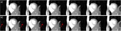 |
7 | Auto-calibrated Multiband Retrospective GRE Cine at 5T with Improved Reconstruction Video Permission Withheld
Yuan Zheng1, Lele Zhao2, Zhongqi Zhang1, Junpu Hu2, Yu Ding1, and Jian Xu1
1UIH America, Inc, Houston, TX, United States, 2Shanghai United Imaging Healthcare Co. Ltd, Shanghai, China
GRE sequences benefit from increased SNR at high field and do not suffer from banding artifacts / SAR issues as bSSFP sequences. We have implemented an autocalibrated multiband retrospective GRE Cine sequence at 5 T. The autocalibrated multiband acquisition increases the efficiency of the sequence by reducing the scan time and simplifying the multiband imaging workflow. A compressed sensing reconstruction with implicit phase interpolation and temporal TV periodic boundary condition were implemented to further improve image quality. We have evaluated this application on volunteers and achieved high quality GRE Cine images.
|
||
4777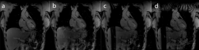 |
8 | Development of a Prospectively Motion Corrected Free-breathing FLASH Sequence
Graeme Harris1, Stephen Jermy1,2, and Ernesta Meintjes1,2
1Department of Human Biology, Division of Biomedical Engineering, University of Cape Town, Cape Town, South Africa, 2Cape Universities Body Imaging Center, University of Cape Town, Cape Town, South Africa
Respiratory motion of the heart is a fundamental challenge to cardiac MR imaging (CMR), frequently compensated for with breath-holding and acceptance-window methods. These methods are not always viable and result in inefficient acquisitions, creating longer scan times1. Here, an adaptive Kalman-filter with a control system was implemented in a FLASH sequence to update the position of the slice during the imaging segment based on repeated navigator measurements acquired during the non-imaging segment. This predictive tracking results in a free-breathing sequence which is less strenuous for subjects, more efficient and reduces scan times.
|
||
4778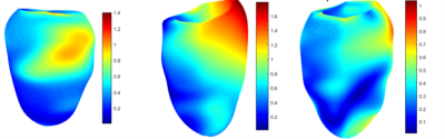 |
9 | Myocardial stress field from combined CMR and FEM in ALL survivors
Mehdi Ghafarinatanzi1
1Polytechnique Montréal, Montreal, QC, Canada
The objective of this study is to investigate the myocardial 3D strain stress field of the patient-specific left ventricles in children with acute lymphoblastic leukemia (ALL) after treatment. The virtual field-finite element approach is applied to the 3D mesh generated tetrahedral for each patient-specific geometry over the cardiac cycle. The results illustrate the difference in distribution stress and displacement field for each LV subject.
|
||
4779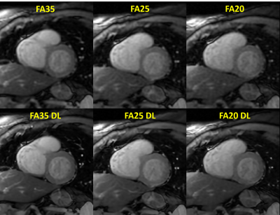 |
10 | Improved myocardial T1 mapping accuracy with Deep Learning reconstruction of low flip angle MOLLI series Video Permission Withheld
Gaspar Delso1, Pablo García-Polo2, Margarita Gorodezky3, Ben Ariff4, Vicente Martínez de Vega5, and Javier Urmeneta6
1GE Healthcare, Barcelona, Spain, 2GE Healthcare, Madrid, Spain, 3GE Healthcare, London, United Kingdom, 4Imperial College Healthcare NHS Trust, London, United Kingdom, 5Servicio de diagnóstico por la imagen, Hospital Universitario Quirónsalud, Madrid, Spain, 6Servicio de cardiología, Hospital Universitario Quirónsalud, Madrid, Spain In this study, we explore the possibility of leveraging Deep Learning regularized reconstruction to enable lower flip angle MOLLI acquisition for myocardial T1 mapping. It has been shown in the past that lowering flip angle helps reduce various artifact sources, at the cost of lower signal to noise ratio. Regularized reconstruction can effectively manage image noise as well as increase feature sharpness. This hypothesis has been tested on a group of clinical patients referred for a cardiac MR exam. |
||
4780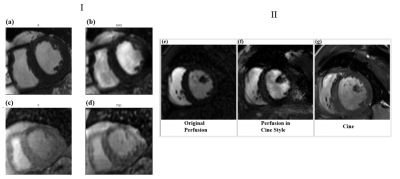 |
11 | Neural style transfer: Applications in cardiac MR image registration
Alper Ozan Turgut1, Matthew Van Houten2, Junyu Wang2, Xue Feng2, and Michael Salerno3
1School of Medicine, University of Virginia, Charlottesville, VA, United States, 2Biomedical Engineering, University of Virginia, Charlottesville, VA, United States, 3Department of Medicine and Radiology, Stanford University, Stanford, CA, United States
Contrast-enhanced cardiac magnetic resonance (CMR) stress perfusion imaging shows excellent utility in evaluating coronary artery disease1. Registering perfusion CMR image series is difficult due to the varying image contrast. Neural style transfer is a deep learning method used to transfer the “style” of one domain to another while preserving the content. Two neural style transfer networks were implemented in Python using TensorFlow and PyTorch. Training of each network was done using three, slice matched patient profiles and cine-like perfusion images were generated and registered. This method is compared to a KL-transform based registration approach.
|
||
4781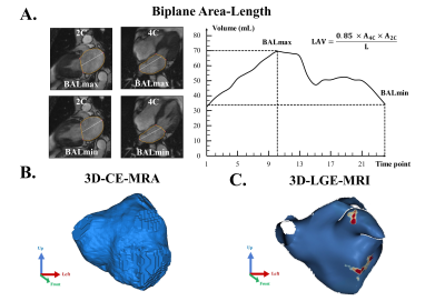 |
12 | Biplane Area-length Method Underestimates 3D Left Atrial Volume in Patients with Atrial Fibrillation
Anthony Maroun1, Justin Baraboo1, Suvai Gunasekaran1, Julia Hwang1, Sophia Liu1, Daniel Kim1, Philip Greenland2, Rod Passman3, Bradley Allen1, Michael Markl1, and Maurice Pradella1,4
1Department of Radiology, Northwestern University Feinberg School of Medicine, Chicago, IL, United States, 2Department of Preventive Medicine, Northwestern University Feinberg School of Medicine, Chicago, IL, United States, 3Department of Cardiology, Northwestern University Feinberg School of Medicine, Chicago, IL, United States, 4Department of Radiology, University Hospital Basel and University of Basel, Basel, Basel, Switzerland
The biplane area-length method is routinely used in clinical settings for left atrial volume (LAV) estimation. This technique is time-resolved but relies on geometrical assumptions of an ellipsoidal shape to estimate the LAV. In contrast, quantification derived from 3D segmentations on late gadolinium-enhanced magnetic resonance imaging and contrast-enhanced magnetic resonance angiography are static but do not rely on geometric assumptions. We compared the LAV estimation from these 3 methods in patients with atrial fibrillation and found a significant underestimation by the biplane area-length technique, indicating that this method may not capture the entire LAV in patients with complex anatomy.
|
||
4782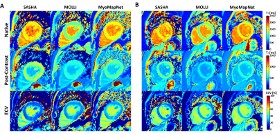 |
13 | Improving Accuracy of Myocardial T1 Estimation in MyoMapNet
Rui Guo1, Zhensen Chen2, Amine Amyar1, Hossam El-Reiwady1, Patrick Pierce1, Beth Goddu1, and Reza Nezafat1
1Department of Medicine (Cardiovascular Division), Beth Israel Deaconess Medical Center and Harvard Medical School, Boston, MA, United States, 2Institute of Science and Technology for Brain-Inspired Intelligence, Fudan University, Shanghai, China MOLLI T1 mapping is routinely used for myocardial tissue characterization. Despite excellent precision, MOLLI has great T1 underestimation and a long breath-holding time. We previously developed a deep learning-based approach (MyoMapNet) for myocardial T1 mapping using four T1-weighted images. MOLLI T1 was used to train a fully connected neural network (FC), which resulted in a similar accuracy error as MOLLI. In this study, numerically simulated and phantom signals with the presence of different B0, flip angles, and heart rates were used to improve MyoMapNet T1 accuracy. Evaluation showed that MyoMapNet T1 could be improved and had higher precision than SASHA. |
||
 Back to Meeting Home
 Back to the Program-at-a-Glance
The International Society for Magnetic Resonance in Medicine is accredited by the Accreditation Council for Continuing Medical Education to provide continuing medical education for physicians.
 Back to Meeting Home
 Back to the Program-at-a-Glance
 View the Presentation