Online Gather.town Pitches
Perfusion & Permeability II
Joint Annual Meeting ISMRM-ESMRMB & ISMRT 31st Annual Meeting • 07-12 May 2022 • London, UK

Online Gather.town Pitches
Perfusion & Permeability II
| Booth # | ||||
|---|---|---|---|---|
4572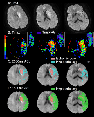 |
1 | Arterial spin labeling estimation of penumbral tissue in acute ischemic stroke
Jinhao Lyu1 and Xin Lou1
1Chinese PLA General Hospital, Beijing, China
Penumbral tissue identification is critical for the treatment decision for patients with acute ischemic stroke. In the present study, we aimed to assess the impact of arterial spin labeling (ASL) at different post-labeling delays (PLDs) in penumbral tissue quantification, and compare their performances in patients’ selection for endovascular treatment with dynamic susceptibility contrast as reference. The results showed ASL appears to persistently overestimate penumbral tissue in acute ischemic stroke. PLD shows substantial impact on quantification of hypoperfusion volume. Long PLD, 2500ms for example, is preferable in penumbral tissue detection and patients’ selection.
|
||
4573 |
2 | Detection of crossed cerebellar diaschisis in subacute ischemic stroke using 3D PCASL with two post-labeling delay time method Video Permission Withheld
Yuelei Lyu1, Hua Gu1, Yanping Xue1, Lizhi Xie2, Tao Jiang1, and Qi Yang1
1Beijing Chao-Yang Hospital, Capital Medical University, Beijing, China, 2MR Research China, GE Healthcare, Beijing, China This study investigated the feasibility of 3D PCASL to detect crossed cerebellar diaschisis (CCD) in patients with unilateral supratentorial subacute stroke using two PLD method and correlated the ischemic perfusion with CCD to identify the relevant imaging factors of CCD development. 3D PCASL can detect CCD in patients of supratentorial subacute stroke. Patients of supratentorial subacute stroke experience late-arriving flow while CCD+ patients suffer late arriving flow in the affected contralateral cerebellum. CCD+ patients may have a propensity towards more hemodynamic compromise and compensatory collateral flow in the cerebellum. |
||
4574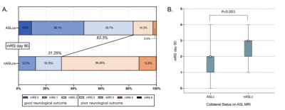 |
3 | Collateral Circulation Detected by Arterial Spin Labeling Can Predict Outcome in Acute Ischemic Stroke Video Permission Withheld
Sangni Liu1, Dandan Fan1, and Chunming Xie1
1Department of Neurology, Affiliated ZhongDa Hospital, School of Medicine, Southeast University, Nanjing, Jiangsu, China
Robust collateral circulation is strongly associated with better outcomes in acute ischemic stroke. In this prospective study, collateral circulation was defined as arterial transit artifact in Arterial Spin Labeling(ASL) images. Patients with ASL collaterals represented higher rate of good outcome and there were significant differences for percent changes of mRS score and NIHSS score between groups. Furthermore, the presence of ASL collaterals could predict good outcome, even after controlling for baseline NIHSS score. Taken together, ASL collaterals provide a powerful tool to predict clinical outcome for acute ischemic stroke with 90 days follow-up.
|
||
4575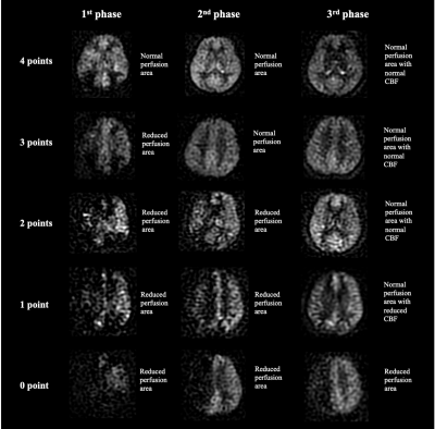 |
4 | Multi-delay arterial spin labeling perfusion map may predict cerebral hyperperfusion after carotid endarterectomy Video Permission Withheld
Xiaoyuan Fan1, Tianye Lin1, Juan Wei2, Jianxun Qu2, Zhentao Zuo1, and Feng Feng1
1Peking Union Medical College Hospital, Beijing, China, 2GE healthcare, Beijing, China Preoperative measurement of cerebrovascular reactivity using acetazolamide or CO2 induced SPECT is the gold standard for predicting cerebral hyperperfusion (CH) after carotid endarterectomy (CEA). However, it is often impractical to perform SPECT examinations on all patients. Arterial spin labeling (ASL) is a non-invasive technique to image the brain perfusion. Based on multi-delay ASL perfusion map, we extracted two hemodynamic parameters (cerebral blood flow and arterial transit time) and established a five-scale scoring system for the prediction of CH. We found in patients with carotid stenosis, multi-delay ASL is a practical and reliable tool for the prediction of CH after CEA. |
||
4576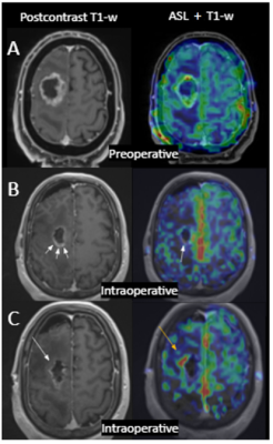 |
5 | Clinical utility of intraoperative Arterial Spin Labeling for resection control in brain tumor surgeries, a 3T study.
Marta Calvo-Imirizaldu1, Verónica Aramendia-Vidaurreta1,2, Marta Vidorreta3, Reyes García-Eulate1,2, Pablo Domingez Echeverri1,2, Josef Pfeuffer4, Bartolome Bejarano5, Lain Hermes Gonzalez-Quarante5, Antonio Martinez-Simon6, and María Fernández-Seara1,2
1Radiology, Clínica Universidad de Navarra, Pamplona, Spain, 2IDISNA, Pamplona, Spain, 3Siemens Healthcare, Madrid, Spain, 4Application Development, Siemens Healthcare, Erlangen, Germany, 5Neurosurgery, Clínica Universidad de Navarra, Pamplona, Spain, 6Anesthesia and Intensive Care, Clínica Universidad de Navarra, Pamplona, Spain
ASL has shown potential to depict residual tumor compared to anatomical imaging in previous intraoperative MRI (iMRI) study performed at 1.5T. However, the technique has not been evaluated at higher field, where field inhomogeneities could compromise the labeling efficiency. We aimed to assess feasibility and utility of iMRI-ASL at 3T. To that end, a PCASL sequence was evaluated in 10 patients. In one patient ASL depicted an additional high CBF focus indicating neovascularization (known to correlate with higher grade component) that wasn’t depicted in the anatomical images, favoring the use of ASL in the iMRI setting to achieve maximal resection.
|
||
4577 |
6 | Does arterial spin labeling provide insights into long-term effects of radiation therapy in the healthy brain?
Andrea Kronfeld1, Alexandra Russo2, Nicole Henninger2, Arthur Wingerter2, Moritz Brunk2, Kaml Khawaja1, Andrei Tropine1, Abdulghani Moussa Pacha1, Violeta Dimova3, Federico Marini4,5, Doris Leithner6, Sabine Nospes7, Roman Kloeckner8, Felix Hahn8, Marcus Stockinger9, Heinz Schmidberger9, Marc Brockmann1, Marie Astrid Neu2, Joerg Faber2, and Yasemin Tanyildizi1
1Neuroradiology, University Medical Center of the Johannes Gutenberg University Mainz, Mainz, Germany, 2Pediatric Hematology, Oncology & Hemostaseology, University Medical Center of the Johannes Gutenberg University Mainz, Mainz, Germany, 3Neurology, University Medical Center of the Johannes Gutenberg University Mainz, Mainz, Germany, 4Center for Thrombosis and Hemostasis, University Medical Center of the Johannes Gutenberg University Mainz, Mainz, Germany, 5Institute of Medical Biostatistics, Epidemiology and Informatics, University Medical Center of the Johannes Gutenberg University Mainz, Mainz, Germany, 6Diagnostic and Interventional Radiology, University Hospital Frankfurt, Frankfurt, Germany, 7Otolaryngology, Head and Neck Surgery, University Medical Center of the Johannes Gutenberg University Mainz, Mainz, Germany, 8Diagnostic and Interventional Radiology, University Medical Center of the Johannes Gutenberg University Mainz, Mainz, Germany, 9Radiation Oncology, University Medical Center of the Johannes Gutenberg University Mainz, Mainz, Germany
28 patients after earlier (1-45 years) whole brain radiation and 22 healthy controls were examined by arterial spin labeling to discover insights ASL could give in long-term effects of radiation therapy in the healthy brain. Relative cerebral blood flow and relative bolus arrival time showed increased values compared to the healthy control, which could be a sign for increased permeability and thickening of the vessels, two well described effects of radiation therapy ASL gives insight into.
|
||
4578 |
7 | A study on MRI arterial spin labeling in predicting early response to chemoradiotherapy in patients with advanced nasopharyngeal carcinoma
zongqiong sun1 and Weiqiang Dou2
1Department of radiology, Affiliated hospital of Jiangnan university, Wuxi, China, 2GE healthcare,MR research China, Beijing, China
This study mainly investigated the feasibility of 3D pseudo-continuous arterial spin labeling (3D-pCASL) imaging in predicting the treatment response to chemoradiotherapy (CRT) for advanced nasopharyngeal carcinoma (ANPC) patients. 3D-pCASL derived blood flow (BF)s in tumors were respectively obtained for patients in response group (RG) and no-response group (NRG). The resultant pre-treatment BF showed significantly different values between RG and NRG patients. Combining with clinical information, BF also showed robust prediction between RG and NRG patients. Therefore, 3D-pCASL could be considered an effective method in tumor response prediction to CRT for ANPC patients before clinical treatment.
|
||
4579 |
8 | ATP-A safe stressor for stress perfusion cardiac magnetic resonance
Huihui Kong1, Jing An2, Jiaxin Cao3, Jinfan Tian4, Jingwen Yong4, Xiantao Song4, and Yi He3
1Radiology, Beijing Friendship Hospital, Beijing, China, 2Siemens Healthcare, Beijing, China, 3Beijing Friendship Hospital, Beijing, China, 4Beijing Anzhen Hospital, Beijing, China
This study investigated the side effects and safety of ATP as a stressor for stress perfusion cardiac magnetic resonance. The result showed the high safety of stress perfusion with ATP and side effects were mild and resolved shortly after receiving ATP. Furthermore, it was also observed splenic switch-off that can be used to assess stress adequacy undergoing stress-CMR with ATP.
|
||
4580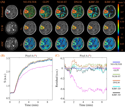 |
9 | Comparison of different denoising approaches for DCE-MRI
Zejun Wang1, Bao Wang2, Ziyi Huang3, Yingchao Liu4, and Ruiliang Bai1,5
1Key Laboratory of Biomedical Engineering of Ministry of Education, College of Biomedical Engineering and Instrument Science, Zhejiang University, Hangzhou, China, 2Department of Radiology, Qilu Hospital of Shandong University, Jinan, China, 3College of Life Sciences, Zhejiang University, Hangzhou, China, 4Department of Neurosurgery, Shandong Provincial Hospital Affiliated to Shandong First Medical University, Jinan, China, 5Department of Physical Medicine and Rehabilitation, Interdisciplinary Institute of Neuroscience and Technology, The Affiliated Sir Run Run Shaw Hospital, School of Medicine,, Zhejiang University, Hangzhou, China
The physiological parameters estimated from pharmacokinetic modeling of DCE-MRI are usually biased by the non-white, spatially-dependent noise. In this study, we compared several state-of-arts denoising approaches, including gaussian low pass filter (GLPF), the dynamic nonlocal mean (DNLM), the nonlocal mean based on spatiotemporal patches (NLM-ST), 2D and 3D kinetics-induced bilateral filter (KIBF). Our results reveal that the 3D KIBF can reduce the noise significantly and reserve subtle information best.
|
||
4581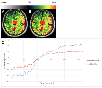 |
10 | Revealing Occult Hemodynamic Signatures from the Acetazolamide-Augmented Cerebral BOLD Response
Siddhant Dogra1, Xiuyuan Wang2, Jelle Veraart1, Alejandro Gupta1, Koto Ishida3, Deqiang Qiu4, and Seena Dehkharghani1,3
1Department of Radiology, New York University Langone Health, New York, NY, United States, 2Department of Radiology, Weill Cornell Medicine, New York, NY, United States, 3Department of Neurology, New York University Langone Health, New York, NY, United States, 4Department of Radiology, Emory University, Atlanta, GA, United States
Acetazolamide-augmented blood-oxygen-level-dependent (ACZ-BOLD MRI) provides robust estimation of cerebrovascular reactivity (CVR). Past approaches are limited to extraction of terminal CVR at arbitrarily selected times in the pharmacologic response, due to low signal-to-noise characteristics. We previously introduced a computational framework that pre-conditions the BOLD response for dynamic analysis and extraction of maximal, rather than terminal augmentation profiles, and novel hemodynamic signatures such as ACZ-BOLD time-to-maxima (Tmax). Several consistent and sometimes paradoxical patterns have emerged, the presentation of which we considered timely for this potentially novel hemodynamic neuroimaging biomarker.
|
||
4582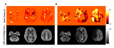 |
11 | Correlation Analysis between BOLD Delay and Cerebral Blood Flow: A Study in Moyamoya Patients Video Permission Withheld
Zhangxuan Hu1, Zhe Zhang2,3, Yuanren Zhai4, Long Qian1, Binbin Sui2,3, and Dong Zhang4
1GE Healthcare, Beijing, China, 2Tiantan Neuroimaging Center of Excellence, Beijing Tiantan Hospital, Capital Medical University, Beijing, China, 3China National Clinical Research Center for Neurological Diseases, Beijing, China, 4Department of Neurosurgery, Beijing Tiantan Hospital, Capital Medical University, Beijing, China
It has been shown that the BOLD delay calculated from resting-state fMRI can reveal perfusion information. In this study, the correlation between BOLD delay and CBF measured from ASL were explored. The results showed the inter-subject correlation between BOLD delay and CBF values. Meanwhile, the inter-group differences of the correlation coefficients between Moyamoya patients and healthy controls were also compared.
|
||
4583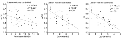 |
12 | Are cerebral microvascular perfusion and diffusion dynamics associated with neurological function in acute ischemic stroke?
Fei Chen1, Zhenyu Dai1, Lizheng Yao1, Congsong Dong1, Haicun Shi2, and Weiqiang Dou3
1Department of Radiology, the Yancheng School of Clinical Medicine of Nanjing Medical University, Yancheng, China, 2Department of Neurology, the Yancheng School of Clinical Medicine of Nanjing Medical University, Yancheng, China, 3GE Healthcare, MR Research China, Beijing, China
This work explored the association of cerebral microvascular perfusion and diffusion dynamics measured by intravoxel incoherent motion (IVIM) imaging with initial neurological function and clinical outcome in acute stroke patients. Significant negative correlations were revealed between IVIM derived diffusion dynamics parameters and initial neurological function as well as clinical outcome for patients with acute ischemic stroke. However, no similar statistical correlation was found for parameters related to microvascular perfusion. IVIM may be therefore suggested as an effective non-invasive method for evaluating the initial neurological function and clinical outcome in acute ischemic stroke.
|
||
4584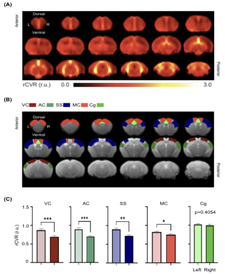 |
13 | Characterization of mouse cerebrovascular reactivity using task-free resting-state fMRI
Yixi Xue1, Russell W. Chan1,2, Sarah Y. Wu1, Emily L. Tse1, Muneeb A. Faiq1, Thajunnisa A. Sajitha1, and Kevin C. Chan1,3
1Department of Ophthalmology, New York University Grossman School of Medicine, New York, NY, United States, 2Neuroscience Institute, New York University Grossman School of Medicine, New York, NY, United States, 3Department of Radiology, New York University Grossman School of Medicine, New York, NY, United States
The recent development of relative cerebrovascular reactivity (rCVR) mapping derived from task-free resting-state blood-oxygenation-level-dependent fMRI resolves issues of experimental complications from hypercapnic challenge or pharmacological administrations required in conventional CVR mapping. While human clinical rCVR studies have begun, there lacks rCVR characterization in healthy adult rodents before it can be applied to preclinical models of neuropathology for translational research. This study demonstrated the feasibility of rCVR mapping using resting-state fMRI at 7 Tesla in mice. The results also revealed potential rCVR lateralization in cortical regions and heterogenous vascularization along the septotemporal axis of the hippocampus.
|
||
4585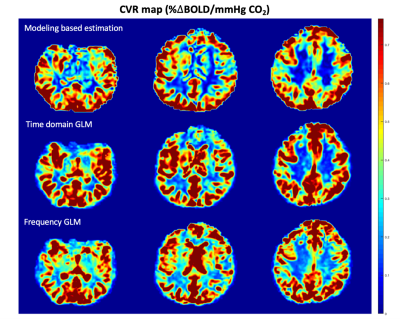 |
14 | Cerebrovascular Reactivity Mapping using General Linear Model in Frequency Domain
Botian Xu1, Chau Vu1, Matthew Borzage2, and John Wood2
1University of Southern California, Los Angeles, CA, United States, 2Children's Hospital Los Angeles, Los Angeles, CA, United States
Cerebrovascular reactivity (CVR), measuring the ability of vessel dilation or constriction, is an important indicator of cerebrovascular health. As a parameter describing the hemodynamic changes, CVR requires a physiological challenge as the stimulus and monitors the response of the brain metabolism. We propose a frequency-based approach, which applies general linear model (GLM) on the magnitude of the stimulus and response spectrum to avoid challenges with signal alignment and improve robustness to noise in CVR estimation.
|
||
4586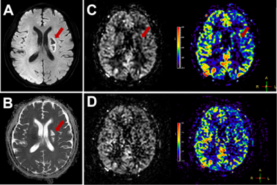 |
15 | Improve the Image Quality of Arterial Spin Labeling with Echo Planar Imaging with Compressed SENSE
Chunqiu Su1, Xiance Zhao2, Jilei Zhang2, Xunning Hong1, and Shanshan Lu1
1Department of Radiology, The First Affiliated Hospital of Nanjing Medical University, Nanjing, China, 2Philips Healthcare, Shanghai, China
Arterial spin labeling (ASL) is a non-invasive MRI perfusion technique that can quantify tissue blood flow, which is meaningful for clinical applications. However, ASL perfusion images always have a limited signal-to-noise ratio (SNR). We aimed to using the echo planar imaging with compressed SENSE (EPICS) to obtain high-quality brain ASL images. Eleven healthy subjects and four patients with intracranial infarction were enrolled, and underwent ASL imaging with and without EPICS. The results demonstrated the feasibility of EPICS to increase the quality of ASL images.
|
||
 Back to Meeting Home
 Back to the Program-at-a-Glance
The International Society for Magnetic Resonance in Medicine is accredited by the Accreditation Council for Continuing Medical Education to provide continuing medical education for physicians.
 Back to Meeting Home
 Back to the Program-at-a-Glance
 View the Presentation