Online Gather.town Pitches
Vascular Imaging & Stroke III
Joint Annual Meeting ISMRM-ESMRMB & ISMRT 31st Annual Meeting • 07-12 May 2022 • London, UK

Online Gather.town Pitches
Vascular Imaging & Stroke III
| Booth # | ||||
|---|---|---|---|---|
4587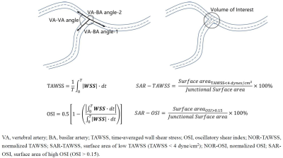 |
1 | The impact of hemodynamic patterns and geometric features of vertebra-basilar system on atherosclerotic plaque occurrence
Rui Shen1, Hualu Han1, Jinmei Zheng2, Huiyu Qiao1, Zihan Ning1, Xinyu Tong3, Yunjing Xue4, and Xihai Zhao1
1Center for Biomedical Imaging Research, Tsinghua University, Beijing, China, 2Fujian Medical University Union Hospital, Fuzhou, Fujian Province, China, 3Department of Biomedical Engineering, School of Life and Science, Beijing Institute of Technology, Beijing, China, 4Department of Radiology, Fujian Medical University Union Hospital, Fuzhou, Fujian, China
This study investigated the relationship between the geometry of vertebra-basilar system and hemodynamic measurements and presence of BA plaques. We found that geometric measurements and most hemodynamic features were significantly correlated with the occurrence of BA plaque (mean curvature, r=0.368; NOR-TAWSS-NF, r=-0.312; SAR-OSI-NF, r=0.290; NOR-TAWSS-nNF, r=-0.387; NOR-OSI-nNF, r=0.440). There were significant differences in mean curvature, hemodynamic characteristics of Newtonian flow and non-Newtonian flow models between patients with and without BA plaque. Our study indicated that geometric features and non-Newtonian flow model-based hemodynamic measurements are risk factors of BA plaque.
|
||
4588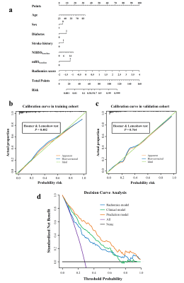 |
2 | High prediction accuracy for functional outcome of acute ischemic stroke using nomogram based on clinical factors and radiomics features
Yiran Zhou1, Guiling Zhang1, Wenzhen Zhu1, and Weiyin Vivian Liu2
1Tongji Hospital affiliated to Tongji Medical College of Huazhong University of Science & Technology, Wuhan, China, 2GE Healthcare, Beijing, China
To develop a nomogram for accurate prediction of acute ischemic stroke (AIS) outcome, 522 AIS patients were retrospectively selected in this study. Radiomics features were extracted from DWI and ADC maps and radiomics score was established. Logistic regression analysis was implemented to sift independent clinical factors and construct prediction model. Finally, the nomogram incorporated age, sex, stroke history, diabetes, baseline modified Rankin Scale, baseline National Institutes of Health Stroke Scale score and radiomics score yield the great predictive performance with an AUC-ROC of 0.868 in training cohort and 0.890 in validation cohort, the AUC-PR reached 0.733 and 0.787 respectively.
|
||
4589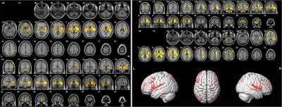 |
3 | Imaing the brain microstructures in patients of severe enlarged perivasclar spaces in the basal ganglia region using diffusion kurtosis imaging Video Permission Withheld
Yuelei Lyu1, Shuna Yang1, Chen Zhang2, Qinglei Shi2, Guang Yang3, Wenli Hu1, and Qi Yang1
1Beijing Chao-Yang Hospital, Capital Medical University, Beijing, China, 2MR Scientific Marketing, Siemens Healthineers, Beijing, China, 3Shanghai Key Laboratory of Magnetic Resonance, School of Physics and Electronic Science, East China Normal University, Shanghai, China
This study used DKI to investigate possible microstructural alterations of brain tissues associated with plenty of EPVS in the basal ganglia (BG) in patients of SVD. Our preliminary results revealed that patients with EPVS in the BG showed significant reductions in kurtosis parameters from DKI and elevated diffusion parameters derived from DKI in bilateral putamen. We also found abnormal diffusion parameters in mainly bilateral BG, limbic system, temporal lobe,and other scatter brain regions. Our data suggests patients with plenty of EPVS in the BG might undergo microstructure impairments in the regions above, especially bilateral insular and BG.
|
||
4590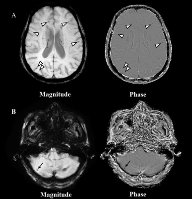 |
4 | Association between antiplatelet therapy and cerebral microbleeds determined by susceptibility weighted imaging in stroke-free pupolation Video Permission Withheld
Miaoxin Yu1, Yanan Jia2, Dandan Yang3, Runhua Zhang1, Yong Jiang1, Guitao Zhang1, Huiyu Qiao4, Hualu Han4, Rui Shen4, Zihan Ning4, Xihai Zhao4, and Gaifen Liu1
1Department of Neurology, Beijing Tiantan Hospital, Capital Medical University; China National Clinical Research Center for Neurological Diseases, Beijing, China, 2Department of Neurology, The Third Hospital of Hebei Medical University, Shijiazhuang, Hebei, China, 3Department of Radiology, Beijing Geriatric Hospital, Beijing, China, 4Center for Biomedical Imaging Research, Department of Biomedical Engineering, Tsinghua University, Beijing, China
In this cross-sectional, observational study, we investigated the association between antiplatelet therapy and cerebral microbleeds (CMBs) in community-based participants without stroke. The presence, count, and location of CMBs were evaluated by susceptibility weighted imaging. Antiplatelet therapy included aspirin use alone, clopidogrel use alone, cilostazol use alone, and dual antiplatelet therapy with aspirin and clopidogrel. We found that antiplatelet therapy was independently associated with CMBs at any location, particularly lobar CMBs, and aspirin use was a risk factor for any CMB and lobar CMBs. Our study suggests that optimizing antiplatelet treatment strategies in stroke-free population may decrease the risk of CMBs.
|
||
4591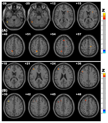 |
5 | Altered Spontaneous Brain Activity Related to Neurologic Dysfunction in Patients with Cerebral Small Vessel Disease: A resting-state study
Mengmeng Feng1, Hongwei Wen2,3, Haotian Xin1, Changhu Liang4, and Lingfei Guo4
1Shandong Provincial Hospital, Jinan, China, 2Key Laboratory of Cognition and Personality (Ministry of Education), Chongqing, China, 3Southwest University, Chongqing, China, 4Shandong Provincial Hospital Affiliated to Shandong First Medical University, Jinan, China
Cerebral small vessel disease (CSVD) patients can present structural and functional abnormalities. We divided 102 subjects into three groups and obtained information about altered spontaneous brain activity using amplitude of low-frequency fluctuation (ALFF), fractional ALFF (fALFF) and regional homogeneity (ReHo) methods based on resting-state functional MRI. Compared with controls and the CSVD without cerebral microbleeds (CSVD-n) group, the CSVD with cerebral microbleeds (CSVD-c) group showed significantly increased ALFF/ReHo values and decreased fALFF values in some brain regions, which are correlated to abnormal clinical characteristics. These results expounded the underlying neurophysiological mechanisms in CSVD patients.
|
||
4592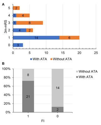 |
6 | Arterial transit artifact could prognose stroke patients beyond 24 hours from last known well
Di Wu1, Shun Zhang2, Weiyin Vivian Liu3, and Wenzhen Zhu4
1Radiology, Tongji Hospital, Tongji Medical College, Huazhong University of Science and Technology, Wuhan, Hubei, China, 2Tongji Hospital, Tongji Medical College, Huazhong University of Science and Technology, Wuhan, Hubei, China, 3GE Healthcare, Beijing, China, Beijing, China, 4Tongji Hospital, Tongji Medical College, Huazhong University of Science and Technology, Wuhan, China
The study aimed to assess the correlation of arterial transit artifact (ATA) with clinical outcomes and collateral circulation status in stroke patients beyond 24 hours from last known well. ATA is assessed by 3-dimentional pseudo-continuous ASL (3D-pCASL), collateral status by magnetic resonance angiography (MRA) using a grading system from 0 to 3, and clinical outcomes by 3-month modified Ranking Scale (3m-mRS). ATA was present in about half of the enrolled patients, most of whom had large artery disease. Though the correlation between ATA and the degree of collateral supply was not statistically significant, ATA could predict the favorable clinical outcomes.
|
||
4593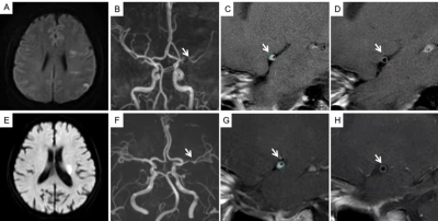 |
7 | Preliminary study on plaque characteristics in young adults with symptomatic intracranial atherosclerotic stenosis
Ling Li1, XiaoLing Zhang1, Xiaoyan Lei1, Min Tang1, Xuejiao Yan1, and Kai Ai2
1Department of MRI, Shaanxi Provincial People's Hospital, Xi'an, China, 2Philips Healthcare, Xi'an, China
This study used high-resolution magnetic resonance imaging (HRMRI) to explore plaque characteristics of young adults with symptomatic intracranial atherosclerotic stenosis (sICAS). Compared with old patients, young patients with sICAS have a smaller Maximum wall thickness and a greater ability to reconstruct, and are more prone to positive remodeling, which may cover up some patients with atherosclerotic stenosis. The results show great clinical importance for a better understanding of youth intracranial arterial plaque features and highlights the importance of HRMRI.
|
||
4594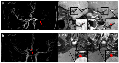 |
8 | MRI Radiomics approach for Identification of high-risk intracranial plaque basing on machine learning
Haining Wei1, Fang Wu2, Jie Lu2, and Rui Li1
1Tsinghua University, Beijing, China, 2Xuanwu Hospital, Capital Medical University, Beijing, China
Intracranial atherosclerotic disease (ICAD) is one of the main causes of ischemic stroke. Increasing evidence supports that vulnerable plaque is correlated with risk for stroke, which reveals the importance of intracranial plaque risk identification. Radiomics is an automated and repeatable approach for extracting massive features for medical imaging. However, few articles have focused on radiomic-based studies of intracranial plaque of basilar artery and middle cerebral artery. In this study, we propose to build a high-risk intracranial plaque model using radiomics features and machine learning to differentiate symptomatic plaque from asymptomatic plaque, which is helpful in guiding clinical management.
|
||
4595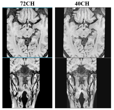 |
9 | A 72-channel coil system for MR vessel wall imaging of intracranial and carotid arteries at 3 T
Yingchao Tan1,2,3, Qiaoyan Chen1,2, Qian Zou3, Yanling Chen3, Junyu Chen3, Xin Liu1,2, Hairong Zheng1,2, and Ye Li1,2
1Paul C. Lauterbur Research Center for Biomedical Imaging, Shenzhen Institutes of Advanced Technology, Chinese Academy of Sciences, Shenzhen, China, 2Key Laboratory for Magnetic Resonance and Multimodality Imaging of Guangdong Province, Shenzhen, China, 3Shanghai United Imaging Healthcare, Shanghai, China
MR vessel wall imaging of intracranial and carotid arteries is still challenging because of the small cross-sectional size of the vessel wall and the cord, and susceptibility effects, especially in the carotid and the spinal cord. In this study, a 72-channel coil system that consists of a 64-channel head coil combined with an 8-channel carotid coil was proposed and characterized in its performance by comparison with a 40-channel coil system. As a result, the proposed 72-channel coil system provides improved performance in SNR, parallel imaging capability, and image quality.
|
||
4596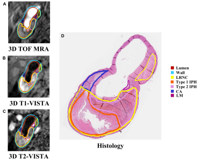 |
10 | Accuracy of Characterizing Carotid Vulnerable Atherosclerotic Plaque by 3D MR Vessel Wall Imaging: A Histological Validation Study
Chenlin Du1, Zihan Ning1, Huiyu Qiao1, Shuo Chen1, Tao Wang2, Jingli Cao3, Huo Ran4, Dongye Li5, Chunjiang Hu1, Shuwan Yu1, Hualu Han1, Rui Shen1, Dandan Yang1,6,7, Cancheng Liu8, Peng Wu9, and Xihai Zhao1
1Center for Biomedical Imaging Research, Department of Biomedical Engineering, Tsinghua University, Beijing, China, 2Department of Neurosurgery, Peking University Third Hospital, Beijing, China, 3China National Clinical Research Center for Neurological Disease, Beijing Tiantan Hospital, Capital Medical University, Beijing, China, 4Department of Radiology, Peking University Third Hospital, Beijing, China, 5Department of Radiology, Sun Yat-Sen Memorial hospital, Sun Yat-Sen University, Guangzhou, China, 6Beijing Institute of Brain Disorders, Laboratory of Brain Disorders, Ministry of Science and Technology, Collaborative Innovation Center for Brain Disorders, Capital Medical University, Beijing, China, 7Department of Radiology, Beijing Geriatric Hospital, Beijing, China, 8Thorough Images, Beijing, China, 9Philips Healthcare, Shanghai, China
This study investigated the accuracy of 3D multi-contrast MR vessel wall imaging (VWI) in characterizing carotid vulnerable plaque compositions including lipid-rich necrotic core (LRNC), intraplaque hemorrhage (IPH) and calcification (CA) validated by histology. Good agreements were found between MR and histology in identifying LRNC (κ=0.67), IPH (κ=0.66), and CA (κ=0.62) after excluding histological sections with the plaque components <1.77 mm2. Moderate agreements were reached in quantifying plaque compositions with r values ranged from 0.46 to 0.61 (LRNC: r=0.52; IPH: r=0.6; CA: r=0.46). Our study demonstrated that 3D multi-contrast MR VWI is capable of accurately characterizing carotid vulnerable atherosclerotic plaques.
|
||
4597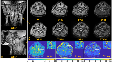 |
11 | 3D Large-Coverage Simultaneous QUantitative T1-T2-T2* Mapping (LC-SQUMA) of Carotid Vessel Wall
Huiyu Qiao1, Qiansu Yang2, Shuo Chen1, Zihan Ning1, Hualu Han1, Rui Shen1, Peng Wu3, Huijun Chen1, and Xihai Zhao1
1Center for Biomedical Imaging Research, Department of Biomedical Engineering, School of Medicine Tsinghua University, Beijing, China, 2Center of Medicine Clinical Research, Department of Pharmacy, Medical Supplies Center of PLA General Hospital, Beijing, China, 3Philips Healthcare, Shanghai, China MR T1-, T2-, and T2*- mapping have been used to characterize carotid plaques. In this study, we realized a 3D Large-Coverage (longitudinal coverage: 160 mm) Simultaneous QUantitative T1-T2-T2* Mapping (LC-SQUMA) sequence with 0.8 mm isotropic spatial resolution. The LC-SQUMA sequence showed excellent agreements with reference imaging in measuring T1 (R2=0.96), T2 (R2=0.85) and T2* (R2=0.90) values. No significant difference was found in measuring T1, T2 and T2* values (all P>0.05) of cervical muscle between LC-SQUMA sequence and clinical referenced quantitative imaging sequences. The 3D LC-SQUMA sequence is feasible in the large-coverage carotid vessel wall quantitative imaging with isotropic spatial resolution. |
||
4598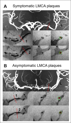 |
12 | Quantitative analysis of Symptomatic and Asymptomatic Atherosclerotic Middle Cerebral Artery Plaques Using 7T Magnetic Resonance Imaging Video Permission Withheld
Xiaoyan Bai1, Qingle Kong2, Zhiye Li1, Pingping Fan1, and Binbin Sui3
1Department of Radiology, Beijing Tiantan Hospital, Capital Medical University, Beijing Neurosurgical Institute, Beijing, China, Beijing, China, 2MR Collaboration, Siemens Healthineers Ltd., Beijing, China, Beijing, China, 3Tiantan Neuroimaging Center of Excellence, China National Clinical Research Center for Neurological Diseases, Beijing, China, Beijing, China A total of 40 MCA atherosclerotic plaques of 34 patients (20 symptomatic and 20 asymptomatic plaques) were quantitatively analyzed of plaque features including plaque burden, length, remodeling index, plaque distribution, and surface morphology using 7T vessel wall Magnetic Resonance Imaging (VW-MRI). Compared with the asymptomatic plaques, symptomatic MCA plaques had larger plaque length and higher plaque burden. Superior-wall distribution and irregular plaque surface were observed more commonly on the symptomatic MCA plaques. 7T VW-MRI is a promising method to evaluate the atherosclerotic plaque features at MCAs. |
||
4599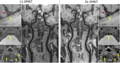 |
13 | Deep Learning regularized SPIRiT reconstruction accelerates joint intracranial and carotid vessel wall imaging into 3.5 minutes
Sen Jia1, Jing Cheng1, Zhuoxu Cui2, Lei Zhang1, Haifeng Wang1, Xin Liu1, Hairong Zheng1, Hongying Zhang3, and Dong Liang1,2
1Paul C. Lauterbur Research Center for Biomedical Imaging, Shenzhen Institutes of Advanced Technology, Shenzhen, China, 2Research Center for Medical AI, Shenzhen Institutes of Advanced Technology, Shenzhen, China, 3Northern Jiangsu People's Hospital, Yangzhou, China
Deep learning regularized SPIRiT reconstruction is developed by unrolling the conventional L1-SPIRiT optimization solved by the projection onto convex sets (POCS) iteration into a multi-layer convolutional neural network. The learnable network regularization with 3D convolution improved the reconstruction accuracy and efficiency compared with the iterative L1-SPIRiT with 2D sparsity regularization in a fixed transform domain. The simplicity of POCS iteration also benefits the design complexity of the DL-SPIRiT network. The proposed DL-SPIRiT could accelerate the joint intracranial and carotid vessel wall imaging of isotropic 0.6 mm resolution by 8-fold, leading to a scan time of only 3.5 minutes.
|
||
4600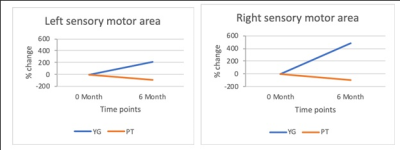 |
14 | Using fMRI to compare physiotherapy and yoga-induced changes in post-stroke rehabilitation Video Permission Withheld
Dushyant Kumar1, Rajesh Mishra1, Chahat Kumar2, Priyanka Bhagat2, Padma Srivastava2, and Rama Jayasundar1
1NMR, All India Institute of Medical Sciences, Delhi, India, 2Neurology, All India Institute of Medical Sciences, Delhi, India
This study has compared using fMRI, the changes effected by physiotherapy and yoga in post-stroke recovery. Ischemic stroke patients (n=20) with motor deficits were divided into two groups - yoga and physiotherapy. Each group practiced yoga or physiotherapy for one hour daily for six months, under the supervision of certified yoga trainers and physiotherapists, respectively. Pre- and post- (6 months) intervention evaluations were carried out using fMRI at 3T and NIHSS scores. Results show significant improvement in the NIHSS scores and BOLD activity in the affected sensory motor region of the stroke patients in the yoga group compared to physiotherapy.
|
||
4601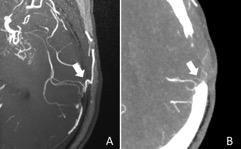 |
15 | High-resolution compressed sensing TOF-MRA might show better performance than CTA for evaluating revascularization in Moyamoya patients Video Permission Withheld
Shujing Ren1, Wei Wu2, Chunqiu Su1, Qianmiao Zhu2, Michaela Schmidt3, Yi Sun4, Christoph Forman3, Peter Speier3, Xunning Hong1, and Shanshan Lu1
1Department of Radiology, The First Affiliated Hospital of Nanjing Medical University, Nanjing, China, 2Department of Neurosurgery, The First Affiliated Hospital of Nanjing Medical University, Nanjing, China, 3Siemens Healthcare, Erlangen, Germany, 4Siemens Healthcare, Shanghai, China
It is important for MMD patients to receive repeated life-time follow-up examinations for evaluation of bypass patency and collateral formation from neovascularization after bypass surgery. In this study we evaluated the clinical value of high-resolution (0.4 × 0.4 × 0.4 mm3 isotropic) CS TOF-MRA for assessment of STA-MCA bypass and neovascularization by using CTA as the reference. We found that high-resolution CS TOF-MRA could provide better visualization for both STA-MCA bypass and neovascularization than traditional CTA within a clinically reasonable time in MMD patients after surgical revascularization.
|
||
4602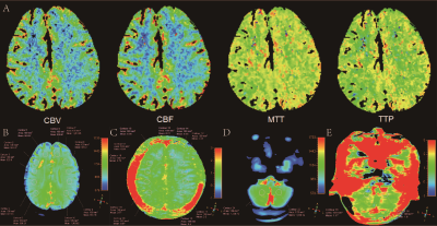 |
16 | Assessment of cerebrovascular related tissue pH in patients with moyamoya disease: an amide proton transfer weighted imaging study
Chao Xia1,2, Jiaxin Zeng1,2, Xia Wei1,2, Ziyu Li1,2, Yuan Sun1,2, Na Hu1,2, Yi Liu3, Kai Ai4, and Su Lui1,2
1Huaxi MR Research Center (HMRRC), Functional and Molecular Imaging Key Laboratory of Sichuan Province, Department of Radiology, West China Hospital, Sichuan University, Chengdu, China, 2Research Unit of Psychoradiology, Chinese Academy of Medical Sciences, Chengdu, China, 3Department of Neurosurgery, West China Hospital, Sichuan University, Chengdu, China, 4Philips Healthcare, Xi'an, China
The amide proton transfer weighted (APTw) imaging was used to investigate the collateral circulation change and pH alteration in patients with moyamoya disease (MMD). Thirteen patients underwent computed tomography perfusion (CTP), digital subtraction angiography (DSA), and APTw imaging. We analyzed the difference in APTw values between the cerebral and cerebellar hemispheres. Then subgroup analysis was conducted according to the stage of preinfarction period based on CTP. The results revealed that APTw values were significantly lower in cerebral hemispheres than those in cerebellar hemispheres. However, no significant difference in APTw values among patients with different stages of preinfarction period was found.
|
||
4603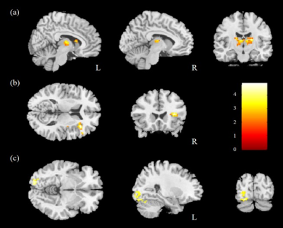 |
17 | Mapping functional connectivity and brain network changes in hypertensive using resting-state fMRI Video Permission Withheld
Yu-Chen Chuang1,2, Wen-Chau Wu1,3, Vincent Chin-Hung Chen4,5, Yen-Hsuan Hsu6, and Jun-Cheng Weng2,5,7
1Institute of Medical Device and Imaging, Graduate Institute of Clinical Medicine, National Taiwan University, Taipei city, Taiwan, 2Department of Medical Imaging and Radiological Sciences, Graduate Institute of Artificial Intelligence, Chang Gung University, Taoyuan, Taiwan, 3Department of Medical Imaging, National Taiwan University Hospital, Taipei, Taiwan, 4School of Medicine, Chang Gung University, Taoyuan, Taiwan, 5Department of Psychiatry, Chang Gung Memorial Hospital, Chiayi, Taiwan, 6Department of Psychology, National Chung Cheng University, Chiayi, Taiwan, 7Medical Imaging Research Center, Institute for Radiological Research, Chang Gung University and Chang Gung Memorial Hospital at Linkou, Taoyuan, Taiwan Although understanding of the relationship between cardiovascular disease and brain dysfunction has improved significantly over the past decades, it remains unclear whether hypertension can be a potential risk factor for cognitive impairment. This study tried to use resting-state fMRI to explore the association between hypertensive (HTN) and brain function alterations. We found significant differences in the DMN subsystems in HTN group compared with control group. We also observed higher assortativity in HTN group. |
||
 Back to Meeting Home
 Back to the Program-at-a-Glance
The International Society for Magnetic Resonance in Medicine is accredited by the Accreditation Council for Continuing Medical Education to provide continuing medical education for physicians.
 Back to Meeting Home
 Back to the Program-at-a-Glance
 View the Presentation