Online Gather.town Pitches
Vascular Imaging & Stroke IV
Joint Annual Meeting ISMRM-ESMRMB & ISMRT 31st Annual Meeting • 07-12 May 2022 • London, UK

Online Gather.town Pitches
Vascular Imaging & Stroke IV
| Booth # | ||||
|---|---|---|---|---|
4888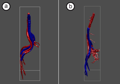 |
1 | Contrast-free MR Angiography and Venography for Visualization of Iliac Vein Compression Syndrome in less than 10 minutes
Vadim Malis1, Jirach Kungsamutr2, Won Bae1, Xiaowei Zhang1, Yoshimori Kassai3, Albert Hsiao1, John S Lane4, and Mitsue Miyazaki1
1Radiology, UC San Diego, San Diego, CA, United States, 2Bioengineering, UC San Diego, San Diego, CA, United States, 3Canon Medical Systems Corp., Tochigi, Japan, 4Surgery, UC San Diego, San Diego, CA, United States
Iliac vein compression syndrome, known as May-Thurner syndrome (MTS), is a condition when the right common iliac artery constrains the flow in the left common iliac vein. Currently, ultrasound and CT venography (CTV) are the imaging modalities of choice for the diagnosis. We propose a framework to visualize iliac vein and artery utilizing contrast free MR venography and MR angiography in less than 10 minutes.
|
||
4889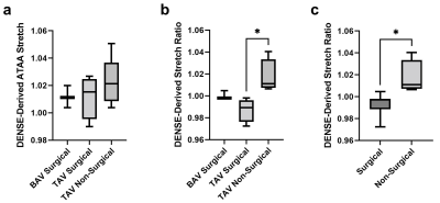 |
2 | Imaging Ascending Thoracic Aortic Aneurysm Wall Stretch: A Comparison to Biaxial Mechanical Testing
Huiming Dong1, Henrik Haraldsson1, Joseph Leach1, Ang Zhou1, Megan Ballweber1, Chengcheng Zhu2, Yue Xuan3, Zhongjie Wang3, Michael Hope1, Liang Ge3, Frederick H. Epstein4, David Saloner1, Elaine Tseng3, and Dimitrios Mitsouras1
1Department of Radiology and Biomedical Imaging, University of California, San Francisco, San Francisco, CA, United States, 2Department of Radiology, University of Washington, Seattle, WA, United States, 3Department of Surgery, University of California, San Francisco, San Francisco, CA, United States, 4Department of Biomedical Engineering, University of Virginia, Charlottesville, VA, United States
Ascending thoracic aortic aneurysm (aTAA) can result in life-threatening rupture or dissection. Displacement encoding with stimulated echoes (DENSE) is a non-invasive phase-contrast MRI technique that can measure aTAA wall deformation during the cardiac cycle. This study investigated DENSE-derived aTAA wall stretch in patients and found that the ratio between aTAA stretch and descending aorta stretch was different in patients who met surgical repair criteria from those who did not. Moreover, mechanical properties of aTAA specimens from patients who underwent surgery correlated significantly with in vivo DENSE measurements. Our findings suggest DENSE as a potential imaging marker for understanding aTAA progression.
|
||
4890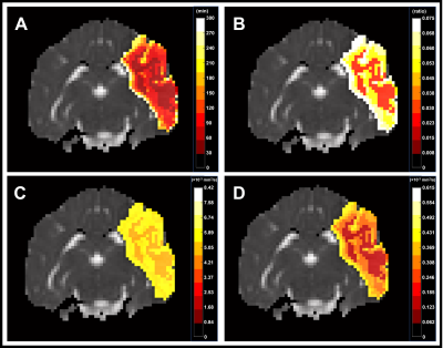 |
3 | Predicting penumbral tissue death using intravoxel incoherent motion MRI in a canine large vessel occlusion model
Mohammed Salman Shazeeb1, Robert King1, Zeynep Vardar1, Josephine Kolstad1, Vania Anagnostakou1, Nils Henninger1, and Matthew Gounis1
1University of Massachusetts Medical School, Worcester, MA, United States
In acute ischemic stroke due to large vessel occlusion (LVO), information about the penumbral tissue can be vital in making decisions on how to treat ischemic stroke patients in the clinic. This study investigated the use of intravoxel incoherent motion (IVIM) MRI in a canine LVO model to quantify the perfusion information in penumbral tissue prior to infarction. The IVIM parameters were assessed to predict the onset of penumbral tissue death. Of the 3 IVIM parameters, f showed the best utility in predicting penumbral tissue death.
|
||
4891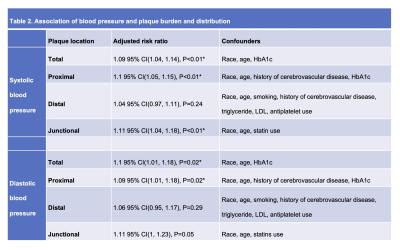 |
4 | Hypertension is Associated with Intracranial Atherosclerotic Plaque Burden and Distribution: a MR Vessel Wall Imaging Study
Jiayu Xiao1, Jae W Song2, Steven Y Cen1,3, Fang Wu4, Xiao Liu5, Tao Jiang5, Konrad H Schlick6, Debiao Li7, Shlee S Song6, Qi Yang5, and Zhaoyang Fan1,8,9
1Radiology, Keck School of Medicine, Los Angeles, CA, United States, 2Radiology, Hospital of the University of Pennsylvania, Philadelphia, PA, United States, 3Neurology, Keck School of Medicine, Los Angeles, CA, United States, 4Radiology, Xuanwu Hospital, Beijing, China, 5Radiology, Beijing Chaoyang Hospital, Beijing, China, 6Neurology, Cedars-Sinai Medical Center, Los Angeles, CA, United States, 7Biomedical Imaging Research Institute, Cedars-Sinai Medical Center, Los Angeles, CA, United States, 8Radiation Oncology, Keck School of Medicine, Los Angeles, CA, United States, 9Biomedical Engineering, University of Southern California, Los Angeles, CA, United States
Hypertension is one of the most important modifiable risk factor for atherosclerosis. However, the association between blood pressure and intracranial plaque remains unclear. In this study, we investigated the impact of hypertension on intracranial plaque burden and distribution in 174 patients with recent ischemic stroke. Systolic blood pressure and diastolic blood pressure were independently associated with a higher total plaue burden as well as higher plaque counts at the proximal and junctional locations. These findings suggest hypertension may influence not only the count burden but also the distribution of intracranial plaque development.
|
||
4892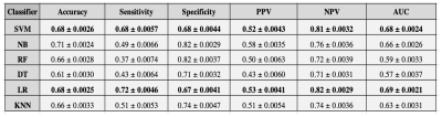 |
5 | Predicting Hematoma Expansion after Spontaneous Intracranial Hemorrhage Through a Magnetic Resonance-Based Radiomics Model
Samantha E Seymour1, Ryan A Rava1, Mitchell T Chudzik1, Kenneth V Snyder1,2,3, Muhammad E Waqas1,3, Jason M Davies1,2,3,4, Elad E Levy1,2,3, Adnan E Siddiqui1,2,3, Xiaoliang E Zhang5, and Ciprian E Ionita1,2,3,6
1Canon Stroke and Vascular Center, Buffalo, NY, United States, 2Jacobs School of Medicine and Biomedical Sciences, Buffalo, NY, United States, 3Department of Neurosurgery, University at Buffalo, Buffalo, NY, United States, 4Department of Bioinformatics, University at Buffalo, Buffalo, NY, United States, 5Department of Biomedical Engineering, University at Buffalo, Buffalo, NY, United States, 6Department of Biomedical Engineerinng, University at Buffalo, Buffalo, NY, United States
Intracranial hemorrhage (ICH) is bleeding within the cranium and occurs within the brain tissue, ventricles, and intracranial space. Hematoma expansion following an ICH has been related to increased mortality and morbidity inpatients. To detect ICH patients at risk, machine learning models can be used to predict whether or not hematoma expansion will occur. This study aims to assess the feasibility of machine learning prediction models using a radiomics approach. The highest sensitivity results indicated as 95% confidence intervals are 0.68 ± 0.004 and 0.72 ± 0.004, were achieved by support vector machine and logistic regression classifier models, respectively.
|
||
4893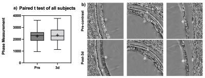 |
6 | Detecting Residual Iron Contrast after Ferumoxytol Infusion in Cerebral Vasculature Using High-Resolution SWI at 7T
Li Jiang1, Yongsheng Chen2, Marco Muccio1, Chenyang Li1, Sagar Buch2, E Mark Haacke2, and Yulin Ge1
1NYU Grossman School of Medicine, New York, NY, United States, 2Wayne State University, Detroit, MI, United States
Ultra-small superparamagnetic iron oxides (USPIO) enhanced MRI has shown its great potential in detecting vascular abnormality in the brain on T2*-weighted sequences for better showing both macro- and micro-vasculature. In this study, we investigated the quantitative phase changes after Ferumoxytol infusion and examined its residual contrast in brain vasculature at 3-, 7- and 14-day follow-up SWI scans. To quantitatively measure the Ferumoxytol-induced susceptibility, the veins parallel to the main field were chosen for measurement. After measuring the intensity on high-pass filter phase images, the paired t-test showed a significant difference between pre-contrast and post-3d values from the follow-up visits.
|
||
4894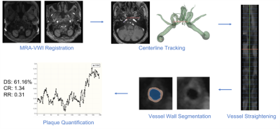 |
7 | Vessel Wall Imaging-Dedicated Automatic Processing Pipeline (VWI-APP): Towards Efficient and Reliable Intracranial Plaque Quantification
Hanyue Zhou1,2, Jiayu Xiao2, Dan Ruan1,3, and Zhaoyang Fan2,4,5
1Bioengineering, University of California, Los Angeles, Los Angeles, CA, United States, 2Radiology, University of Southern California, Los Angeles, CA, United States, 3Radiation Oncology, University of California, Los Angeles, Los Angeles, CA, United States, 4Radiation Oncology, University of Southern California, Los Angeles, CA, United States, 5Biomedical Engineering, University of Southern California, Los Angeles, CA, United States
We propose an automatic pipeline for atherosclerosis plaque analysis based on magnetic resonance (MR) vessel wall imaging (VWI). The pipeline consists of five modules, i.e., 1) MR angiography (MRA) to VWI registration, 2) vessel centerline tracking, 3) vessel straightening and cross-sectional slices generating, 4) deep learning -based vessel wall and lumen segmentation, and 5) plaque quantification. The pipeline achieved good to excellent reliability for the clinically relevant plaque measures and largely reduced the analysis time required for physicians.
|
||
4895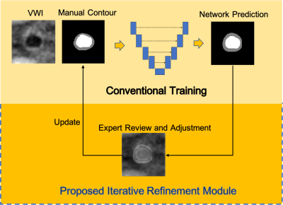 |
8 | Iterative Refinement of Expert Contours for Improved Ground-truth Quality in Intracranial Vessel Segmentation Neural Network Training
Hanyue Zhou1,2, Jiayu Xiao2, Dan Ruan1,3, and Zhaoyang Fan2,4,5
1Bioengineering, University of California, Los Angeles, Los Angeles, CA, United States, 2Radiology, University of Southern California, Los Angeles, CA, United States, 3Radiation Oncology, University of California, Los Angeles, Los Angeles, CA, United States, 4Radiation Oncology, University of Southern California, Los Angeles, CA, United States, 5Biomedical Engineering, University of Southern California, Los Angeles, CA, United States
With a typical slice-by-slice labeling fashion, the manual contouring process is subject to large intra-observer variation, especially for small-sized intracranial arteries. We propose an iterative refinement approach for ground truth contours with the help of deep neural networks for intracranial lumen and vessel wall segmentations. We demonstrate that the approach improved the smoothness of the predicted contours and feature quantification, which can potentially boost the robustness of a neural network as a consequence of the reduced uncertainty in expert labels.
|
||
4896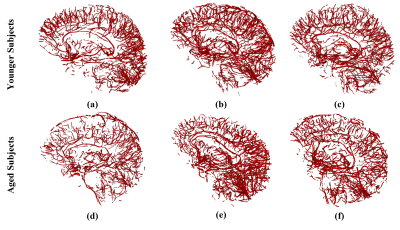 |
9 | Vessel Density Mapping of Brain Small Vessels on 3D High Resolution Black Blood MRI
Mona Sharifi Sarabi1, Samantha J Ma1,2, Danny JJ Wang1, and Yonggang Shi1
1University of Southern California, Los Angeles, CA, United States, 2Siemens Medical Solutions USA, Inc., Los Angeles, CA, United States
High resolution 3D black blood MRI with sub-millimeter spatial resolution has been proposed for visualizing small vessels of the brain. Here we present and evaluate a novel approach for mapping brain vessel density from 3D black blood images acquired at 3T. Using automated vessel segmentation and non-linear registration, localized detection and quantification of small vessel density is demonstrated to be feasible. This framework can potentially serve as a useful tool for detection and monitoring of localized vascular changes in aging and neurovascular disorders.
|
||
4897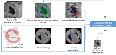 |
10 | Carotid atherosclerotic plaque segmentation in multi-weighted MRI using a two-stage neural network model
Ran Li1,2, Jie Zheng1,2, Pamela Woodard1,2, and Jha Abhinav1,2
1Mallinckrodt Institute of Radiology, Washington University in St. Louis, St. Louis, MO, United States, 2Department of Biomedical Engineering, Washington University in St. Louis, St. Louis, MO, United States
Our objective of this study is to accurately segment and classify carotid atherosclerotic plaque components in a completely automated manner, based on multi-weighted MRI. Specifically, we segmented each pixel using a two-module neural network model. Furthermore, we generated segmentation uncertainty maps with a Bayesian method to evaluate the inherent uncertainty of this segmentation task.
|
||
4898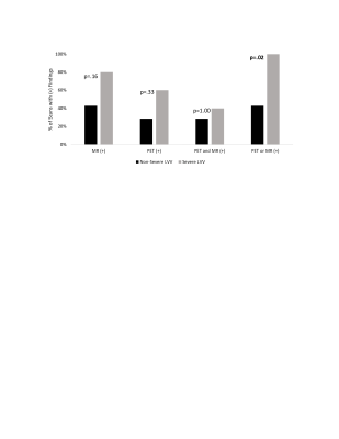 |
11 | Combined Modality PET/MR For the Detection of Severe Large Vessel Vasculitis
John Cerne1, Sophia Liu1, Muhammad Umair1, Ashitha Pathrose1, Hatice Savas1, Lisa Wilsbacher1, Jackson E Moore1, Michael Markl1, James Carr1, and Ryan Avery1
1Northwestern University, Chicago, IL, United States
Combined modality PET/MR may be a sensitive modality to detect severe large vessel vasculitis. To distinguish severe large vessel vasculitis, a qualitative review of PET imaging findings appears more useful than standardized uptake values of a patient’s highest-uptake vessel. Inflammatory marker levels and trends, as well as single-modality scan findings, were not significantly different between severe and non-severe large vessel vasculitis patients.
|
||
4899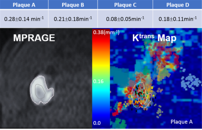 |
12 | Dynamic 3D T1 MRI method for T1-based Quantification of DCE for the Measurements of kinetic Parameters on Carotid Atherosclerotic Plaques
Seong-Eun Kim1, John A Roberts1, J. Scott MacNally1, Mathew Alexander 1, Gerald S Treiman2,3, Hediyeh Baradaran1, and Dennis L Parker1
1UCAIR, Department of Radiology and Imaging Sciences, University of Utah, Salt Lake City, UT, United States, 2Department of Surgery, University of Utah, Salt Lake City, UT, United States, 3VASLCHCS, Salt Lake City, UT, United States
This work introduced a reliable, comprehensive carotid DCE technique, including a high spatial and temporal resolution 3D dynamic T1 sequence and retrospective reconstruction methods to measure kinetic parameters on carotid atherosclerotic plaque. This technique can be used to evaluate the correlation between kinetic parameters and ischemic stroke. We believe that plaque kinetic parameters can be used in subsequent clinical trials as a changeable predictor of ischemic stroke risk. Clinicians could then use carotid DCE to monitor early response to medical therapy, and thereby improve detection and treatment of unstable carotid plaque and prevention of stroke.
|
||
 Back to Meeting Home
 Back to the Program-at-a-Glance
The International Society for Magnetic Resonance in Medicine is accredited by the Accreditation Council for Continuing Medical Education to provide continuing medical education for physicians.
 Back to Meeting Home
 Back to the Program-at-a-Glance
 View the Presentation