Online Gather.town Pitches
MSK IV
Joint Annual Meeting ISMRM-ESMRMB & ISMRT 31st Annual Meeting • 07-12 May 2022 • London, UK

| Booth # | ||||
|---|---|---|---|---|
4615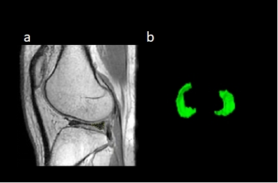 |
1 | Feasibility of synthetic PD-weighted images on degenerative OA in comparison to structural ZTE-MRI
Huizheng Wang1, Weiyin Vivian Liu2, Xingyao Yu3, Wen Chen4, Ling Sang4, and Kejun Wang4
1Department of Radiology, Taihe Hospital, Jinzhou Medical University Union Training Base, Shiyan,Hubei, China, 2GE Healthcare, Beijing, China, 3Biomedical Engneering College,Hubei University of Medicine, Shiyan,Hubei, China, 4Department of Radiology, Taihe Hospital, Hubei University of Medicine, Shiyan,Hubei, China
Image follow-up for intervention effectiveness of osteoarthritis is necessary to avoid or slow down the development of joint space stenosis, bone sclerosis and osteophyte formation. Compared to ZTE-MRI with the feature of nominal zero echo time, MAGIC, synthetic MRI, offers not only anatomical structure images but also T1, T2, and PD maps. We found a significant correlation between joint space value and meniscus volume in reflection of antagonism effect of joint space and surrounding tissues. Proton density weighted synthetic MR images (PD-MAGIC) offered as good as ZTE-MR structure images and might supplement quantitative information clinically to improve follow-up diagnostic performance.
|
||
4616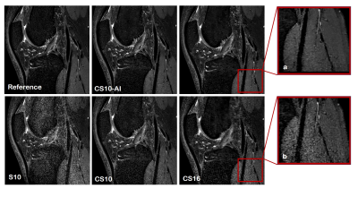 |
2 | Accelerated 3D PDWI in knee imaging: Quality and efficiency of a deep learning-based Compressed SENSE reconstruction
Lin Mu1, Ying Qiu2, Yun Pei3, Yi Zhu4, and Ke Jiang4
1Radiology, The First Hospital of Jilin University, Changchun, China, 2The First Hospital of Jilin University, Changchun, China, 3College of Electronic Science and Engineering, Jilin University, Changchun, China, 4Philips Healthcare, Beijing, China
The use of three dimensional (3D) volumetric acquisition in clinical settings has been limited due to long scan time. A deep learning-based reconstruction algorithm allows shortening of scan time and provide comparable overall image quality when compared with standard sequences. Adaptive-CS-Net, a deep neural network previously introduced at the 2019 fast MRI challenge, was expanded and presented here as a Compressed-SENSE Artificial Intelligence (CS-AI) reconstruction. The purpose of the study is to determine the feasibility of 3D PDWI accelerated with CS-AI for evaluating the knee image quality and compared with SENSE and standard Compressed-SENSE.
|
||
4617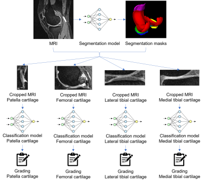 |
3 | Knee Osteoarthritis: Automatic Grading with Deep Learning
Junru Zhong1, Yongcheng Yao1, Sheheryar Khan2, Fan Xiao1, Dόnal G. Cahill1, James F. Griffith1, and Weitian Chen1
1Department of Imaging and Interventional Radiology, The Chinese University of Hong Kong, Sha Tin, NT, Hong Kong, 2School of Professional Education & Executive Development, The Hong Kong Polytechnic University, Kowloon, Hong Kong We present a deep learning-based knee osteoarthritis grading system that automatically provides a binary classification for cartilage degeneration. The system was trained on MRI data sets applying MOAKS grading from the Osteoarthritis Initiative (OAI). The proposed method achieved an accuracy of 0.75 to 0.83, despite being conducted on highly imbalanced data sets. Significant improvement in accuracy is expected with more balanced data sets. |
||
4618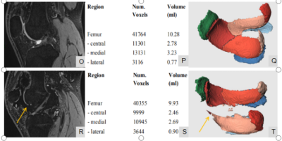 |
4 | Quantitative evaluation of cartilage damage in hemophiliac arthropathy using automatic segmentation of 3D high resolution MRI images
Jiajia Li1, Shufang Wei1, Xianchang Zhang2, Jing An3, Esther Raithel4, and Yinghui Ge1
1Fuwai Central China Cardiovascular Hospital, Zhengzhou, China, 2MR Collaboration, Siemens Healthineers Ltd, Beijing, China, 3Siemens Shenzhen Magnetic Resonance Ltd, Shenzhen, China, 4Siemens Healthcare GmbH, Erlangen, Germany
Hemophilic arthropathy (HA) is a serious complication of haemophilia, characterized by bone and cartilage damage due to recurrent joint bleeding. Existing evidence showed HA has a fast disease progression. MRI-based evaluation plays an important role in the detection, categorization and staging of soft tissue and osteochondral changes in HA, which is important for assessing and monitoring treatment outcomes. However, currently semi-quantitative scoring system based on conventional MRI images cannot give an objective assessment of knee cartilage damage. To this end, this study for the first time used an automatic segmentation algorithm to quantitatively evaluate the morphological change of knee cartilage in HA.
|
||
4619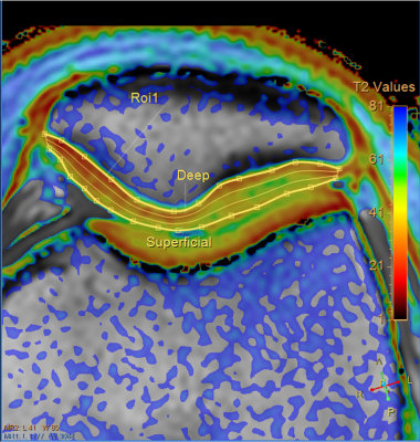 |
5 | T2 map segmentation into the deep, intermediate, and superficial layers when staging of patellar cartilage chondromalacia
Elena Voronkova1,2, Petr Menshchikov3,4, Ilya Melnikov1, Andrei Manzhurtsev1,4, Maxim Ublinskii1, Denis Vorobyev1, Dmitriy Kupriyanov3, and Tolib Akhadov1
1Clinical and Research Institute of Emergency Pediatric Surgery and Trauma, Moscow, Russian Federation, 2National Research Nuclear University MEPhI, Moscow, Russian Federation, 3Philips Healthcare, Moscow, Russian Federation, 4Institute of Biochemical Physics, Russian Academy of Sciences, Moscow, Russian Federation
This paper has demonstrated the benefits of segmentation into the deep, intermediate, and superficial layers in analysis of T2 maps of patellar cartilage with chondromalacia. In the deep and intermediate layers, the Т2 values were significantly different in all the patients groups, while the T2 values in the superficial layer did not depend on the disease grade. Compared with the whole cartilage assessment, this approach increased the sensitivity and specificity of chondromalacia staging by 17% and therefore can significantly increase the clinical efficiency of T2 mapping.
|
||
4620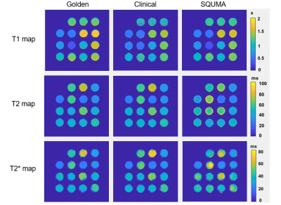 |
6 | In vivo Feasibility of Three-Dimensional Simultaneous Quantitative T1-T2-T2* Mapping (SQUMA) for Knee Cartilage
Renchi Ma1, Huiyu Qiao2, Xinrong Qiao1, Huijun Chen2, Xihai Zhao2, and Zhuozhao Zheng1
1Department of Radiology, Beijing Tsinghua Changgung Hospital, School of Medicine, Tsinghua University, Beijing 102218, China, Beijing, China, 2The Center for Biomedical Imaging Research, Department of Biomedical Engineering, School of Medicine, Tsinghua University, Beijing 100084, China, Beijing, China
T1, T2, and T2* mapping provide valuable information for evaluating joint cartilage. However, few studies simultaneously quantified T1, T2, and T2* map to evaluate knee cartilage. In this study, a three-dimensional (3D) simultaneous quantitative T1-T2-T2* mapping (SQUMA) sequence is utilized in knee cartilage. Excellent correlation and good consistency in measuring T1, T2 and T2* values were found between 3D SQUMA sequence and referenced sequences in phantom studies. The T1, T2 and T2* values of knee cartilage were 949.90±82.59 ms, 36.85±2.25 ms and 27.89±5.74 ms, respectively, with good repeatability. 3D SQUMA sequence is feasible for in vivo evaluation of knee cartilage.
|
||
4621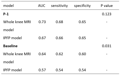 |
7 | Can infrapatellar fat pad predict the incidence of knee osteoarthritis by using deep learning based on MRI? Data from osteoarthritis initiative
Keyan Yu1,2, Chuanyang Zheng3, Jiaping Hu1, Lijie Zhong1, Xiaodong Zhang1, and Qi Dou3
1Department of Medical Imaging, The Third Affiliated Hospital of Southern Medical University, Guangzhou, China, 2Department of Radiology, Peking University Shenzhen Hospital, Shenzhen, China, 3Department of Computer Science & Engineering, The Chinese University of Hong Kong, Hong Kong, China
Infrapatellar fat pad (IPFP) is an important risk factor for the incident of radiographic knee osteoarthritis (iROA)1, 2. However, the potential of being an independent biomarker to predict iROA is untapped. Deep learning (DL) is a set of algorithms that enable computers to discover complicated patterns in large data sets3. In this study, we train a DL model to predict iROA with auto-segmented IPFP, comparing it to the DL model set up with corresponding whole knee MR images (MRI). The results reveal that IPFP alteration can predict iROA independently comparably to the whole knee MRI at one year before iROA.
|
||
4622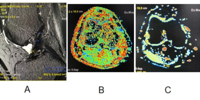 |
8 | Application of MR diffusion tensor imaging (DTI) in acute anterior cruciate ligament (ACL) injury
Xiaoxuan Wang1, Xiaowen Ma1, Xiaocheng Wei2, and Haiyan Li1
1Department of Magnetic Resonance Imaging, Hong Hui Hospital of Xi’an Jiaotong University, Xi’an, China, 2GE Healthcare, Beijing, China
In this study, we aim to investigate whether DTI is superior to conventional MRI in evaluating acute ACL injury. It was concluded that the mean FA values is lower and the mean ADC values is higher in the ACL injury group than control group. The sensitivity, specificity, accuracy and Kappa value of DTI were higher than those of conventional MRI.
|
||
4623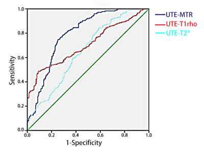 |
9 | In vivo preliminary assessment of knee cartilage degeneration by Quantitative Ultrashort Echo Time MRI
Xiaolian Su1, Guangyu Tang1, and Pu-Yeh Wu2
1Department of Radiology, Shanghai tenth People’s Hospital, Tongji University School of Medicine, Shanghai, China, shanghai, China, 2GE Healthcare, Beijing, China, Beijing, China
Recent advances in UTE-MRI techniques development allow for quantitative measurements of tissues with very short T2. However, in vivo experiments are needed to extend its application in clinical practice. In this in vivo study, we found that both UTE-MTR and UTE-adiabatic-T1rho values indicated a moderate correlation with MOAKS Grading. Meanwhile, both of them had moderate diagnostic efficacy for the mild cartilage degeneration (MOAKS = 1). These findings suggested that quantitative UTE-MRI techniques have a great potential as imaging biomarkers for early OA diagnosis in clinic.
|
||
4624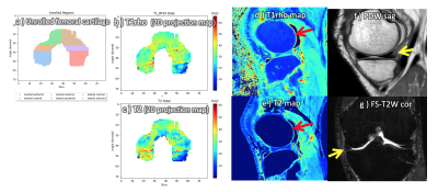 |
10 | MIXTURE-DOSMA: Initial clinical research of a comprehensive and multi-parametric quantitative 3D knee MR exam in patients with knee joint pain.
Takayuki Sakai1, Jihun Kwon2, Masami Yoneyama2, Daichi Murayama1, Quin Lu3, Arjun Desai4, Akshay S Chaudhari5, Tosiaki Miyati6, Shigehiro Ochi1, and Atsuya Watanabe7,8
1Radiology, Eastern Chiba Medical Center, Chiba, Japan, 2Philips Japan, Tokyo, Japan, 3Philips Healthcare NA, San Francisco, CA, United States, 4Electrical Engineering, Stanford University, Stanford, CA, United States, 5Radiology, Stanford University, Stanford, CA, United States, 6Faculty of Health Sciences, Institute of Medical, Pharmaceutical and Health Sciences, Kanazawa University, Kanazawa, Japan, 7General Medical Services, Chiba University Graduate School of Medicine, Chiba, Japan, 8Orthopaedic Surgery, Eastern Chiba Medical Center, Chiba, Japan
Multi-Interleaved X-prepared TSE with inTUitive RElaxometry (MIXTURE) is a recently developed 3D magnetization-prepared turbo spin-echo sequence. MIXTURE provides 3D high-resolution isotropic knee morphometry and T1ρ relaxometry in a clinically reasonable scan time. In this study, we integrated MIXTURE with the into a Deep-learning, Open-Source Medical Analysis (DOSMA) framework for deep learning-powered musculoskeletal MR image and evaluated its clinical usefulness in patients with knee joint pain. We show that the combination of MIXTURE and DOSMA enables a comprehensive whole knee MR exam including 3D isotropic submillimeter imaging, T2- and T1ρ-mapping, and standardized image analysis. The clinical insight MIXTURE-DOSMA brings is demonstrated.
|
||
4625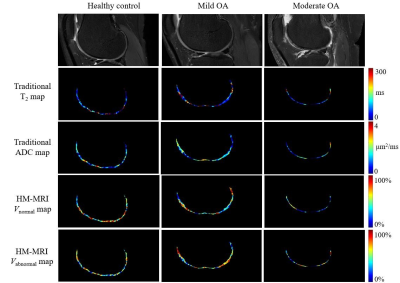 |
11 | Evaluate Articular Cartilage in Early-Stage Knee Osteoarthritis with a Two-Compartment Hybrid Multidimensional MR Imaging Method
Peng Luo1, Wentao Hu2, Guanwu Li1, and Yongming Dai2
1Department of Radiology, Yueyang Hospital of Integrated Traditional Chinese and Western Medicine, Shanghai University of Traditional Chinese Medicine, Shanghai, China, 2United Imaging Healthcare, Shanghai, China Evaluation of early-stage knee osteoarthritis (OA) has long been of wide concern, while MRI has proved its value in this field by the richness of contrasts and non-invasive nature. However, MRI detection for composition change in OA still calls for deeper researches. ADC and T2 measured by conventional MRI are only an average of intra-voxel components and assumed to be independent. However, by performing a hybrid multidimensional MR imaging (HM-MRI) method, an implied correlation between ADC and T2 in articular cartilage has emerged. Moreover, Vnormal might offer extra information in detecting cartilage degeneration at early stage of OA than conventional ADC/T2 parameters. |
||
4626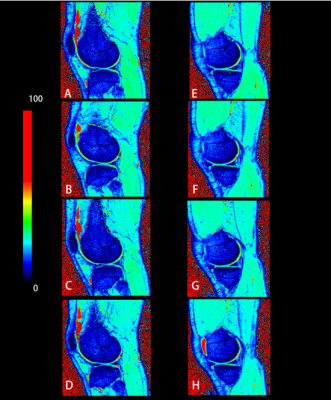 |
12 | Reversible Changes in knee articular cartilage after half marathon: an MRI T2* study in heathy amateur runners
Yanjing Zhang1,2, Li Zhang3,4, Wanzhen Yao1,2, Siyu Dai1,2,5,6, Dingbo Shu1,2, Jianping Ding2,5,7, and Mengxiao Liu8
1Department of Radiology, The Affiliated Hospital of Hangzhou Normal University, Hangzhou, China, 2Institute of Sport Medicine, Hangzhou Normal University, Hangzhou, China, 3Department of Radiology, The Children’s Hospital, Zhejiang University School of Medicine, Hangzhou, China, 4National Clinical Research Center for Child Health, Hangzhou, China, 5School of Clinical Medicine, Hangzhou Normal University, Hangzhou, China, 6Faculty of Medicine, The Chinese University of Hong Kong, HongKong, China, 71 Department of Radiology, The Affiliated Hospital of Hangzhou Normal University, Hangzhou, China, 8MR scientific Marketing, Diagnostic Imaging, Siemens Healthineers Ltd, Shanghai, China
This research investigated the difference in T2* value of knee cartilage between amateur runners and non-exercisers, and examined the trends in the T2* value of knee cartilage in amateur runners before and after a half marathon. The results showed that the T2* value in lateral femorotibial joint of amateur runners was lower than non-exercisers. And the T2* value increased significantly after half-marathon running, and returned to the baseline level after 3 days. This indicates that long-term running may be beneficial to joint health; the effect of a half-marathon exercise on cartilage is reversible.
|
||
4627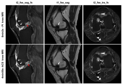 |
13 | AI-assisted compressed sensing (ACS) in accelerating knee MR imaging: A pilot study in adults Video Permission Withheld
Xu Xu1, Chunchao Xia2, Wen Zeng2, Wanlin Peng2, Yongming Dai3, Ke Xue3, Zhenlin Li2, Zhenlin Li2, and Zhenlin Li2
1Department of Radiology, west China hospital of Sichuan University, Chengdu, China, 2west China hospital of Sichuan University, Chengdu, China, 3MR Collaboration, Central Research Institute, United Imaging Healthcare, Shanghai, China In knee MRI, patients with pain or limited motion can have difficult to endure a long-time imaging position. At the same time, the relatively high spatial resolution and good image quality were required in such musculoskeletal MRI. Therefore, shortening scan time and improving the image quality is the aim of current knee MRI.Our study compared conventional parallel imaging (PI) and artificial intelligence assisted compressed sensing (ACS) in terms of imaging quality and diagnostic performance in knee MRI, and achieved a 5-minute comprehensive examination of the knee, without compromising either image quality or visualization of anatomical and pathological structures. |
||
4628 |
14 | Periodic assessment of four horns of knee meniscus using MR T2mapping images in volunteers before and after amateur marathons
Li Jujia1, Jian Zhao1, Xuesong Zhang1, Ping Zhang1, Congcong Ren1, Yan Zeng1, Ranxu Zhang1, Ming Wang1, and Xiaoyue Zhou2
1The Third Hospital of Hebei Medical University, Shijiazhuang, China, 2Siemens Healthineers, Ltd., Shanghai, 201318, China., Shanghai, China
T2 mapping can evaluate the dynamic changes of the meniscal microstructure. The study was designed to evaluate the T2 values of the menisci of amateur marathon volunteers at three different timings. Compared with the T2 values of menisci one week before running, the values 12 h after running increased significantly (P <0.05), and the values 2 months after running did not change significantly (P > 0.05). The result suggests that marathons do not necessarily cause irreversible knee damage.
|
||
4629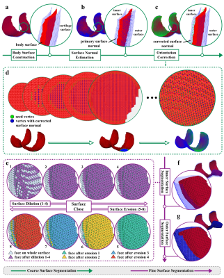 |
15 | A Systematic Cartilage Surface Segmentation Method for Cartilage Thickness Mapping Video Permission Withheld
Yongcheng Yao1, Dόnal G. Cahill1, James F. Griffith1, and Weitian Chen1
1The Chinese University of Hong Kong, New Territories, Hong Kong
Thickness mapping techniques are important in the management of osteoarthritis. Since most thickness mapping approaches require two surfaces as inputs, surface segmentation is important for articular cartilage thickness mapping. In this work, a systematic approach is proposed for automatic articular cartilage surface segmentation from a volume segmentation mask, which includes cartilage surface reconstruction, body surface construction, surface normal estimation, directional voxels searching, restricted surface dilation, and surface close operation. The proposed method can be used for reliable cartilage thickness mapping.
|
||
 Back to Meeting Home
 Back to the Program-at-a-Glance
The International Society for Magnetic Resonance in Medicine is accredited by the Accreditation Council for Continuing Medical Education to provide continuing medical education for physicians.
 Back to Meeting Home
 Back to the Program-at-a-Glance
 View the Presentation