Online Gather.town Pitches
MSK I
Joint Annual Meeting ISMRM-ESMRMB & ISMRT 31st Annual Meeting • 07-12 May 2022 • London, UK

| Booth # | ||||
|---|---|---|---|---|
3081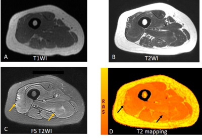 |
1 | Assessment of idiopathic inflammatory myopathy using muscle T2 mapping segmented by Deep Anatomical Federated Network (DAFNE)
Fengdan Wang1, Francesco Santini2, Jinxia Zhu3, Tom Hilbert4,5,6, Tobias Kober4,5,6, and Zhengyu Jin1
1Radiology, Peking Union Medical College Hospital, Beijing, China, 2University Hospital Basel, Basel, Switzerland, 3Siemens Healthcare Ltd., Beijing, China, 4Siemens Healthcare AG, Lausanne, Switzerland, 5Lausanne University Hospital and University of Lausanne, Lausanne, Switzerland, 6École Polytechnique Fédérale de Lausanne (EPFL), Lausanne, Switzerland
Idiopathic Inflammatory Myopathy (IIM) is a group of immune-mediated myopathies, with high morbidity and mortality. Prior work showed that dedicated accelerated T2 mapping of bilateral thighs can be performed within 3 minutes at high resolution and can detect IIM-induced muscle inflammation. However, manual drawing methods to delineate regions of interest are subject to sampling errors. This study investigated the first utility of a fully automated deep-learning method for segmentation of T2 maps in 64 patients with IIM against healthy controls, with results confirming its feasibility, accuracy, and efficiency.
|
||
3082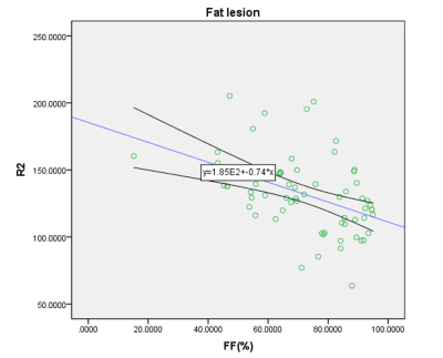 |
2 | Quantification of bone marrow component change using chemical shift-encoded MRI in Ankylosing Spondylitis patients in different stages
Liping Ma1, Long Qian2, Zhibin Zeng1, Cuiyun Sheng1, Yang Wang1, Gen Li3, and Gaoyun Wu1
1Department of Radiology, Huazhong University of Science and Technology Union Shenzhen Hospital, Shenzhen, China, 2MR Research, GE Healthcare, Bejing, China, 3Department of Emergency, Huazhong University of Science and Technology Union Shenzhen Hospital, Shenzhen, China
Quantification of fat fraction (FF) and R2* in areas of bone marrow edema (BME) and fat metaplasia in different stages Ankylosing Spondylitis (AS). 75 AS in 3 different stage (early acute group, late acute group, stable group), 42 BME and 39 fat metaplasia were detected. Difference of FF, R2* in BME and fat metaplasia in different stages were compared and relationship between FF and R2* were analyzed. Result showed R2* in fat metaplasia, but not in BME, is significantly negative with FF. FF and R2* measurements can quantitatively analyze bone marrow component change in AS.
|
||
3083 |
3 | 3D texture analysis of MRI relaxation time maps for assessment of repair cartilage with treatment of mesenchymal progenitor cells
Xinxin Zhao1, Jingjing Ruan1, Jia Li2, Chengxiang Dai3, and Yan Zhou1
1Department of Radiology, Ren Ji Hospital, School of Medicine, Shanghai Jiao Tong University, No. 160, Pujian Road, Shanghai, 200127, China., Shanghai, China, 2Department of Rheumatology, Ren Ji Hospital, School of Medicine, Shanghai Jiao Tong University, No. 160, Pujian Road, Shanghai, 200127, China., Shanghai, China, 3Cellular Biomedicine Group, Inc., No. 85 Faladi Road, Building 3, Zhangjiang, Pudong New Area, Shanghai, 201210, China., Shanghai, China We used 3D texture analysis of MRI relaxation times, combined with clinical outcomes, to evaluate the potential of repair cartilage with allogeneic human adipose-derived mesenchymal progenitor cells in patients with KOA. Significant differences were observed in texture RLM parameters of T1rho and T2 maps in patients. We also found correlations between WOMAC pain scores and texture parameters, suggesting the spatial heterogeneity of relaxation time maps maybe associated with clinical scores. As conclusion, texture analysis has potential applications in understanding mechanism of stem cells repairing cartilage and assessing response to treatment. |
||
3084 |
4 | Repeatability and feasibility of amide proton transfer imaging in musculoskeletal tumors
Ming Wen1, Yujin Zhang1, Jianling Cui1, and Zhiwei Shen2
1The Third Hospital, Hebei Medical University, Shijiazhuang, China, 2Philips healthcare,Beijing,China, Beijing, China
Amide proton transfer (APT) imaging is a noninvasive emerging molecular MRI technique. Currently, APT was mainly used for the diagnosis, classify and recurrence prediction of head glioma. The application in other tissue such as musculoskeletal disease is limited. The feasibility and the repeatability of APT imaging in body or other tissues is necessary for its clinical practice. In this study, we explorer the scan-rescan repeatability of APT imaging in musculoskeletal (MSK) tumor on a clinical 3.0T MR scanner, and further to differentiate malignant from benign solid tumors in the study.
|
||
3085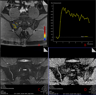 |
5 | The diagnostic value of dynamic contrast enhancement (DCE-MR) and ZOOmitDWI quantitative parameters for the activity of sacroiliac arthritis
Qingqing Zhu1 and Mengxiao Liu2
1department of radiology, Zhejiang University School of Medicine Sir Run Run Shaw Hospital, Hangzhou, China, 2MR scientific Marketing, Diagnostic Imaging, Siemens Healthineers Ltd, Shanghai, China
The purpose of this study was to evaluate the relationship between magnetic resonance dynamic contrast-enhanced imaging and quantitative parameters of the ZOOMit-DWI technique and the activity of sacroiliac arthritis. Dynamic enhanced MRI microvascular permeability parameters can visually reflect the microcirculatory perfusion status of the subchondral bone marrow region of the sacroiliac joint, and its diagnostic efficacy is better than DWI-ADC. Sacral Ktrans values and sacral iAUC values are highly correlated with ASDAS-CRP scores, which can be used as important quantitative indicators for assessing the inflammatory activity of the sacroiliac joint.
|
||
3086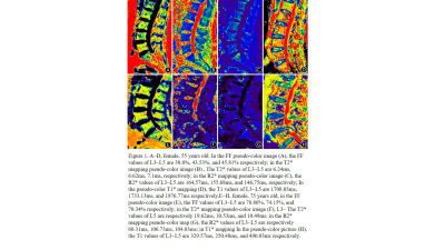 |
6 | Feasibility Study of T2* Mapping, R2* Mapping and T1 Mapping Technology in the Quantitative Evaluation for Primary Osteoporosis of Lumbar Spine
Yuan Li1, Guanhua Yang1, Wei Jing1, Shimin Lan1, and Xiuzheng Yue2
1The First People's Hospital of Shizuishan, Shizuishan, China, 2Philips Healthcare, Beijing, China
The aim of this study was to investigate the feasibility of T2* mapping, R2* mapping and T1 mapping techniques for quantitative assessment of primary osteoporosis of the lumbar spine.The differences and correlation between T2* mapping, R2* mapping and T1 mapping values of patients with osteoporosis group and the normal population were analyzed. The statistical results of our data showed that the T2 values in the osteoporotic group of L3 and L5 vertebrae were greater than the normal group, and the R2* values and T1 values were less than the normal group.
|
||
3087 |
7 | CSE-MRI Detects the Changes in Bone Marrow Fat after Weight Loss Induced by Healthy Low-carbohydrate Diet
Lin Gengyun1, Wu Junying1, Wen Zhibo1, and Zhao Chen2
1Zhujiang Hospital, Guangzhou, China, 2Philips Healthcare, Guangzhou, China
Obesity is a global health condition that leads to many kinds of diseases. Bone marrow fat (BMF) is considered as the third largest fat depot in human body. It has been proved that BMF is closely related with energy storage, bone metabolism, endocrine function and hematologic diseases. To understand the relation between BMF and weight loss is crucial within obesity related diseases scenario. Few studies have been published focusing on BMF variation so far. In this study, chemical shift encoding-based water-fat MRI (CSE-MRI) was employed to demonstrate the positive correlation between BMF and parts of the weight loss indicators.
|
||
3088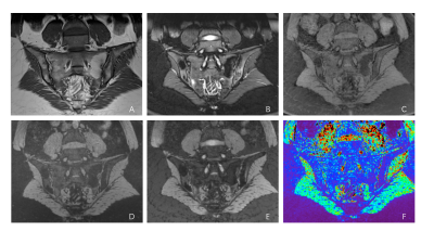 |
8 | Three-dimensional ultrashort echo time MRI to evaluate sacroiliac joint in patients with ankylosing spondylitis Video Permission Withheld
Cui Ren1, Qing Li2, Stefan Sommer3,4,5, Qiao Zhu1, and Hui Shu Yuan1
1Radiology, Peking University Third Hospital, Beijing, China, 2MR Collaborations, Siemens Healthineers Ltd., Shanghai, China, 3Siemens Healthcare, Zurich, Switzerland, 4Swiss Center for Musculoskeletal Imaging (SCMI), Balgrist Campus, Zurich, Switzerland, 5Advanced Clinical Imaging Technology (ACIT), Siemens Healthcare AG, Lausanne, Switzerland To assess the diagnostic performance of three-dimensional ultrashort echo time sequence (3D-UTE) in bone erosion detection of the sacroiliac joint (SIJ) in patients with clinically confirmed ankylosing spondylitis (AS) and to test whether SIJ cartilage T2* values might help in identification of patients with AS. |
||
3089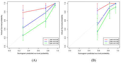 |
9 | The potential role of MRI-based bone marrow Radiomics in survival prediction of newly diagnosed multiple myeloma
Yang LI1 and Nan Hong1
1Peking university people's hospital, Beijing, China
Multiple myeloma (MM) is the second most common hematologic malignancy. Despite the more effective therapies were introduced, this incurable disease remains highly heterogeneous in clinical outcome. Accurate predicative markers for prognosis are needed to develop appropriate treatment in newly diagnosed MM.
|
||
3090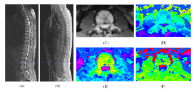 |
10 | Intravoxel Incoherent Motion-Diffusion Weighted Imaging derived parameters for survival prediction of multiple myeloma
Yang LI1 and Nan Hong1
1Peking university people's hospital, Beijing, China
The prognostic biomarkers for multiple myeloma (MM) was urgently needed, and this study was the first to confirm the value of Intravoxel Incoherent Motion-Diffusion Weighted Imaging (IVIM-DWI) for predicting progression-free survival in patients with MM.Purpose: To evaluate the prognostic ability of Intravoxel Incoherent Motion-Diffusion Weighted Imaging (IVIM-DWI) derived parameters in newly diagnosed multiple myeloma (MM).
|
||
3091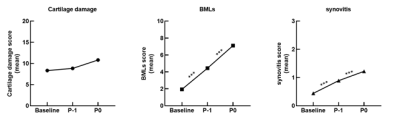 |
11 | The Trajectory of Knee Structures Before The Onset of Rheumatoid Arthritis: A 4-Year Longitudinal Magnetic Resonance Imaging Study
Qiaoling Zhong1, Xintao Zhang1, Zhongping Zhang2, and Xiaodong Zhang1
1Department of Medical Imaging, The Third Affiliated Hospital of Southern Medical University, Guangzhou, China, 2Philips Healthcare, Guangzhou, China
The knee joint is one of the most commonly affected large joint in rheumatoid arthritis (RA). Its progression often causes joint destruction and deformity. The cartilage injury, bone marrow lesions and synovitis scores of the knees at baseline, P-1 (1 year prior to P0), and P0 (time of onset of RA) were evaluated using the MOAKS score. The generalized estimation equation (GEE) was used to compare the MR scores of knee joints at three time points to observe the longitudinal changes. The changes of bone marrow lesions and synovitis scores in knee joints were significant before the occurrence of RA.
|
||
3092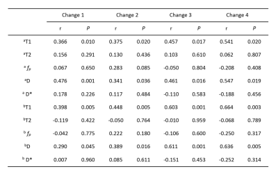 |
12 | Evaluation of Multimodal MRI in RA Efficacy Based on Quantitative Bone Marrow Edema
Wenzhao Yuan1, Yi Dai2, Yiwu Lei3, Fang Qin4, Huiting Zhang5, Zisan Zeng3, and Liling Long1
1The First Affiliated Hospital of Guangxi Medical University, Nanning, China, 2Radiology, The Second Affiliated Hospital of Guangxi Medical University, Nanning, China, 3Radiology, The First Affiliated Hospital of Guangxi Medical University, Nanning, China, 4Rheumatology, The First Affiliated Hospital of Guangxi Medical University, Nanning, China, 5Siemens Healthcare Ltd., Wuhan, China
This study evaluated the efficacy of MRI bone marrow edema imaging for rheumatoid arthritis (RA). Wrist joints of RA patients were examined at baseline and follow-up with T1 mapping, T2 mapping and IVIM, and their clinical indicator DAS28-ESR was also collected. Results showed that the changes of both T1 and D values had statistically positive correlations with DAS28-ESR. And the results showed that T1 and D values of bone marrow edema area in lunate bone, triangle bone and capitate bone can significantly reflect the curative effect of RA, and the change rate of T1 can predict the curative effect.
|
||
 Back to Meeting Home
 Back to the Program-at-a-Glance
The International Society for Magnetic Resonance in Medicine is accredited by the Accreditation Council for Continuing Medical Education to provide continuing medical education for physicians.
 Back to Meeting Home
 Back to the Program-at-a-Glance
 View the Presentation