Online Gather.town Pitches
MSK I
Joint Annual Meeting ISMRM-ESMRMB & ISMRT 31st Annual Meeting • 07-12 May 2022 • London, UK

| Booth # | ||||
|---|---|---|---|---|
3093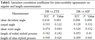 |
1 | Feasibility of ZTE-MRI on wrist in comparison with routine MR imaging and radiography
Xingyao Yu1, Weiyin Vivian Liu2, Ling Sang3, Qianqian Feng3, Kejun Wang3, and Lin Xu3
1Biomedical Engineering College, Hubei University of Medicine, Shiyan, Hubei, China, 2GE Healthcare, Beijing, China, 3Taihe Hospital, Hubei University of Medicine, Shiyan Hubei, China
ZTE-MRI have high feasibility of angle measurements such as femoral neck-shaft angle as well as bone erosions on temporomandibular joint in reflection of altered bone morphometry and function. In our study, X-ray taken as reference, good consistency of ulnar deviation angle and the length of ulnar styloid process was shown between each two modalities, but better consistency between ZTE-MRI processed with average intensity projection and X-ray. Significantly different number of bone erosions between ZTE-MRI and T1 weighted images were found. To sum up, ZTE-MRI might have potential in elevating diagnosis efficiency of detecting altered bone morphometry and function.
|
||
3094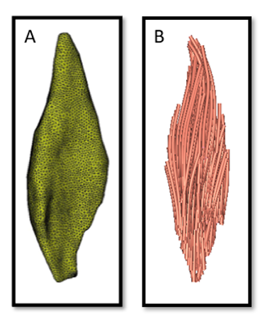 |
2 | Three-dimensional architecture and diffusion properties of the medial gastrocnemius muscle in human infants in vivo
Brian V. Y. Chow1,2, Bart Bolsterlee1,3, Catherine Morgan4, Caroline Rae1, David I. Warton5,6, Iona Novak4,7, Suzanne Davies1, Ann Lancaster1, Gordona C. Popovic5,8, Rodrigo R. N. Rizzo1,2, Claudia Y. Rizzo1, Maria Kyriagis9, Melissa Smith1, Erin Muling1, and Robert D. Herbert1,2
1Neuroscience Research Australia, Sydney, Australia, 2School of Medical Sciences, University of New South Wales, Sydney, Australia, 3Graduate School of Biomedical Engineering, University of New South Wales, Sydney, Australia, 4Cerebral Palsy Alliance Research Institute, The University of Sydney, Sydney, Australia, 5School of Mathematics and Statistics, University of New South Wales, Sydney, Australia, 6Evolution & Ecology Research Centre, University of New South Wales, Sydney, Australia, 7Faculty of Medicine and Health, The University of Sydney, Sydney, Australia, 8Stats Central, Mark Wainwright Analytical Centre, University of New South Wales, Sydney, Australia, 9Rehab2Kids, Sydney Children’s Hospital, Sydney, Australia Diffusion tensor magnetic resonance imaging (DT-MRI) was used to investigate the three-dimensional architecture and diffusion properties of medial gastrocnemius muscles in living human infants aged 2-3 months. Mean muscle volume, physiological cross-sectional area and fascicle length in infants were 1.8%, 3.8% and 47.2% of values previously obtained in 8 adult muscles. Radial diffusivity in infant muscle was half that in adult muscle, presumably because infant muscle fibres have much smaller transverse dimensions. |
||
3095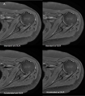 |
3 | Shoulder PROPELLER MRI with Deep learning-based reconstruction: image quality and agreement between standard and accelerated sequence Video Permission Withheld
Jisook Yi1, Ho-Joon Lee1, Seok Hahn1, Joonsung Lee2, Xinzeng Wang3, and Maggie Fung4
1Radiology, Haeundae Paik Hospital, Inje University College of Medicine, Busan, Korea, Republic of, 2GE Healthcare, Seoul, Korea, Republic of, 3GE Healthcare, Houston, TX, United States, 4GE Healthcare, New York, NY, United States Although the shorter scan time enabled by DLRecon has potential to reduce motion related image degradation, it cannot remove or mitigate such artifacts. The periodically rotated overlapping parallel lines with enhanced reconstruction (PROPELLER) or similar techniques can reduce motion artifacts in shoulder MRI, however usually takes a longer time to acquire. Recently, DLRecon for PROPELLER has been developed. Accelerated PROPELLER sequence with DLRecon showed improved image quality and comparable interobserver agreement for shoulder pathology compared to standard PROPELLER acquisition with conventional reconstruction, despite the shorter scan time (45% reduction). |
||
3096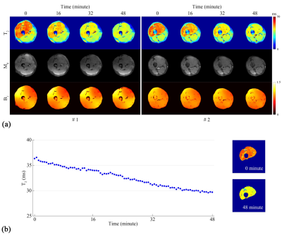 |
4 | Dynamic T2 mapping of human skeletal muscles via multiple overlapping-echo detachment planar imaging
Yang Qizhi1, Wang Xiaoyin2, Cai Shuhui1, He Hongjian2, Cai Congbo1, Wu Zhigang3, and Zhong Jianhui2,4
1Department of Electronic Science, School of Electronic Science and Engineering, Xiamen University, Xiamen, Fujian, China, 2Center for Brain Imaging Science and Technology, College of Biomedical Engineering and Instrumental Science, Zhejiang University, Hangzhou, Zhejiang, China, 3MSC Clinical & Technical Solutions, Philips Healthcare, Shenzhen, Guangdong, China, 4Department of Imaging Science, University of Rochester, Rochester, NY, United States
Magnetic resonance (MR) parametric mapping and MR dynamic imaging are both valuable clinical tools. Nevertheless, quantitative imaging and high time resolution usually cannot be achieved simultaneously with conventional dynamic MR imaging methods. Herein, the multiple overlapping-echo detachment imaging (MOLED) method with short echo time (TE), dubbed MOLED-short, is proposed to fetch the dynamic variation of T2 value on in vivo extremities with high temporal resolution (about 110 ms). The robustness of MOLED-short was demonstrated. MOLED-short can capture immediate T2 changes of muscles and may provide new insights into the metabolic responses of musculatures to human activities.
|
||
3097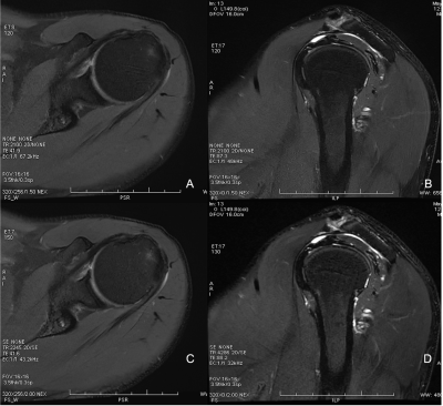 |
5 | Clinical Feasibility Study of Accelerated 2D Magnetic Resonance shoulder Imaging Using Deep Learning-based Algorithm Video Not Available
Jing Liu1, Ke Xue2, Yongming Dai2, Peng Wu2, and Jianxing Qiu1
1Peking University First Hospital, Beijing, China, 2MR Collaboration, Central Research Institute,United Imaging Healthcare, Shanghai, China
Deep Learning-based magnetic resonance imaging (DL-MRI) could accelerate 3D MRI scanning time. In this study, we investigate the feasibility of DL-MRI in 2D shoulder MRI. Totally 20 consecutive patients were enrolled for both conventional MRI and DL-MRI. Both qualitative and quantitative analyses were conducted to compare the image quality and lesion diagnosis on conventional MRI and DL-MRI. And our results revealed that DL-MRI was valuable for improving the overall workflow of shoulder MRI with scanning time saved and image quality improved.
|
||
3098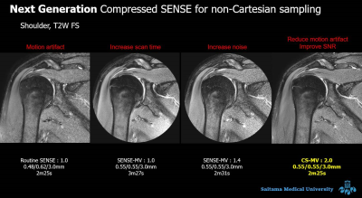 |
6 | Efficacy of motion-free MRI of the shoulder using Compressed SENSE MultiVane Video Not Available
Shinji Saruya1, Masami Yoneyama2, Yasutomo Katsumata2, Kaiji Inoue1, Eito Kozawa1, and Mamoru Niitsu1
1Saitama Medical University, iruma-gun, Japan, 2Philips Japan, minato-ku, Japan
Combining Compressed SENSE (CS), an acceleration technique that combines with compressed sensing and SENSE, with MultiVane achieves comparable scan time to Cartesian scan and increases motion robustness while maintaining similar image quality to Cartesian scan without increasing streak artifacts. We compared this newly developed CS-MultiVane with conventional acquisition method such as routine SENSE, SENSE-MuliVane with reduction factor = 1.0 and SENSE-MuliVane with reduction factor = 1.4 in terms of the overall image quality, overall degree of artifacts and signal-to-noise ratio (SNR). In conclusion, CS-MultiVane acquisition method is feasible for MRI of the shoulder.
|
||
3099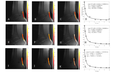 |
7 | Monitoring changes of Achilles Tendon in amateur Marathon athletes by using component Analysis UTE-T2* technology: a preliminary study Video Permission Withheld
Dantian Zhu1, Yijie Fang1, Wenhao Wu1, Wenjun Yu1, Wei Li1, Yajun Ma2, and Shaolin Li1
1Department of Radiology, Fifth Affiliated Hospital, SUN Yat-Sen University, Zhuhai, China, 2Department of Radiology, University of California, San Diego, CA, United States
Long-distance running is a common cause of Achilles tendinopathy. A fast, reliable, and non-invasive magnetic resonance imaging (MRI) technique to track the early changes in tendon is of critical importance for effective clinical intervention and evaluation that can prevent the progression of Achilles tendinopathy. This study aims to evaluate UTE-T2* in the detection of changes in the Achilles tendons of amateur marathon runners before and after long-distance running.
|
||
3100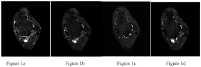 |
8 | Acceleration of 3D Ankle Joint T2WI-SPAIR Images with Compressed SENSE to display the anterior talofibular ligament Video Not Available
Xiaoyan ZHANG1, Peiqi MA1, Yushan YUAN1, Zhongqiu WANG1, Bin Peng1, and Xiuzheng YUE2
1Fuyang People's Hospital, Fuyang Anhui, China, 2Philips Healthcare, Beijing, China
Compressed SENSE(CS) is a new techonolgy that can be effective accelerat 3D acquisitions and reduce scan times.In this study ,we discuss the value of the compressed sensing technology’s three-dimensional MR to display the anterior talofibular ligament (ATFL), and compare the impact of different acceleration factors on imaging. We find that when the acceleration factor(AF) of CS is six, scan times was reduced by 60 percent without significant degradation of image quality.
|
||
3101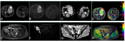 |
9 | Preliminary assessment of 3D APT-weighted combined with diffusion weighted imaging in characterization of bone and soft tissue tumors Video Permission Withheld
Ying Li1, Jingliang Cheng1, Wenhua Zhang1, and Liangjie Lin2
1The First Affiliated Hospital of Zhengzhou University, Zhengzhou, China, 2Philips Healthcare, Beijing, 100102, China, Beijing, China
Amide proton transfer weighted imaging (APTWI) is a noninvasive emerging molecular MRI technique based on chemical exchange saturation transfer (CEST) between amide protons of proteins and polypeptides and free water protons. This work investigated and evaluated the ability of APT parameter in distinguishing benign from malignant bone and soft tissue tumors. Results indicated that APTWI combined with diffusion weighted imaging showed a significantly improved differentiation between benign and malignant tumors.
|
||
3102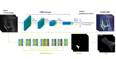 |
10 | Predicting Knee Osteoarthritis Progression with an Interpretable Deep Learning Approach on MRIs: Data from the Osteoarthritis Initiative
Jiaping Hu1, Chuanyang Zheng2, Lijie Zhong1, Keyan Yu1, Yanjun Chen1, Zhao Wang2, Zhongping Zhang3, Qi Dou2, and Xiaodong Zhang1
1Department of Medical Imaging, The Third Affiliated Hospital of Southern Medical University, GuangZhou, China, 2Department of Computer Science & Engineering, The Chinese University of Hong Kong, HongKong, China, 3Philips Healthcare, GuangZhou, China
Identifying knee osteoarthritis progressors is significant. MRIs can reflect the structures of the knee. However, currently no tool could rapidly and objectively predict knee osteoarthritis progression based on MRI. Therefore, we applied deep learning algorithms on MRIs of the whole knee to predict progression at three time points. The Gradient-weighted Class Activation Maps were employed for interpretability, and the highlighted infrapatellar fat pad (IPFP) was segmented for progression prediction. We showed that the deep learning framework performed well on discrimination of progressors, especially at 24th months, and that the infrapatellar fat pad plays an important role in predicting progression.
|
||
3103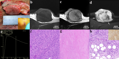 |
11 | Differential Diagnosis of Dermatofibrosarcoma Protuberans: Clinical Values of High-resolution DCE-MRI
QIUYU YU1, YANG YANG1, RENJUN HUANG1, YONGGANG LI1, NAIHUI ZHOU2, PING LI1, PENG WU3, and MENGXIAO TANG4
1Radiology, The First Affiliated Hospital of Soochow University, SUZHOU, China, 2Dermatology, The First Affiliated Hospital of Soochow University, SUZHOU, China, 3Philips Healthcare, SHANGHAI, China, 4Radiology, Sir Run Run Shaw Hospital, School of Medicine, Zhejiang University, HANGZHOU, China
Dermatofibrosarcoma protuberans (DFSP) is a type of intermediately malignant cutaneous spindle cell neoplasms, which is easy to be confused with several benign ones even after needle biopsy, especially cellular fibrous histiocytoma (cFH), resulting in inadequate excision and local recurrence. We found that as a novel skin imaging technique, high-resolution (HR) DCE MRI could distinguish DFSP. The features include infiltration of surrounding fat, ill-defined margins and large quantitative parameters. Both DFSP and cFH have type-III time-signal intensity curves (TICs). In contrast, other confused lesions presented type-II-or-I TIC. The recommendation of preoperative HR-MRI could assist dermatologists to perform surgical plan more confidently.
|
||
3104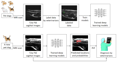 |
12 | Deep Learning for Veterinary MRI: Automated Detection of Intervertebral Disc Herniation in Pet Dogs
Guoxiong Deng1,2,3, Shoujin Huang1, Ziran Chen1, Lifeng Mei1, Jianzhong Li3, Ruixiang Jiang3, WenYue Xiao3, Dexing Wei3, Yan Kang1,2, and Mengye Lyu1,2
1College of Health Science and Environmental Engineering, Shenzhen Technology University, Shenzhen, China, 2College of Applied Sciences, Shenzhen University, Shenzhen, China, 3Shenzhen GoldenStone Medical Technology Co. , Ltd, Shenzhen, China The automated detection of Intervertebral disc (IVD) herniation in animal MRI may facilitate veterinary diagnosis, yet it is rarely studied due to the lack of training data and the challenges from inter-breed variations. Here, we constructed a dog spinal cord MRI dataset with bounding box annotations of herniated discs, and conducted experiments using a number of well-known deep learning models. We demonstrated that automated detection of animal IVD herniation was feasible and in general two-stage detection models such as Faster R-CNN outperformed one-stage models. |
||
3105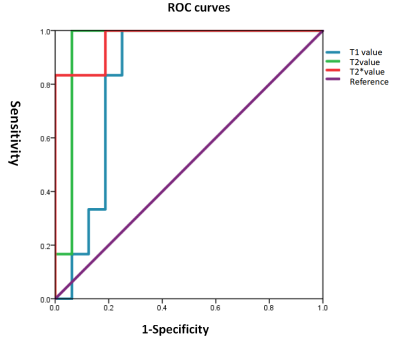 |
13 | QMRI for assessment of anterior cruciate ligament mucoid degeneration and injury Video Not Available
Guangtao Fan1, Yudan Li1, Fenglin Xue1, Yilong Huang1, Yanlin Li 1, Guoliang Wang1, Tianfu Qi1, Lisha Nie2, and Bo He1
1the First Affiliated Hospital of Kunming Medical University, Kunming, China, 2GE Healthcare, MR Research, Beijing, China
The study aims to explore the difference in relaxation time between mucoid degeneration (MD) and injured anterior cruciate ligament (ACL), and evaluate the performance of QMRI (Quantitative magnetic resonance imaging) to identify the grade of MD and injured ACL. It was concluded that T2 * value of the ACL-MD group was higher than that of the injured group, T1, T2 and T2* could be used to quantitatively identify different grades of ACL-MD (T2* may deliberately has the highest diagnostic efficacy), and the same goes for T1 to evaluate ACL injury.
|
||
3106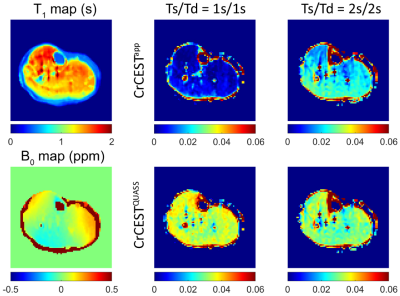 |
14 | Fast and accurate creatine chemical exchange saturation transfer muscle imaging with quasi-steady state post-processing algorithm at 3T
Zhou Liu1,2, Qian Yang1, Honghong Luo1, Dehong Luo1, Long Qian3, Xin Liu2, Hairong Zheng2, Phillip Zhe Sun4, and Yin Wu2
1National Cancer Center/National Clinical Research Center for Cancer/Cancer Hospital & Shenzhen Hospital, Chinese Academy of Medical Sciences and Peking Union Medical College, Shenzhen, China, 2Shenzhen Institute of Advanced Technology, Chinese Academy of Sciences, Shenzhen, China, 3GE Healthcare, Beijing, China, 4Emory University School of Medicine, Atlanta, GA, United States
Although CrCEST MRI is increasingly employed in muscle imaging, its measurement depends on the RF saturation duration (Ts) and relaxation delay (Td). This study evaluated the quasi-steady-state (QUASS) algorithm for fast and robust CrCEST measurement. Briefly, CrCEST experiments were performed at 3T under two representative imaging conditions. Both phantom and volunteer experiments showed that the routine CrCEST increased significantly with Ts and Td and were significantly smaller than the corresponding QUASS indices. In comparison, the QUASS CrCEST meaurement showed little dependence on the scan protocol, establishing the robustness and accuracy of QUASS CrCEST muscle MRI.
|
||
 Back to Meeting Home
 Back to the Program-at-a-Glance
The International Society for Magnetic Resonance in Medicine is accredited by the Accreditation Council for Continuing Medical Education to provide continuing medical education for physicians.
 Back to Meeting Home
 Back to the Program-at-a-Glance
 View the Presentation