Online Gather.town Pitches
MSK V
Joint Annual Meeting ISMRM-ESMRMB & ISMRT 31st Annual Meeting • 07-12 May 2022 • London, UK

| Booth # | ||||
|---|---|---|---|---|
 |
4939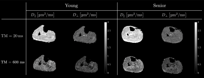 |
1 | Time-Dependent Diffusion Tensor Imaging and Modeling of Aging Muscles: Correlations to Muscle Strain, Histology and Physical Assessment
Vadim Malis1, Usha Sinha2, Edward Smitaman1, Jed Keenan Obra3, Henning T Langer3, Agata A Mossakowski3, Keith Baar3, and Shantanu Sinha1
1Radiology, UC San Diego, San Diego, CA, United States, 2Physics, San Diego State University, San Diego, CA, United States, 3Physiology and Membrane Biology, UC Davis, Davis, CA, United States
Diffusion modeling (RPBM) of the time dependence of the skeletal muscle diffusion eigenvalues was applied to probe tissue changes with age. λ2 (TM=30 ms) was significantly lower in seniors; permeability and the residence time from RPBM were significantly positively correlated with grip size and 200 m walk. Significant negative correlations of λ1 and λ2 to age and to physical λ2 / λ3 to collagen matrix angle and to systolic/diastolic blood pressure were seen. λ2 and λ3 were significantly positively correlated to the projection of the strain on the diffusion eigenvector corresponding to the secondary eigenvalue.
|
|
4940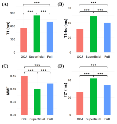 |
2 | Quantitative Ultrashort Echo Time (UTE) imaging of Osteochondral Junction Video Permission Withheld
Alecio F. Lombardi1,2, Zhao Wei1, Dina Moazamian1, Saeed Jerban1, Hyungseok Jang1, Nicole Le1, Jiang Du1, Christine B. Chung1,2, Eric Y. Chang1,2, and Ya-Jun Ma1
1Department of Radiology, University of California, San Diego, CA, United States, 2Radiology Service, Veterans Affairs, San Diego Healthcare System, San Diego, CA, United States
MRI-based compositional imaging of cartilage has higher sensitivity to detect early changes in osteoarthritis. The osteochondral junction (OCJ) is of particular interest since it has been associated with early changes and progression of cartilage degeneration. However, conventional MRI sequences cannot differentiate with high contrast the OCJ region for quantification. Here we present quantification of T1, T1rho, T2*, and macromolecular fraction (MMF) of deep and superficial layers knee cartilage using 3D ultrashort echo time (UTE) MRI cones sequences and correlate the values from the OCJ with the degree of cartilage degeneration according to the MRI Osteoarthritis Knee Cartilage Score (MOAKS).
|
||
4941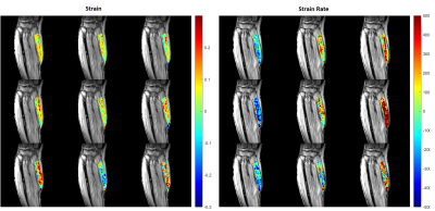 |
3 | Study of Aging Muscles using 3D Strain and Strain Rate Projected along Muscle Fiber DTI Eigenvector Directions
Brandon Cunnane1, Vadim Malis2, Usha Sinha1, Ryan Hernandez1, John Hodgson3, and Shantanu Sinha2
1Physics, San Diego State University, San Diego, CA, United States, 2Radiology, UC San Diego, San Diego, CA, United States, 3Inteegrative Biology and Physiology, UC Los Angeles, Los Angles, CA, United States
Feasibility is shown of studying aging muscles using rapid, compressed sensing VE-PC technique at relatively high isometric forces to calculate projections of 3D strain and strain rate tensor along fiber eigenvectors calculated from DTI.
|
||
4942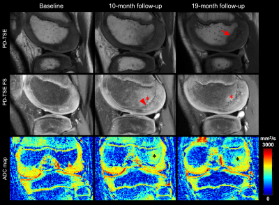 |
4 | High Prevalence of Abnormal 3T MRI Findings in Clinically Asymptomatic Siblings of Patients with Osteochondritis Dissecans of the Knee
Abdul Wahed Kajabi1,2, Stefan Zbyn1,2, Marc A. Tompkins3, Bradley J. Nelson3, Kevin G. Shea4, Cathy S. Carlson5, and Jutta M. Ellermann1,2
1Center for Magnetic Resonance Research, University of Minnesota, Minneapolis, MN, United States, 2Department of Radiology, University of Minnesota, Minneapolis, MN, United States, 3Department of Orthopedic Surgery, University of Minnesota, Minneapolis, MN, United States, 4Stanford Children's Hospital, Stanford University, Palo Alto, CA, United States, 5Department of Veterinary Medicine, University of Minnesota, St. Paul, MN, United States
This study examined seven clinically asymptomatic siblings of patients with Osteochondritis Dissecans (OCD) utilizing 3T MRI, including quantitative apparent diffusion coefficient (ADC) mapping to evaluate morphological integrity of the epiphyseal cartilage, integrity of secondary physis and trabecular bone quality in the distal femoral condyles of bilateral knees. A high prevalence (86%) of morphological MRI abnormalities that can be seen in early OCD and increased diffusivity (high quantitative ADC values) in trabecular bone were detected. The high prevalence of early signs of OCD in clinically asymptomatic siblings of OCD patients supports evidence of a genetic predisposition for the disease.
|
||
4943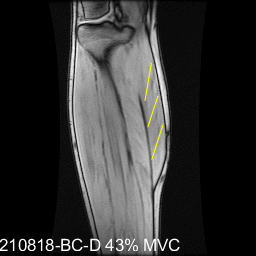 |
5 | 2D Fiber Strains Correlated to DTI in the Medial Gastrocnemius under Isometric Contractions at Different Foot Positions
Brandon Cunnane1, Usha Sinha1, Vadim Malis2, Ryan Hernandez1, John Hodgson3, and Shantanu Sinha2
1Physics, San Diego State University, San Diego, CA, United States, 2Radiology, UC San Diego, San Diego, CA, United States, 3Inteegrative Biology and Physiology, UC Los Angeles, Los Angles, CA, United States
Study of muscle fiber strains at different foot positions can reveal the dependence of muscle force on muscle architecture. Prior studies of isometric contraction in calf muscle using dynamic MRI revealed in the medial gastrocnemius (MG), strain heterogeneity along and across fibers, constant in-plane areas, and gear ratios decreasing with unloading. This study examines correlation of fiber strains in the MG at three-foot positions and two sub-maximal isometric contractions with fiber directions extracted from DTI. The plantarflexed foot position had the highest normalized fiber strain (to force and to torque) while the dorsiflexed position had the lowest normalized fiber strains.
|
||
4944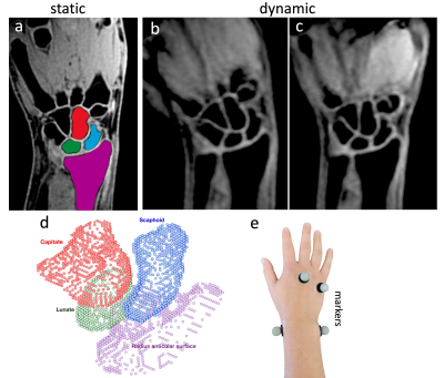 |
6 | Correlation of 4D MRI and Motion Capture during Dynamic Wrist Movements
Mohammad Zarenia1, Samantha R. Schwartz2, Joshua M. Leonardis2, Alyssa J. Schnorenberg2, Volkan E. Arpinar1, Brooke A. Slavens2, and Kevin M. Koch1
1Radiology, Medical College of Wisconsin, Milwaukee, WI, United States, 2Department of Rehabilitation Sciences & Technology, University of Wisconsin-Milwaukee, Milwaukee, WI, United States
4D MRI and external motion capture systems were utilized to track unconstrained movement of wrist carpal bones. Using slab-to-volume registration of dynamic MRI to static MRI reference images, wrist kinematic profiles for 19 healthy subjects were computed and compared to gold-standard motion analysis metrics during ulnar/radial deviation and flexion/extension motions. The agreement of kinematic measures derived using the intrinsic (i.e. carpal bone volume) MRI-based approach and external (sensor-based) motion capture methods provided validation of the deployed MRI-based kinematic profiling methodology.
|
||
4945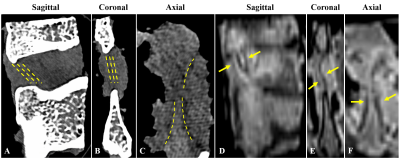 |
7 | Correlation between T1, T1rho, T2*, MMF, and Biomechanics in Human Interspinous Ligament Using quantitative Ultrashort Echo Time (UTE) MRI Video Permission Withheld
Alecio F. Lombardi1,2, Saeed Jerban1, Ya-Jun Ma1, Micah Blais3, Robert K. Eastlack3, Gregory M. Mundis Jr.3, Jiang M. Du1, Eric Y. Chang1,2, and Bahar K. Shahidi4
1Department of Radiology, University of California, San Diego, CA, United States, 2Radiology Service, Veterans Affairs, San Diego Healthcare System, San Diego, CA, United States, 3Department of Orthopedic Surgery, Scripps Clinic, San Diego, CA, United States, 4Department of Orthopedic Surgery, University of California, San Diego, CA, United States This study aimed to quantify T1, T1rho, T2*, and MMF values of the human interspinous ligament using 3D ultrashort echo time (UTE) cones MRI and correlate them with biomechanical properties. We found a significant negative correlation between T1 and elastic modulus; significant moderate to strong negative correlations between T1rho and both tensile stress and maximum load; and significant moderate to strong positive correlations between MMF and both tensile stress and maximum load. There was an overall trend towards negative correlation between T2* and biomechanical properties. |
||
4946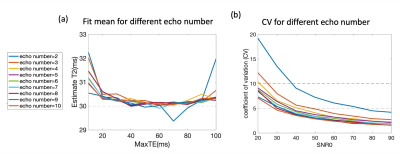 |
8 | Single and Multi-channel evaluations of factors that affect cartilage T1ρ and T2 quantification
zhiyuan zhang1,2,3, Jeehun Kim2,3,4, Richard Lartey2,3, Carl S. Winalski2,3,5, and Xiaojuan Li2,3,5
1Biomedical Engineering, Case Western Reserve University, Cleveland, OH, United States, 2Program of Advanced Musculoskeletal Imaging (PAMI), Cleveland Clinic, Cleveland, OH, United States, 3Department of Biomedical Engineering, Lerner Research Institute, Cleveland Clinic, Cleveland, OH, United States, 4Department of Electrical, Computer, and Systems Engineering, Case Western Reserve University, Cleveland, OH, United States, 5Department of Diagnostic Radiology, Imaging Institute, Cleveland Clinic, Cleveland, OH, United States
MR T1ρ and T2 have been suggested as promising imaging markers for detecting early cartilage degeneration in osteoarthritis1. However, one hurdle of applying such quantitative MRI to large clinical trials and clinical practice is the lack of standardization of data acquisition and processing. In this study, we evaluated multiple factors that affect cartilage T1ρ and T2 quantification including maximum TE, number of echoes, SNR, and fitting methods for single and multi-channel situations with simulations, phantoms and human data. Such evaluations will help to provide useful guidance in standardizing data acquisition and processing for cartilage T1ρ and T2 imaging.
|
||
4947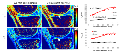 |
9 | Dynamic T1ρ and T2 mapping of Articular Cartilage Biochemical Recovery Post-exercise with 3T MRI
Jason Matakas1, Steven Shamah1, Esther Rong1, Jenna Le1, Karen Sperling1, Can Wu2, and Qi Peng1
1Department of Radiology, Albert Einstein College of Medicine and Montefiore Medical Center, Bronx, NY, United States, 2Memorial Sloan Kettering Cancer Center, New York, NY, United States
Osteoarthritis (OA) is a leading cause of disability characterized by proteoglycan loss and collagen matrix disruption in the cartilage. Quantitative T1ρ and T2 mapping obtained in a static setting have been proposed to detect the biochemical changes associated with OA, which however lack the functional information of the cartilage. We here present a novel dynamic approach to elucidate biochemical recovery of knee cartilage after stair-climbing exercise with high-spatiotemporal-resolution simultaneous 3D T1ρ and T2 mapping. It could serve as an innovative, clinically feasible imaging biomarker to evaluate both biochemical and functional properties of cartilage for early OA diagnosis and prognosis.
|
||
4948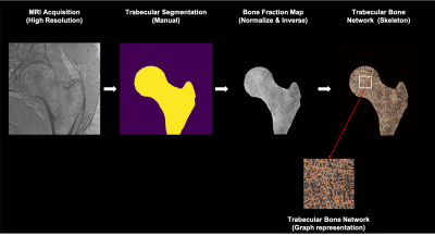 |
10 | 3T MRI Graph Analysis of the Proximal Femur trabecular network in Osteoporosis Subjects with Compared to Those without Prior Fragility Fractures
Anmol Monga1, Dimitri Martel1, Stephen Honig2, and Gregory Chang1
1Radiology, NYU Langone Health, New York, NY, United States, 2Osteoporosis Center, Hospital for Joint Diseases, NYU Langone Health, New York, NY, United States
Detecting microstructural information in bones leads to improved diagnosis, monitoring, and understanding of osteoporosis in patients. Recent advances allow for comprehensive and automated mining of quantitative image features to capture disease characteristics. Among these, graph and network analysis permit to characterize and assess network as the trabecular one and been previously applied in the wrist. In this study, we aim to apply the graph and network analysis method to the femoral trabecular bone network of osteoporosis subjects with and without previous fragility fracture history and assess features associated with fracture.
|
||
4949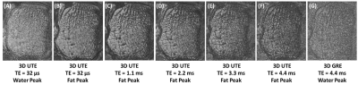 |
11 | Trabecular bone imaging using 3D ultrashort echo time (UTE) Cones at the fat peak frequency: feasibility study
Saeed Jerban1, Michael Song2, Amir M Afsahi1, Dina Moazamian1, Michael Carl2, Yajun Ma1, Alecio Lombardi1, Christine B Chung1, Eric Y Chang1,3, and Jiang Du1
1Department of Radiology, University of California, San Diego, San Diego, CA, United States, 2General Electric Healthcare, San Diego, CA, United States, 3Radiology Service, VA San Diego Healthcare System, San Diego, CA, United States
High susceptibility levels at the marrow/bone interface may significantly reduce T2* of marrow, leading to trabecular bone volume overestimation when imaged using conventional MRI sequences. The presence of fat in bone marrow further complicates trabecular bone imaging due to chemical shift artifacts. In this study, an ultrashort echo time MRI (UTE-MRI) technique focused on the fat peak frequency was investigated to image trabecular bone ex vivo and in vivo. This technique was shown to improve trabecular bone imaging by minimizing chemical shift artifacts as well as susceptibility related short T2* effects, thereby providing more accurate estimation of trabecular bone structure.
|
||
4950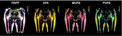 |
12 | 3T Chemical Shift encoded MRI detects Bone Marrow Adipose Tissue changes In Osteoporosis with Fragility Fracture
Dimitri Martel1, Benjamin Leporq2, Anmol Monga1, Stephen Honig3, and Gregory Chang1
1Radiology, NYU Langone Health, New York, NY, United States, 2Université de Lyon; CREATIS CNRS UMR 5220, Inserm U1206, INSA-Lyon, Villeurbanne, France, 3Osteoporosis Center, Hospital for Joint Diseases, NYU Langone Health, New York, NY, United States
Osteoporosis (OP) is associated with low bone mass and deterioration of bone tissue microarchitecture leading to increased bone fragility and fracture risk. There are increasing pieces of evidence that bone marrow adipose tissues (BMAT) play a significant role in the pathophysiology of osteoporosis. Our aim was to assess the proximal femur BMAT composition in OP without (OP) and with history of fragility fracture (OP-Fx) and compare it to naive controls using Chemical Shift Encoded MRI.
|
||
4951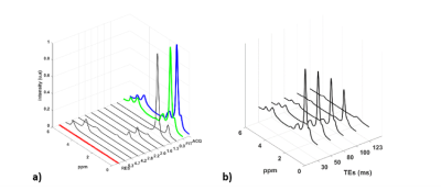 |
13 | Fatty Acid Composition of Proximal Femur Bone Marrow in Subjects with Systemic Lupus Erythematous Using 3T Magnetic Resonance Spectroscopy
Dimitri Martel1, Amit Saxena2, H. Michael Belmont2, Stephen Honig3, Anmol Monga1, and Gregory Chang1
1Department of Radiology, NYU Langone Health, New York, NY, United States, 2Department of Rheumatology, NYU Langone Health, New York, NY, United States, 3Osteoporosis Center, Hospital for Joint Diseases, NYU Langone Health, New York, NY, United States
Systemic Lupus Erythematosus (SLE) is a chronic, inflammatory, multisystem disease predominantly affecting young women. Patients with SLE have a significantly worse health-related quality of life compared to healthy subjects or patients with other chronic diseases. MSK manifestations in SLE patients are common, including reductions in bone quality. Studies using magnetic magnetic resonance spectroscopy (MRS) have shown that change in bone marrow fat amount and composition is associated with decreased bone quality. The aim of our study was to use MRS to assess differences of BMAT in SLE without treatment (n=28), SLE treated (n=15) and controls (n=21).
|
||
4952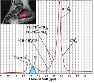 |
14 | Bone-marrow fatty-acids profiles from women calcaneus by 1H-MR Spectroscopy to search early osteoporotic biomarkers
Daniele Mattioli1, Vincenzo Vinicola2, Marco Montuori3, Umberto Tarantino4, and Silvia Capuani5
1Physics Dpt., Sapienza University of Rome, Rome, Italy, 2Center for Prevention, Diagnosis and Treatment of Osteoporosis, IRCCS Santa Lucia Foundation, Rome, Italy, 3Institute for Complex Systems, National Research Council CNR-ISC, Rome, Italy, 4Department of Orthopaedics and Traumatology, Tor Vergata University of Rome, Rome, Italy, 5Institute for Complex Systems, National Research Council CNR-ISC, Roma, Italy Changes occurring in the bone-marrow fatty-acids (BMFA) could be precursors of the bone mineral loss due to the development of osteoporosis. In this view, early biomarkers of osteoporosis among BMFA could be highlighted by quantifying fatty-acids with 1H-MRS in young, premenopausal, postmenopausal women with normal t-score and in osteoporotic women. In this study, L13, L09, fUFA, and fPUFA together with TL quantification obtained in women calcanei could be early osteoporosis markers. The present work confirms the potential of MRS to investigate BMFA metabolic variations due to postmenopause and to the development of osteoporosis. |
||
4953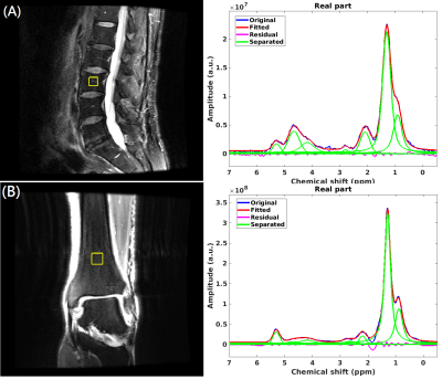 |
15 | Bone marrow fat quantification on vertebral and lower extremities using in-vivo MR spectroscopy: diabetes patients v.s healthy control
Po-hung Wu1, Kaipin Xu2, Gabby Joseph1, Yan Li1, Xiaojuan Li2, Thomas Link1, and Galateia Kazakia1
1Radiology and Biomedical Imaging, University of California - San Francisco, San Francisco, CA, United States, 2Biomedical Engineering, Program of Advanced Musculoskeletal Imaging (PAMI), Cleveland Clinic, Cleveland, OH, United States
Patients with type II diabetes (T2D) have increased fracture risk. Increased bone marrow fat is associated with increased fracture risk. We hypothesized that T2D patients will have altered bone marrow fat (BMF) and marrow composition biomarkers. In this study, we performed magnetic resonance spectroscopy (MRS) to quantify BMF and composition biomarkers at the spine in 37 T2D patients and 36 controls, and at the distal tibia in 30 T2D patients 32 controls. The results suggest that T2D may change vertebral marrow composition, particularly in men, which may be a factor in the development of increased bone fragility related to T2D.
|
||
 Back to Meeting Home
 Back to the Program-at-a-Glance
The International Society for Magnetic Resonance in Medicine is accredited by the Accreditation Council for Continuing Medical Education to provide continuing medical education for physicians.
 Back to Meeting Home
 Back to the Program-at-a-Glance
 View the Presentation