Online Gather.town Pitches
Quantitative Neuroimaging & Neurofluids IV
Joint Annual Meeting ISMRM-ESMRMB & ISMRT 31st Annual Meeting • 07-12 May 2022 • London, UK

Online Gather.town Pitches
Quantitative Neuroimaging & Neurofluids IV
| Booth # | ||||
|---|---|---|---|---|
4831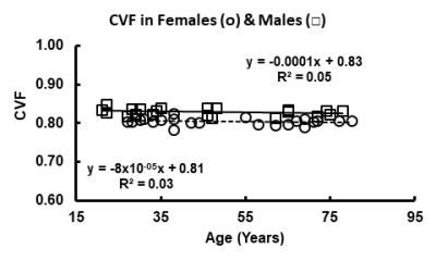 |
1 | Gender Differences in Cell Volume Fraction (CVF) of Human Brains Measured by Quantitative Sodium MR Imaging
Keith R Thulborn1
1Center for Magnetic Resonance Research, University of Illinois at Chicago, Chicago, IL, United States
The cell volume fraction (CVF), derived from tissue sodium concentration as measured by quantitative sodium MR imaging, in the human brain is high (~0.82) and maintained with normal aging despite decreases in brain volume. The gender difference (females: 0.807±0.009; males: 0.827±0.011, p<0.001) is also maintained over normal aging. This difference is posited to reflect the energy saving from higher CVF required for the larger male brain with higher number of neurons and synapses compared to female brains.
|
||
4832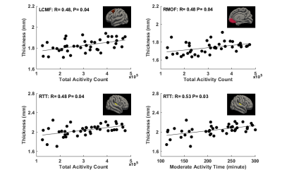 |
2 | Physical Activity during Pregnancy is Associated with Newborn’s Brain Cortical Development
Xiaoxu Na1, Rajikha Raja1, Aline Andres2,3, and Xiawei Ou1,3,4
1Radiology, University of Arkansas for Medical Sciences, Little Rock, AR, United States, 2Pediatrics, University of Arkansas for Medical Sciences, Little Rock, AR, United States, 3Arkansas Children’s Nutrition Center, Little Rock, AR, United States, 4Arkansas Children's Research Institute, Little Rock, AR, United States
This study examined relationships between mother’s physical activity during pregnancy and newborn’s brain cortical development. Healthy pregnant women were recruited and their physical activity level were monitored throughout pregnancy. Their newborns underwent a brain MRI examination at 2 weeks of postnatal age. Brain structural images were post-processed and mean cortical thickness for different brain regions was calculated. We found that at both 1st and 2nd trimester of pregnancy, there were significant positive correlations between mother’s physical activity level and newborn’s brain cortical thickness in multiple regions, suggesting positive impact of mother’s physical activity during pregnancy on offspring brain cortical development.
|
||
4833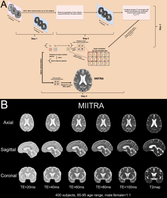 |
3 | Development of high resolution PD-weighted, T2-weighted, and T2 map templates for use in studies on older adults.
ABDUR RAQUIB RIDWAN1, Mohammad Rakeen Niaz1, Yingjuan Wu1, Shengwei Zhang2, David A. Bennett2, and Konstantinos Arfanakis1,2
1Department of Biomedical Engineering, Illinois Institute of Technology, Chicago, IL, United States, 2Rush Alzheimer's Disease Center, Rush University Medical Center, Chicago, IL, United States
Atlas based MRI investigations involving PD-weighted (PDw), T2-weighted (T2w) MRI sequences on older adults typically utilized younger adult templates such as those of the ICBM. Additionally, a thorough quantitative assessment of how available standardized PDw and T2w templates perform on aging population has not yet been performed. Here a new standardized high resolution PDw, T2w and T2 map templates of the MIITRA atlas dedicated for studies on older adults was developed using concepts of super-resolution and sparse-representation and compared to other available standardized templates in terms of image quality, inter-subject spatial normalization accuracy and representativeness of the older adult brain.
|
||
4834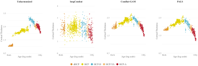 |
4 | Harmonization of Cortical Thickness Measurements Throughout the Human Lifespan
Sahar Ahmad1, Fang Nan2, Ye Wu1, Zhengwang Wu1, Weili Lin1, Li Wang1, Gang Li1, Di Wu3,4, and Pew-Thian Yap1
1Department of Radiology and Biomedical Research Imaging Center (BRIC), The University of North Carolina at Chapel Hill, Chapel Hill, NC, United States, 2Department of Biostatistics, University of Washington, Seattle, WA, United States, 3Department of Biostatistics, Gillings School of Global Public Health, The University of North Carolina at Chapel Hill, Chapel Hill, NC, United States, 4Division of Oral and Craniofacial Health Research, Adams School of Dentistry, The University of North Carolina at Chapel Hill, Chapel Hill, NC, United States
Pooling and integrating diverse imaging data across multiple sites is key to big data analytics in neuroimaging. Data amassed from multiple studies are inevitably heterogeneous due to differences in scanners, acquisition protocols, and post-acquisition image processing pipelines, substantially complicating downstream analyses. Here, we present a harmonization technique for multi-site large-scale longitudinal and cross-sectional data. We demonstrate the utility of our method in removing non-biological variability in cortical thickness measurements of individuals from birth to 100 years of age.
|
||
4835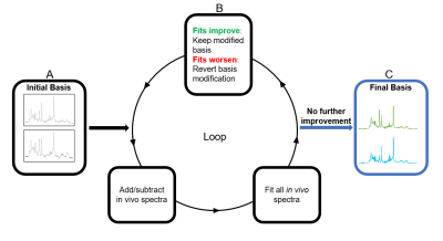 |
5 | Principal Spectra Analysis by Linear Modeling (PSALM)
Alexander Saunders1,2 and Stefan Blüml1,2
1Radiology, Children's Hospital Los Angeles, University of Southern California, Los Angeles, CA, United States, 2Rudi Schulte Research Institute, Santa Barbara, CA, United States
We hypothesized in vivo MR spectra can be sufficiently described by spectra representing predominant cell/tissue types rather than individual metabolites. We sought to extract two putative basis spectra of grey matter and white matter from 584 single-voxel 3T MR spectra using the principal spectra analysis by linear modeling (PSALM) custom algorithm. Two extracted PSALM bases explained >95% of fit variance; principal component analysis required 6-8 components to achieve the same. We found the algorithm produced high signal-to-noise, low linewidth basis spectra that robustly fit in vivo spectra from normal brain.
|
||
4836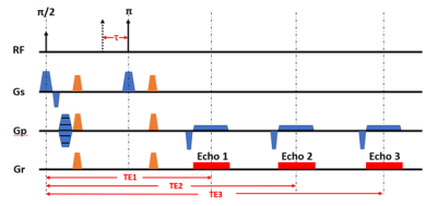 |
6 | Improvement of Accuracy and Stability of Oxygen Extraction Fraction Measurements by Using Triple-Echo Asymmetric Spin Echo Sequence Video Permission Withheld
Jian Shen1 and John Wood1,2
1University of Southern California, Los Angeles, CA, United States, 2Cardiology, Children's Hospital Los Angeles, Los Angeles, CA, United States
Asymmetric Spin Echo (ASE) is a popular technique to obtain quantitative estimates of R2’, cerebral venous blood volume (vCBV), and oxygen extraction fraction (OEF) noninvasively in normal subjects. In this study, We demonstrated the feasibility of using Triple-Echo ASE to induce a more stable and accurate vCBV and OEF estimates with no increase in scan time. In addition, this study investigated the role of diffusion in ASE measurements and provided insights into future acquisition approaches.
|
||
4837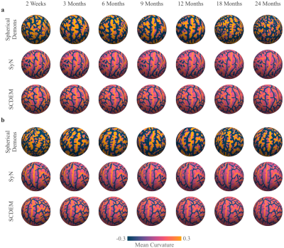 |
7 | Surface-Volume Registration Produces Atlases More Realistic than Either Surface or Volume Registration
Sahar Ahmad1, Zhengwang Wu1, Weili Lin1, Gang Li1, Li Wang1, and Pew-Thian Yap1
1Department of Radiology and Biomedical Research Imaging Center (BRIC), The University of North Carolina at Chapel Hill, Chapel Hill, NC, United States
We show that spatial registration by simultaneously considering both tissue contrast and cortical geometry produces brain atlases that are more reflective of individuals than registration by considering only one of the two factors alone. Registration with tissue contrast alone produces atlases with blurred cortical structures. Registration with cortical geometry alone produces surface atlases that are artificially sharp but do not reflect surface attributes of individuals.
|
||
4838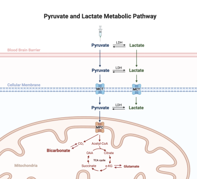 |
8 | Pathway analysis of cerebral bicarbonate production from hyperpolarized [1-13C]pyruvate
Maheen Zaidi1,2,3, Jun Chen1, Junjie Ma1, Xiaodong Wen1, Brenda Bartnik-Olson4, and Jae Mo Park1,5,6
1Advanced Imaging Research Center, UT Southwestern, Dallas, TX, United States, 2Psychology, UT Dallas, Richardson, TX, United States, 3Neurobiology, UT Dallas, Richardson, TX, United States, 4Radiology, Loma Linda University, Loma Linda, CA, United States, 5Radiology, UT Southwestern, Dallas, TX, United States, 6Electrical Engineering, UT Dallas, Richardson, TX, United States Detection of [13C]bicarbonate in the brain from hyperpolarized [1-13C]pyruvate provides a unique opportunity for assessing cerebral mitochondrial function in-vivo. Although decarboxylation of the labeled pyruvate occurs within the mitochondria, the metabolic path of the injected pyruvate could be complex before being transported to intracellular space. In this study, we investigated the likelihood of pyruvate being converted to lactate prior to decarboxylation. |
||
4839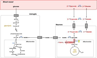 |
9 | Assessment of human brain pyruvate oxidation using functional hyperpolarized 13C MRS
Maheen Zaidi1,2,3, Binu P. Thomas1,4, Junjie Ma1, Salvador Pena1, Jun Chen1, James Ratnaker1, Craig R. Malloy1,4,5, Brenda Bartnik-Olson6, and Jae Mo Park1,4,7
1Advanced Imaging Research Center, UT Southwestern, Dallas, TX, United States, 2Psychology, UT Dallas, Richardson, TX, United States, 3Neurobiology, UT Dallas, Richardson, TX, United States, 4Radiology, UT Southwestern, Dallas, TX, United States, 5Internal Medicine, UT Southwestern, Dallas, TX, United States, 6Radiology, Loma Linda University, Loma Linda, CA, United States, 7Electrical and Computer Engineering, UT Dallas, Richardson, TX, United States
[13C]Bicarbonate production from hyperpolarized [1-13C]pyruvate in the brain is directly related to pyruvate oxidation and, thus serves a potential biomarker of brain function. In this study, we assessed time-wise bicarbonate production in the visual cortex during activation with minimal perturbation of its precursors, pyruvate and lactate, using a multichannel 13C receive array, a spectral-spatial RF pulse that fully excites bicarbonate signals, and dynamic 13C MRS. The real-time changes of bicarbonate production in response to visual stimuli were observed in healthy volunteers.
|
||
4840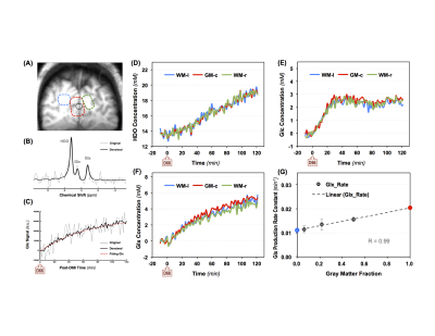 |
10 | Differentiating TCA Cycle Activity of Gray and White Matter in Human Brain at 7T Using High Resolution Dynamic Deuterium MRS Imaging with SPICE Video Permission Withheld
Xiao-Hong Zhu1, Hannes M Wiesner1, Yudu Li2,3, Wei Zhu1, Tao Wang1, Xin Li1, Zhi-Pei Liang2,3, and Wei Chen1
1University of Minnesota, Minneapolis, MN, United States, 2Beckman Institute for Advanced Science and Technology, Urbana, IL, United States, 3Department of Electrical and Computer Engineering, University of Illinois at Urbana-Champaign, Urbana, IL, United States
The emerging deuterium MRS (DMRS) imaging (DMRSI) technology has shown great promise in studying brain glucose metabolism, TCA cycle and glycolytic activity. In particular, its potential as a clinical tool for tumor detection in brain or other organs has attracted a lot of attention. However, the temporal and spatial resolution of dynamic DMRSI is limited by its low detection sensitivity. In this study, we proved that 7T high-resolution dynamic DMRSI combined with SPICE method can not only map the Glx production rate constant, but also distinguish the TCA cycle activity level between human brain gray matter and white matter.
|
||
 |
4841 |
11 | Rapid, high-spatial resolution in vivo diffusion MRI with joint subsampling and reconstruction in k-,q- and RF-space
Gabriel Ramos-Llorden1, Berkin Bilgic1,2, and Susie Y. Huang1,2
1Athinoula A. Martinos Center for Biomedical Imaging, Massachusetts General Hospital, Harvard Medical School, Charlestown, MA, United States, 2Harvard-MIT Division of Health Sciences and Technology, Cambridge, MA, United States
In this work, we accelerate the high-SNR, high-spatial resolution in vivo diffusion MRI technique gSlider by applying subsampling jointly in the k,-q, and RF-encoding space. Good reconstruction quality is achieved if complementary information from the multi-dimensional space k-,q-, and RF-space is combined in a complementary fashion and the reconstruction problem is solved jointly.
|
|
4842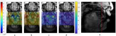 |
12 | Enhancing linguistic research through 2-mm isotropic 3D dynamic speech magnetic resonance imaging
Riwei Jin1, Brad Sutton1, Ryan Keith Shosted1, Jonghye Woo2, Fangxu Xing2, Jamie Perry3, Imani Gilbert3, and Zhipei Liang1
1University of Illinois Urbana-Champaign, Champaign, IL, United States, 2Massachusetts General Hospital, Boston, MA, United States, 3East Carolina University, Greenville, NC, United States
We are able to push the spatial resolution of dynamic speech magnetic resonance imaging to 2-mm near-isotropic level with 64 mm coverage of 32 3D slice locations that are spaced 2-mm apart with 35 fps. We choose to analyze lingual differences of American English voiced lateral [l] and (central) [t]. Several analysing methods are utilized such as magnitude comparison, t-test and deformation map comparison. The results give us detailed observations of lingual articulatory differences such as tongue grooving, twisting and coarticulation. Through this high spatial and temporal resolution, we demonstrate that this method will show great potentials on linguistic research.
|
||
4843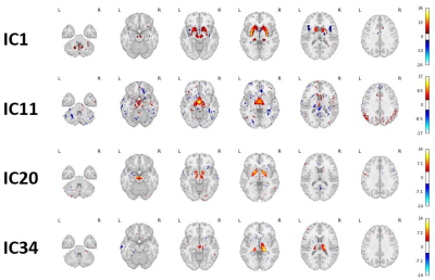 |
13 | Discovery of two novel deep gray matter brain iron networks through an Independent Component Analysis of Quantitative Susceptibility Maps
Jack A Reeves1, Niels Bergsland1,2, Dejan Jakimovski1, Fahad Salman1, Juliane Damm1, Nicklas Meineke1, Michael G Dwyer1,3, Robert Zivadinov1,3, and Ferdinand Schweser1,3
1Buffalo Neuroimaging Analysis Center, Department of Neurology, Jacobs School of Medicine and Biomedical Sciences, University at Buffalo, The State University of New York, Buffalo, NY, United States, 2MR Research Laboratory, IRCCS, Don Gnocchi Foundation ONLUS, Milan, Italy, 3Center for Biomedical Imaging, Clinical and Translational Science Institute, University at Buffalo, The State University of New York, Buffalo, NY, United States
Magnetic resonance imaging (MRI) studies have shown age and neurological disease affect iron concentrations in the deep gray matter (DGM). However, it is unknown whether iron accumulation occurs independently across DGM regions or in inter-regional patterns, i.e. “iron networks”. Here, we identified two highly reproducible, inter-regional DGM iron networks by applying independent component analysis (ICA) to quantitative susceptibility maps (QSM) from healthy volunteers. Areas of network overlap had relatively high iron concentrations and each network was related to separate environmental variables, indicating they have independent mechanisms of iron change. Our results advance the understanding of brain iron physiology.
|
||
4844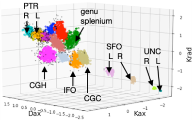 |
14 | Unsupervised learning with multi-parametric diffusion MRI to detect brain injury in concussed athletes
L. Tugan Muftuler1, Timothy B. Meier 1, and Michael A. McCrea1
1Neurosurgery, Medical College of Wisconsin, Milwaukee, WI, United States
Despite high prevalence of concussion in contact sports, there is still a lack of reliable, unbiased biomarkers of brain injury and recovery following sport-related concussion. The changes in imaging biomarkers after concussion are generally subtle such that signs of abnormalities are detected only in group analyses with large cohorts. Although diffusion MRI techniques show promise, detecting injury in individual subjects is still a challenge. The goal of this project is to identify potential signatures of brain injury in an individual subject with concussion using an unsupervised learning method using parameters derived from diffusion kurtosis imaging (DKI).
|
||
4845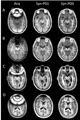 |
15 | Improved Structural Neuro-MRI with Triple TSE-based Synthetic MP-RAGE
Lei Zhang1, Ryan McNaughton2, Hernan Jara2, David N. Kennedy3, Carmen A. Marable1, Ning Hua4, Karl C. K. Kuban4, T. Michael O'Shea1, and Rebecca C. Fry1
1University of North Carolina at Chapel Hill, Chapel Hill, NC, United States, 2Boston University, Boston, MA, United States, 3University of Massachusetts Chan Medical School, Worcester, MA, United States, 4Boston University Medical Center, Boston, MA, United States
Purpose: To develop and evaluate a metal-artifact robust triple turbo-spin-echo (TSE)-based synthetic MP-RAGE method. Methods: T1, T2 and PD maps generated from triple TSE, two MP-RAGE image sets simulated by the Bloch equation, and FreeSurfer brain segmentation were performed on 44 participants at age of fifteen. Results: Reduced artifact and homogenous synthetic MP-RAGE images were produced in orthodontic brace-wearing subjects. Comparable image contrast and FreeSurfer brain segmentation results were observed between synthetic and acquired MP-RAGE in normal subjects. Conclusion: Triple TSE based synthetic MP-RAGE may enhance conventional MP-RAGE in the neuroimaging studies of orthodontic brace-wearing subjects with reduced metal artifacts.
|
||
 Back to Meeting Home
 Back to the Program-at-a-Glance
The International Society for Magnetic Resonance in Medicine is accredited by the Accreditation Council for Continuing Medical Education to provide continuing medical education for physicians.
 Back to Meeting Home
 Back to the Program-at-a-Glance
 View the Presentation