Online Gather.town Pitches
Quantitative Neuroimaging & Neurofluids III
Joint Annual Meeting ISMRM-ESMRMB & ISMRT 31st Annual Meeting • 07-12 May 2022 • London, UK

Online Gather.town Pitches
Quantitative Neuroimaging & Neurofluids III
| Booth # | ||||
|---|---|---|---|---|
4375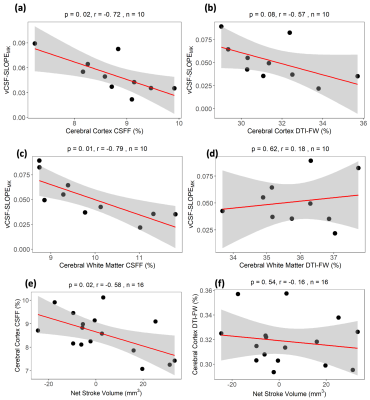 |
1 | T2 relaxometry based CSF fraction (CSFF) mapping is a better biomarker for brain drainage pathology than DTI-based free water (DTI-FW) mapping
Liangdong Zhou1, Thanh Nguyen2, and Yi Li2
1Radiology, Weill Cornell Medicine, New York, NY, United States, 2Weill Cornell Medicine, New York, NY, United States
We compared two free water methods, T2 relaxometry-based CSFF and DTI-based DTI-FW, by associating them with age, cognitive score RAVLT, amyloid beta deposition from PiB SUVr, tau deposition from MK6240, aqueduct CSF flow from PC-MRI and ventricle CSF clearance from MK-PET. Results show that CSFF outperforms DTI-FW in most relation pairs. CSFF works great for quantifying clearance related measures but DTI-FW failed.
|
||
 |
4376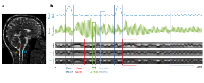 |
2 | Flow Quantification for Cerebrospinal Fluid by Spatial-temporal Perturbation Patterns in SSFP
Yicun Wang1, Peter van Gelderen1, Jacco A. de Zwart1, Pinar S. Özbay1, Hendrik Mandelkow1, Dante Picchioni1, and Jeff H. Duyn1
1AMRI, LFMI, NINDS, National Institutes of Health, Bethesda, MD, United States
We report a fast quantitative CSF flow imaging method based on the spatial-temporal perturbation patterns formed at the transition bands in balanced steady-state-free-precession (SSFP) images when combined with a background magnetic field gradient. By modeling CSF flow as a combination of a pulsatile (AC) and a constant (DC) flow component across the cardiac cycle, a dictionary was generated based on Bloch simulations and used to translate the observed patterns to flow velocities. Monte-Carlo simulations were performed to evaluate the accuracy of the approach. CSF flow resulting from different physiological mechanisms was quantified in selected regions in 12 healthy subjects.
|
|
4377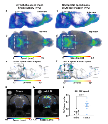 |
3 | Blockade of lymphatic drainage to deep cervical lymph nodes impacts CSF flow and brain morphometry
Sunil Koundal1, Zachary Gursky1, Xinan Chen2, Hedok Lee1, Laura Santambrogio3, Jonathan Kipnis4, Allen Tannenbaum2, and Helene Benveniste1
1Anesthesiology, Yale University, New Haven, CT, United States, 2Department of Applied Mathematics and Statistics, Stony Brook University, Stony Brook, NY, United States, 3Department of Radiation Oncology, Weill Cornell Medicine, New York, NY, United States, 4Department of Pathology and Immunology, Washington University, St. Louis, MO, United States
The altered CNS fluid flow dynamics and homeostasis mechanisms in response to blockade of lymphatic drainage to deep cervical lymph nodes (dcLN) is poorly understood. We used multi-modality MRI and computational fluid dynamics approach to study the same in rats with electrocauterized afferent lymphatic vessels of dcLN. The brain morphometry of Cauterized-dcLN rats showed localized volume expansion in Pons, Hippocampus and Corpus-callosum, while glymphatic speed maps showed hyperdynamic CSF flow in ambient and quadrigeminal cisterns. These results clearly show that the blockade of CSF drainage to dcLN alters CNS fluid homeostasis/flow dynamics, long-term may result in waste accumulation and neurodegeneration.
|
||
4378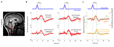 |
4 | Cerebral Blood Flow Autoregulation is a Major Contributor to Flow Dynamics of Cerebrospinal Fluid
Yicun Wang1, Peter van Gelderen1, Jacco A. de Zwart1, Pinar S. Özbay1, Hendrik Mandelkow1, Dante Picchioni1, and Jeff H. Duyn1
1AMRI, LFMI, NINDS, National Institutes of Health, Bethesda, MD, United States
In addition to the well-established origin in cardiac and respiratory cycles, CSF pulsations may also result from the much slower variations in vascular tone associated with cerebral blood flow (CBF) autoregulation. Using a novel quantitative MRI approach, we measured CSF flow velocities and displaced volumes resulting from these three mechanisms in 12 healthy human controls. We found the autoregulatory effects to be a major factor, with associated CSF velocities comparable to and displaced volumes an order of magnitude larger than the cardio-respiratory effects. This may be an important mechanism underlying the CSF oscillations recently observed in resting-state fMRI.
|
||
4379 |
5 | Evaluation of Cerebral Blood Flow and BBB Water Exchange in an Aged African American Cohort
Brandon Ojogho1, Farzan Abdolahi2, Xingfeng Shao1, Samantha Ma3, Chenyang Zhao1, Kay Jann1, Xuejuan Jiang2, and Danny JJ Wang1
1Laboratory of FMRI Technology (LOFT) Mark & Mary Stevens Neuroimaging and Informatics Institute Keck School of Medicine University of Southern California (USC), Los Angeles, CA, United States, 2Department of Ophthalmology and Department of Preventive Medicine Keck School of Medicine University of Southern California (USC), Los Angeles, CA, United States, 3Siemens Healthcare, Los Angeles, CA, United States
This study evaluated CBF and BBB water exchange rate (kw) measured by diffusion prepared pseudo-continuous arterial spin labeling (DP-pCASL) in a cohort of aged African American individuals. The results showed decreasing CBF and kw with age, and higher CBF in females. Both CBF and kw were correlated with picture sequence memory test score. CBF was negatively correlated with white matter hyperintensities (WMH), free water (FW) and peak skeletonized mean diffusivity (PSMD), although age accounted for these correlations. These findings suggest the potential for CBF and kw as biomarkers of cerebral small vessel disease.
|
||
4380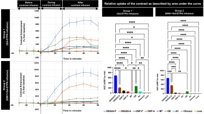 |
6 | Role of insulin resistance in the optic nerve glymphatic system
Muneeb A Faiq1, Anoop Sainulabdeen1, Vishnu Adi1, Thajunnisa A Sajitha1, Sophia Khoja1, Carlos Parra1, Choong H Lee2, Jiangyang Zhang2, and Kevin C Chan1,2
1Department of Ophthalmology, New York University Grossman School of Medicine, NYU Langone Health, New York, NY, United States, 2Department of Radiology, New York University Grossman School of Medicine, NYU Langone Health, New York, NY, United States
Recent evidence suggests the existence of a functional glymphatic system in the optic nerve. Insulin resistance in the central nervous system (CNS-IR) has been implicated in neurodegenerative conditions. Its mechanism remains unclear though one candidate involves dysregulation of the cerebrospinal fluid (CSF). Here, we investigated if CNS-IR affects CSF flow in the visual pathway by measuring the spatiotemporal profiles of CSF dynamics using gadolinium-enhanced MRI following intrathecal administration of the specific insulin receptor blocker, S961. Our results suggested that central insulin function may play an imperative role in CSF dynamics for the homeostasis of the visual system.
|
||
4381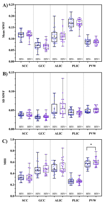 |
7 | Myelin Water Imaging in an HIV Population at Risk of Cerebral Small Vessel Disease
Md Nasir Uddin1, Abrar Faiyaz2, Alan Finkelstein3, and Giovanni Schifitto1,2
1Department of Neurology, University of Rochester, Rochester, NY, United States, 2Department of Electrical and Computer Engineering, University of Rochester, Rochester, NY, United States, 3Department of Biomedical Engineering, University of Rochester, Rochester, NY, United States
HIV-infected individuals are at increased risk of developing cerebral small vessel disease (CSVD). Myelin damage is a feature in CSVD, and can be quantified using myelin water imaging. In this work, we investigated if MWI can be used to quantify myelin related changes in white matter in an HIV cohort at risk of CSVD. Our results lend support to the use of MHI to track myelin related abnormalities in the white matter.
|
||
4382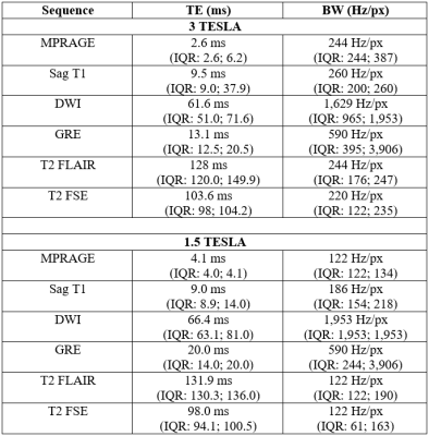 |
8 | Feasibility of 3T imaging in idiopathic normal pressure hydrocephalus patients with Certas Plus Valve: comparison of artifacts 3T vs 1.5T.
Emanuele Camerucci1, Benjamin Elder1, Jeffrey Gunter1, John Huston1, Clifford Jack1, and Petrice Cogswell1
1Mayo Clinic, Rochester, MN, United States
We compared the artifact induced by the Certas Plus at 3.0T vs 1.5T in 9 patients with idiopathic normal pressure hydrocephalus (iNPH) on six sequences (3D MPRAGE, Sagittal T1, axial DWI, axial GRE, axial T2 FLAIR, axial T2 FSE) at three levels (atria of the lateral ventricles, cerebral aqueduct, and cerebellum hemispheres). The 1.5T scans had a significantly greater area of artifact on DWI (all levels) and GRE (cerebellar level) sequences, and a smaller area of artifact on T2 FLAIR (cerebellar level). Post-shunt imaging at 3T with the Certas plus valve is feasible and with lesser artifact than on 1.5T.
|
||
4383 |
9 | R2* changes reflect the kinetics of formalin-based penetration and fixation in post-mortem tissue
Azadeh Nazemorroaya1, Ali Aghaeifar2, Hildegard Schulz3, Klaus Scheffler 3,4, Thomas Shiozawa-Bayer5, Bernhard Hirt5, and Gisela Hagberg3,4
1High Field Magnetic Resonance, Max Planck Institute for Biological Cybernetics, Tuebingen, Germany, 2Wellcome Centre for Human Neuroimaging, London, United Kingdom, 3High Field Magnetic Resonance,, Max Planck Institute for Biological Cybernetics, Tuebingen, Germany, 4Biomedical Magnetic Resonance, University Hospital Tuebingen, Tuebingen, Germany, 5Clinical Anatomy, University Hospital Tübingen, Tuebingen, Germany
Post-mortem brain MRI can yield valuable information. However, tissue preservation requires substitution of the CSF-fluid by fixation agents, which is time-consuming for large samples. R2* maps, dominated by R2-effects, were measured at several timepoints during fixation of pig-brain tissue samples using formalin-based fixatives.
|
||
4384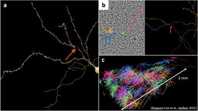 |
10 | A user interface for brain cell analysis of a petascale database in cerebral cortex and its application to numerical simulations of diffusion
Benjamin Sylvanus1, Susie Yi Huang2,3, and Hong Hsi Lee2,3
1Northeastern University, Boston, MA, United States, 2Athinoula A. Martinos Center for Biomedical Imaging, Massachusetts General Hospital, Charlestown, MA, United States, 3Department of Radiology, Harvard Medical School, Boston, MA, United States
The 1.4 petabyte electron microscopy volume with fully segmented cells in the human cerebral cortex is publicly available, serving as a valuable resource of building numerical phantoms of MRI. However, even few neurite interruptions in cell segmentations can lead to substantial biases in numerical simulations of diffusion inside cells. Here we propose a user interface to automatically correct most of the neurite interruptions and manually edit the corrections. Combing the proposed user interface with Trees toolbox and SpinDoctor for quantitative cell analysis and diffusion simulations, we provide a convenient platform to validate biophysical modeling of MRI evaluating tissue microstructure.
|
||
 Back to Meeting Home
 Back to the Program-at-a-Glance
The International Society for Magnetic Resonance in Medicine is accredited by the Accreditation Council for Continuing Medical Education to provide continuing medical education for physicians.
 Back to Meeting Home
 Back to the Program-at-a-Glance
 View the Presentation