Online Gather.town Pitches
Quantitative Neuroimaging & Neurofluids II
Joint Annual Meeting ISMRM-ESMRMB & ISMRT 31st Annual Meeting • 07-12 May 2022 • London, UK

Online Gather.town Pitches
Quantitative Neuroimaging & Neurofluids II
| Booth # | ||||
|---|---|---|---|---|
3586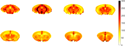 |
1 | A high-resolution whole-brain CBF atlas based on more than 100 adult normal mice
Xiang Chen1, Wenjing Xu1, Xiaoyang Han1, Yuting Zhai1, Jiadi Xu2, and Xiao-Yong Zhang1
1Institute of Science and Technology for Brain-Inspired Intelligence, Fudan University, Shanghai, China, 2F.M. Kirby Research Center for Functional Brain Imaging, Kennedy Krieger Research Institute, Baltimore, MD, United States
Arterial spin labeling (ASL) is a powerful MRI technique used to noninvasively measure cerebral blood flow (CBF). In preclinical studies, rodent brain CBF serves as an important indicator in many brain disease models. In this work, we established a whole-brain CBF atlas based on 102 adult normal mice and profiled CBF distributions in 27 brain regions. We found that there is a high correlation of CBF between the left and right hemispheres. Additionally, the high-resolution mouse CBF atlas can be used to distinguish mice with autism disorder, indicating the potential applications of the atlas in preclinical studies.
|
||
3587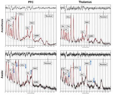 |
2 | Differences in brain metabolites in awake and anesthetized mice revealed by 1H-MRS
Ren Li1, Wenjing Xu1, Zhifeng Liang2, and Xiao-Yong Zhang1
1Institute of Science and Technology for Brain-Inspired Intelligence, Fudan University, Shanghai, China, 2Institute of Neuroscience, CAS Center for Excellence in Brain Sciences and Intelligence Technology, Key Laboratory of Primate Neurobiology, Chinese Academy of Sciences, Shanghai, China
It is important to investigate neural metabolism and brain functions in awake animals for preclinical studies. However, there are few magnetic resonance spectroscopy (MRS) studies in rodents under awake conditions. In this work, we developed an MRI-compatible restraint device for the measurement of brain metabolism in awake mice. Using 1H-MRS, we successfully observed the brain metabolic profile of prefrontal cortex and thalamus in awake mice. In addition, we found metabolic alterations between awake and anesthetized states, indicating that anesthesia has a non-negligible impact on brain metabolism.
|
||
3588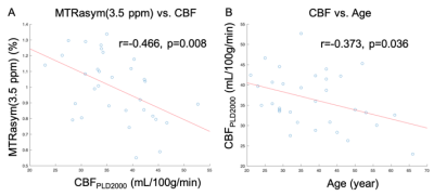 |
3 | Assessment of the correlation between blood perfusion and protein contents in brain of healthy subjects Video Not Available
Fan Yu1, Qiuxuan Li1, Cheng Zhao1, Liangjie Lin2, Jiazheng Wang2, and Jie Lu1
1Radiology and Nuclear medicine, Xuanwu Hospital, Capital Medical University, Beijing, China, 2Philips Healthcare, Beijing, China, Beijing, China
The blood perfusion and protein contents in brain of healthy subjects were measured in this study by 3D pseudo continuous arterial spin labeling and 3D amide proton transfer weighted imaging. Results showed a negative correlation between blood perfusion and the content of mobile cellular proteins and peptides in brain of heathy subjects; and the blood perfusion in normal brain, rather than the protein content, showed a significant dependence to age.
|
||
3589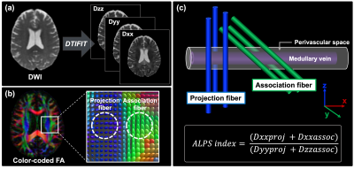 |
4 | Non-invasive MRI measures of glymphatic system activity associated with CSF-Aβ and FDG-PET uptakes in Alzheimer’s disease
Koji Kamagata1, Christina Andica1, Kaito Takabayashi1, Yuya Saito1, Toshiaki Taoka2, Hayato Nozaki1, Junko Kikuta1, Shohei Fujita1, Kouhei Kamiya3, Akihiko Wada1, Toshiaki Akashi1, Masaaki Hori3, Shinji Naganawa2, and Shigeki Aoki1
1Department of Radiology, Juntendo University, Tokyo, Japan, 2Department of Radiology, Nagoya University, Aichi, Japan, 3Department of Radiology, Toho University, Tokyo, Japan
We assessed the non-invasive MRI measurements, such as the diffusivity along the perivascular space (PVS) represented by ALPS index, PVS volume, and fractional volume of free water in the white matter (FW-WM), in cognitively normal subjects and subjects with Alzheimer’s disease (AD) or mild cognitive impairment (MCI). Abnormalities were detected in the subjects with AD and MCI, and theirALPS index and FW-WM values were significantly associated with CSF Aβ levels and FDG PET uptake, as well as with multiple cognitive scores, thus suggesting that a glymphatic system dysfunction could be associated with Aβ deposition and cognitive impairments.
|
||
3590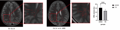 |
5 | The structural and functional change of glymphatic system in children with ADHD
Yingqian Chen1, Shu Su1, Yan Dai1, Long Qian2, Miaomiao Wang3, and Zhiyun Yang1
1First Affiliated Hospital, Sun Yat-sen University, Guangzhou, China, 2GE Healthcare, Beijing, China, 3the first affliated hospital of Xi'an Jiaotong University, Xi'an, China
The newly found structure – glymphatic system, may offer a new point for exploring the pathogenesis of attention deficit hyperactivity disorder (ADHD),. Our study investigated the change of glymphatic system in the treatment-naïve ADHD children by quantitatively measuring the VRS volume and using DTI-ALPS method. As the results showed, ADHD children have enlarged VRS, and the diffusivities along the VRS and ALPS-index were significantly lower in children with ADHD than in TD subjects, suggesting the impaired glymphatic drainage. Our study suggested that the glymphatic system alternation may play a role in the pathogenesis of ADHD and deserves further investigation.
|
||
3591 |
6 | Associations of gestational age, birth indicators, and brain volume in full-term newborns Video Permission Withheld
Linlin Zhu1, Yuying Feng1, Pengxuan Bai1, Yao Ge1, Congcong Liu1, Yichu He2, Feng Shi2, Jian Yang1, Xiaocheng Wei3, and Chao jin1
1The First Affiliated Hospital of Xi’an Jiaotong University, Xi’an, China, 2Shanghai United Imaging Intelligence Co., Shanghai, China, 3GE Healthcare, Beijing, China
Newborn gestational age (GA) and birth indicators (birth weight, birth length and head circumference) have been used as clinical indicators to assess the level of brain development. Cerebral changes are particularly intense during the last weeks of gestation. Previous studies have shown that the total brain volume at birth is about one third of the brain volume of healthy adults[1], but it remains unclear about the effect factors of brain volume. Our studies have shown that compared with other birth indicators, the gestational age of full-term newborns is strongly correlated with brain volume.
|
||
3592 |
7 | Quantitative assessment of neonatal hyperbilirubinemia brain injury based on T1 mapping
Xing li1, Xiaoli Meng2, Wei Zhang1, Huipeng Ren1, Yumiao Zhang1, Xiaohu Wang1, Qing Fan1, Xiaocheng Wei3, and Zhuanqin Ren1
1Radiology department, Baoji Central Hospital, baoji, China, 2Nuclear medicine department, Xijing Hospital of Air Force Military Medical University, Xi'an, China, 3GE Healthcare, MR Research China, Beijing, Beijing, China
In this study, we aim to investigate whether T1 mapping can be used to assess neonatal brain injury. It was concluded that the influence of bilirubin value on the globus pallidus (GP) and the posterior limb of internal capsule (PLIC) was obvious. Among them, the T1 values of GP, putamen (PU) and PLIC decreased with the increase of bilirubin.
|
||
 |
3593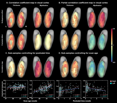 |
8 | Postnatal experience promotes the thickening but not myelination of the neonatal ventral visual cortex
Mingyang Li1, Tingting Liu1, Xinyi Xu1, Qingqing Wen1, Zhiyong Zhao1, Yi Zhang1, Yi-Cheng Hsu2, Yi Sun2, and Dan Wu1
1College of Biomedical Engineering & Instrument Science, Zhejiang University, Zhejiang, China, 2MR Collaboration, Siemens Healthineers Ltd, Shanghai, China
Postnatal experience is important for the development of the visual cortex in animal newborns, but its influence in human infants remains unclear. We collected a dataset from the developing human connectome project, including multi-modal MRI of 783 newborns. We found the cortical thickness (CT) and myelination of ventral visual cortex were significantly increased between 37 and 45 weeks of postmenstrual menstrual age (PMA). Interestingly, the CT but not myelination showed significant correlation with postnatal experience in both the V1 and higher-level visual cortex. Our result suggested that experience plays an important role in early cortical thickening
|
|
3594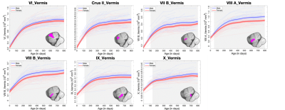 |
9 | Longitudinal Development of Regional Cerebellar Volumes from Birth to 27 Months of Age
Ya Wang1,2, Liangjun Chen2, Yue Sun2, Tengfei Li2, Zhengwang Wu2, Wenhua Huang1, Weili Lin2, Li Wang2, and Gang Li2
1National Key Discipline of Human Anatomy, School of Basic Medical Sciences, Southern Medical University, Guangzhou, China, 2Department of Radiology and Biomedical Research Imaging Center (BRIC), University of North Carolina at Chapel Hill, Chapel Hill, NC, United States
Early cerebellar development in infant brains is very dynamic and highly related to normal cognitive functions and neurodevelopmental disorders, but remains largely unexplored, due to the lack of densely-sampled longitudinal data of early ages. Herein, we unprecedently explored the dynamic developmental trajectories of the volumes in 27 cerebellar lobules based on 511 high-resolution longitudinal structural MRI scans from 235 healthy infants from the Baby Connectome Project (BCP) densely covering the age ranging from birth to 27 months. The trajectories of the cerebellar structures reveal lobule-specific nonlinear developmental patterns and are sexually dimorphic starting from different ages.
|
||
3595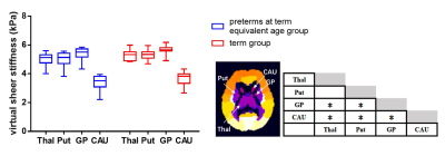 |
10 | Measuring the stiffness of subcortical gray matter structures with virtual MR elastography in preterm and term infants
Miaomiao Wang1, Yao Ge1, Xianjun Li1, Congcong Liu1, and Jian Yang1
1the first affiliated hospital of Xi'an Jiaotong university, Xi'an, China
MR elastography is a promising approach for describing biomechanical properties. Some studies investigated stiffness of subcortical gray matter (SGM), which are critical for cognitive. However, they have not been explored in infants. MRE has some limitations in application of infants, and this study aims to measure SGM stiffness in preterm and term infants using virtual MRE. Our study suggested that virtual shear stiffness of SGM were similar between the preterm at term equivalent and the term, and GP is the stiffest region. This finding suggested that virtual MRE is a sensitive method to characterize the SMG maturity in infants.
|
||
3596 |
11 | Brain structural development on oriental and occidental neonates: a comparison between ITPNBDI and dHCP datasets
Yan Zhang1, Miaomiao Wang1, Congcong Liu1, Xiaoyu Wang1, Yao Ge1, Yichu He2, Feng Shi2, Xianjun Li1, and Jian Yang1
1Department of Radiology, The First Affiliated Hospital of Xi'an Jiaotong University, Xi'an, China, 2Department of Research and Development, Shanghai United Imaging Intelligence Co., Ltd., Shanghai, China Despite studies that have demonstrated population differences due to differences in genetics, culture, and environmental exposures, a comparison of brain structural development between oriental and occidental neonates remains to be fully examined. We compared 108 neonates from ITPNBDI and 170 neonates from dHCP to examine differences in neonatal brain structure. Results demonstrated ITPNBDI neonates held shorter brain length, wider brain width and higher brain height than dHCP neonates. They also had greater total brain volume, greater grey matter and less white matter than dHCP neonates. Specifically, volumes of some brain regions involved in speech perception were different between two datasets. |
||
3597 |
12 | PM2.5 exposure induces structural and functional changes in the mouse brain
Xiaoyang Han1, Yuting Zhai1, and Xiao-Yong Zhang1
1Institute of Science and Technology for Brain-Inspired Intelligence, Fudan University, Shanghai, China
We investigated the structural and functional changes induced by exposure in PM2.5 in the mouse model using MRI at 11.7T. We compared the gray matter volume ratio (GMR) of major brain regions between mice exposed to concentrated PM2.5 (PM) and mice exposed to filtered air (FA) after 30-day and 90-day exposure. Our results demonstrate that exposure to PM2.5 for 90 days may induce brain structural and functional changes.
|
||
3598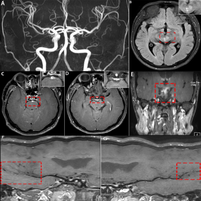 |
13 | Application of high-resolution 3D vessel wall MR(3D VW-MR) imaging in Primary Angiitis of the Central Nervous System Video Not Available
hongwei zhou1, kexin zhao1, and tianjing zhang2
1the First Hospital of Jilin University, Chang Chun, China, 2Philips Healthcare, Guangzhou, China
Primary angiitis of the central nervous system(PACNS) is a kind of rare disease, but the clinical diagnosis is difficult. Recently, high-resolution HR-3D-VW-MRI(3-dimensional vessel wall MR imaging) method has been used to evaluate cerebral vessels because it could directly show the vessel wall as well the lumen; thus it can assist in differentiating various types of vasculopathy. The purpose of our study was to summarize the typical imaging performance of PACNS and evaluate the value of 3D- VW-MRI sequence in demonstrating the detailed information in detection, diagnosis, evaluation, and follow-up for PACNS.
|
||
3599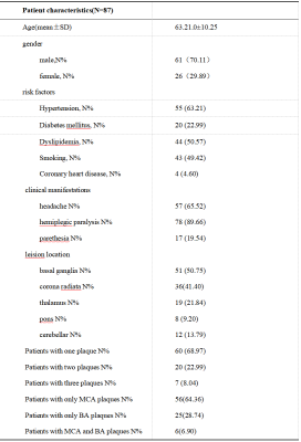 |
14 | The application of High-Resolution Vessel Wall MRI(HR-VW-MRI) in determining the stability of intracranial MCA and BA plaques Video Not Available
hongwei zhou1, kexin zhao1, and tianjing zhang2
1Radiology, the First Hospital of Jilin University, Chang Chun, China, 2Philips Healthcare, Guangzhou, China
The stability of intracranial of middle cerebral artery(MCA) and basilar artery( BA) plaques related to the stroke events is a crucial issue. However, the discrimination of the plaque’s stability could be rather challenging. Compared with traditional imaging methods such as CTA, MRA or DSA, high-resolution vessel wall MR imaging(HR-VW-MRI) method could demonstrate the abnormality of the vessel wall .It could also potentially evaluate the stability of the intracranial artery. This study aims to compare HR-VW-MRI’s characteristic features of MCA plaque with BA plaque, and to figure out the relationship between plaques’ imaging features with the stroke events.
|
||
3600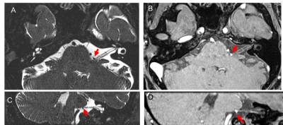 |
15 | Preoperative high-resolution 3D VIBE evaluations of the neurovascular anatomy of patients with microvascular compression for hemifacial spasm
Liu Xiaoming1, Kong Xiangchuang1, Li Qiang2, Sun Peng3, and Liu Dingxi1
1Departments of Radiology, Union Hospital, Tongji Medical College, Huazhong University of Science and Technology, Wuhan, China, Wuhan,Hubei,China, China, 2Departments of Radiology, Union Hospital, Tongji Medical College, Huazhong University of Science and Technology, Wuhan,Hubei,China, China, 3Philips Healthcare, Beijing, China, Beijing,China, China
The main cause of hemifacial spasm is neurovascular compression syndrome. It is very important to preoperative evaluate the relationship between nerve and responsible blood vessel and its compression site. High-resolution 3D MR cisternography techniques were used preoperatively to assess neurovascular anatomy in patients with neurovascular compression syndrome, but the contrast between vessels and cranial nerves at the point of neurovascular contact is limited. In this study, we evaluated 3D VIBE image to could not only clearly show the structure of nerves and blood vessels, but also improve the contrast between blood vessels and nerves or surrounding brain stem.
|
||
3601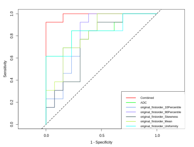 |
16 | The value of first-order features based on ADC map in evaluating the neuroprotective effect of LIPUS for acute traumatic brain injury with rat model Video Not Available
Dan Du1, Lanxiang Liu1, and Qinglei Shi2
1The first hospital of Qinhuangdao, Qinhuangdao, China, 2Scientific Clinical Specialist,Siemens Ltd, Beijing, China
In this study we studied the value of LR model established with first-order features based on ADC map in evaluating the neuroprotective effect of Low-intensity pulsed ultrasound (LIPUS) for acute traumatic brain injury (TBI). The results demonstrated that the model based on the first-order features may have potential value in predicting the therapy effect of LITUS in clinical practice in future.
|
||
3602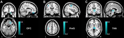 |
17 | Transcriptomic Signatures Associated With Gray Matter Volume Changes In Patients With Functional Constipation
Wangli Cai1, Guangyu Tang1, Jilei Zhang2, Ruiling Zhang1, and Lidi Wan1
1Shanghai Tenth People’s Hospital, Shanghai, China, 2Philips Healthcare, Shanghai, China
Functional constipation (FCon) is a common functional gastrointestinal disorder, with a considerable proportion of patients has anxiety and depression. Neuroimaging studies have shown that the functional/structural abnormalities across patients. We aimed to better understand the relationships between cortical atrophy and clinical observations in FCon, and its relationship with the underlying molecular mechanisms. Based on the densely sampled gene expression data from the Allen Human Brain Atlas, we conducted the transcription-neuroimaging association analysis and found genes associated with the central nervous system and the bowel to understand the molecular functions of brain regions that are vulnerable to cortical atrophy in FCon.
|
||
 Back to Meeting Home
 Back to the Program-at-a-Glance
The International Society for Magnetic Resonance in Medicine is accredited by the Accreditation Council for Continuing Medical Education to provide continuing medical education for physicians.
 Back to Meeting Home
 Back to the Program-at-a-Glance
 View the Presentation