Online Gather.town Pitches
Quantitative Neuroimaging & Neurofluids I
Joint Annual Meeting ISMRM-ESMRMB & ISMRT 31st Annual Meeting • 07-12 May 2022 • London, UK

Online Gather.town Pitches
Quantitative Neuroimaging & Neurofluids I
| Booth # | ||||
|---|---|---|---|---|
2937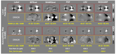 |
1 | A deep learning-based background field removal method for brains containing high susceptibility sources
Xuanyu Zhu1, Yang Gao1, Feng Liu1, Stuart Crozier1, and Hongfu Sun1
1University of Queensland, Brisbane, Australia
Background field removal (BFR) is a critical step in quantitative susceptibility mapping (QSM). Eliminating the background field in brains containing high susceptibility sources, such as intracranial hemorrhages, is challenging due to the relatively large scale of the local field induced from these sources. This study proposed a new deep learning-based method, "BFRnet", and compared it with several conventional BFR methods in processing two simulated and two in vivo brain datasets. The BFRnet method was effective in background field removal for acquisitions of arbitrary orientations and performed significantly better than other methods in the regions with high susceptibility sources.
|
||
2938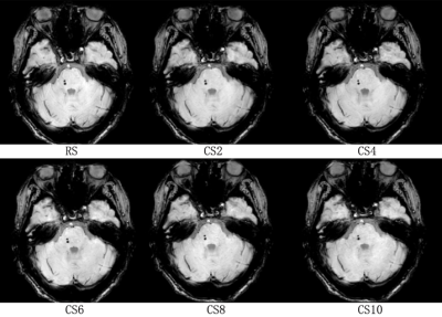 |
2 | Accelerating brain susceptibility weighted imaging using compressed-SENSE Video Not Available
Jinli Ding1, Liangjie Lin2, and Jiazheng Wang2
1Beijing Tiantan Hospital, Capital Medical University, Beijing, China, 2Philips Healthcare, Beijing, China
Susceptibility weighted imaging (SWI), acquiring both magnitude and phase information for tissues using a 3D gradient-recalled echo sequence, is advantageous to detect microhemorrhages and microvasculature and useful in diagnosing small vessel diseases etc., but can be suffering from the long acquisition time. This study evaluated the effectiveness of brain SWI accelerated by compressed SENSE (CS-SENSE), and the CS-SENSE factor 4 is recommended in routine practice. Besides, the CS-SENSE factor 10 maybe a fast surrogate for distinguishing CMBs but sacrificing SN-RN visualizations.
|
||
2939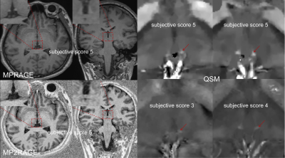 |
3 | Visualizing the habenula using high resolution MPRAGE, MP2RAGE and QSM at 3T MRI Video Not Available
BingYang Bian1, Lei Zhang1, Yueluan Jiang2, ZeChen Yu3, Zhuo Wang1, and HongChao Wang1
1The first hospital of Jilin University, changchun, China, 2MR Scientific Marketing, Siemens Healthineers Ltd., Beijing, China, 3MR CS APP, Siemens Healthineers Digital Technology Co., Harbin, China
MPRAGE, MP2RAGE and QSM can clearly visualize the Hb compared to conventional T1WI. Due to its higher CNR, MP2RAGE and QSM could be considered for structural imaging in Hb morphology study.
|
||
2940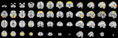 |
4 | Assessment of age-related changes of oxygen extraction fraction in of normal adults using QSM and quantitative BOLD
Jieyu Wang1, Junghun Cho2, Chen Zhang3, Yi Wang2, and Bing Yu4
1the Fourth Affiliated Hospital of China Medical University, Shenyang, China, 2Weill Cornell Medicine, New York, NY, United States, 3MR Scientific Marketing, Siemens Healthineers, Beijing, China, 4Shengjing Hospital of China Medical University, Shenyang, China
With aging, the brain undergoes comprehensive changes in its function and physiology, including oxygen consumption metabolism. In this study of healthy aging from 24-69, oxygen extraction fraction (OEF) mapping was generated using QSM and quantitative BOLD (QSM+qBOLD, QQ) processing of challenge-free 3D multi-echo gradient echo (mGRE). OEF values were found to increase with age only in specific gray matter brain regions (mainly in SomMot and SalVentAttn functional networks) and to decrease with age only in specific white matter brain regions (mainly Callosum ,cingulate,superior frontal blade).
|
||
2941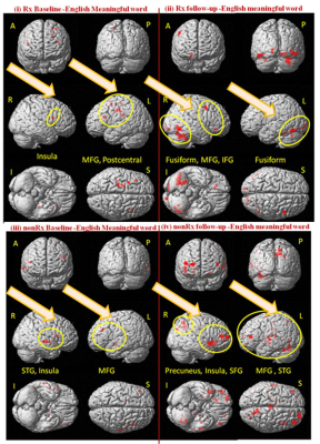 |
5 | Post-Therapy cortical normalization for phonological processing in developmental dyslexia Video Not Available
Sunita Gudwani1,2, Manju Mehta3, Rajesh Sagar4, Madhuri Behari5,6, Vaishna Narang7, Sadanad Sadanad Dwivedi8, N.R. Jagannathan2, and S.Senthil Kumaran9
1Dept. of NeuroRehab, speech therapy and audiology, Escorts Heart Institute and Research Center, New Delhi, India, 2Former Dept. of NMR & MRI Facility, All India Institute of Medical Sciences, New Delhi, India, 3Former Dept. of Psychiatry (Psychology Unit), All India Institute of Medical Sciences, New Delhi, India, 4Dept. of Psychiatry, All India Institute of Medical Sciences, New Delhi, India, 5Dept. of Neurology, Fortis Hospital, Vasant Kunj, New Delhi, India, 6Former Dept. of Neurology, All India Institute of Medical Sciences, New Delhi, India, 7Former Dept. of Linguistics, School of Language, Literature and Culture Studies, Jawaharlal Nehru University, New Delhi, India, 8Dept. of Biostatistics, All India Institute of Medical Sciences, New Delhi, India, 9Dept. of NMR & MRI Facility, All India Institute of Medical Sciences, New Delhi, India
Dyslexia is a neurodevelopmental disorder with complex spectrum and continuum of cognitive deficits leading to academic and emotional life-long burden, so optimal remediation becomes essential and challenging. Pre- and post- functional imaging studies show the recovery changes in brain with rehabilitation, but can fMRI biomarkers be explored and utilized for customizing therapeutic strategies in dyslexia. In present study at baseline we randomly allocated dyslexic children to therapy (Rx) and waitlist (nonRx) groups. Based on the potential (positive and negative) neural-markers at baseline, we tailored cognitive modules for phonological intervention. Remediation resulted in improved performance and cortical normalization
|
||
2942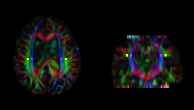 |
6 | Alterations in ALPS index and choroid plexus volume after lumboperitoneal shunt in patients with idiopathic normal pressure hydrocephalus
Junko Kikuta1, Koji Kamagata1, Toshiaki Taoka2, Kaito Takabayashi1, Wataru Uchida1, Akihiko Wada1, Kaito Kawamura3, Chihiro Akiba4, Madoka Nakajima3, Masakazu Miyajima4, Shinji Naganawa5, and Shigeki Aoki1
1Department of Radiology, Juntendo University Graduate School of Medicine, Bunkyo-ku, Japan, 2Department of Innovative Biomedical Visualization, Graduate School of Medicine, Nagoya University, Nagoya, Japan, 3Department of Neurosurgery, Juntendo University Faculty of Medicine, Bunkyo-ku, Japan, 4Department of Neurosurgery, Juntendo Tokyo Koto Geriatric Medical Center, Koto-ku, Japan, 5Department of Radiology, Nagoya University Graduate School of Medicine, Nagoya, Japan
Idiopathic normal pressure hydrocephalus (iNPH) is a condition resulting from impaired cerebrospinal fluid (CSF) absorption and excretion that is characterized by a triad of symptoms comprising cognitive decline, gait disturbance, and urinary incontinence. By improving CSF turnover through shunt surgery, symptoms of iNPH can become less severe. However, many mysterious points still exist in the mechanism of CSF dynamics in patients with iNPH. We examined alterations in ALPS index and choroid plexus volume after lumboperitoneal shunt (LPS) surgeries in patients with iNPH. Our results showed improvements in ALPS index and choroid plexus volume in these patients after LPS.
|
||
2943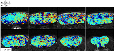 |
7 | Measurement of Cerebrospinal fluid and Interstitial Fluid Flow in Mouse Brain using Q-space Imaging
Satoshi Yatsushiro1,2, Anju Satou3, Mitsunori Matsumae4, Hideki Atsumi4, Tomohiko Horie5, and Kagayaki Kuroda1,3
1Human and Information Science, Tokai university, Hiratsuka, Kanagawa, Japan, 2BioView, Inc., Tokyo, Japan, 3Electrical and Electronic Engineering, Tokai university, Hiratsuka, Kanagawa, Japan, 4Neurosurgery, Tokai university, Isehara, Kanagawa, Japan, 5Radiological Technology, Tokai university hospital, Isehara, Kanagawa, Japan
To visualize cerebrospinal fluid (CSF) and interstitial fluid (ISF) flows, in this study, q-space imaging (QSI) based on 3-dimensional stimulated echo and echo planar imaging (3D STE-EPI) was performed for mouse brain. QSI can measure velocity of flow as µm/s order. The preliminary results showed the CSF and ISF flows in the mouse brain. In addition, the technique seemed to exhibit the CSF/ISF flows along the corpus callosum of the mouse brain. In the future, QSI with STE-EPI may quantitatively reveal the CSF and ISF flows in human brain.
|
||
2944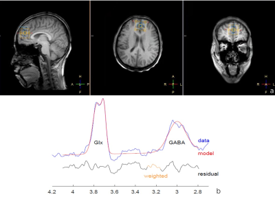 |
8 | Correlation between GABA+ levels in mPFC and topological characters of EEG functional connectivity network during light sleep in young adults
Sihui Zhao1, Yanting Liu1, Yanan Gao1, bing Yu2, Mikkelsen Mark3, Hui Steve3, Edden A.E Richard3, and Chen Zhang4
1China Medical University, Shenyang, China, 2Department of Radiology, Shengjing Hospital of China Medical University, Shenyang, China, 3Russell H. Morgan Department of Radiology and Radiological Science, The Johns Hopkins University School of Medicine, Baltimore, MD, USA F. M. Kirby Center for Functional Brain Imaging, Kennedy Krieger Institute, Baltimore, MD, United States, 4MR Scientific Marketing, Siemens Healthineers, Beijing, China
The aim of our study is to explore the relationship between GABA levels of medial prefrontal cortex (mPFC)and the topological characters of cerebral EEG functional connectivity network in young adults during sleep using Hadamard Encoding and Reconstruction of Mega-Edited Spectroscopy (HERMES) method. Synchronous EEG-MRS data were acquired from 37 young adults. GABA+ level in mPFC and the global topological characters of the EEG functional connectivity network were calculated. The results demonstrated a significant negative correlation between global efficiency of EEG functional connectivity network in delta bandand and GABA+ levels in mPFC during N2 sleep stage.
|
||
2945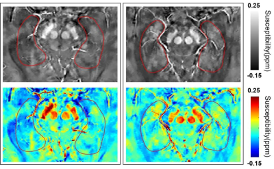 |
9 | Increased Brain Iron Deposition in Moderate and Severe Obstructive Sleep Apnea using Quantitative Susceptibility Mapping
Xinchun Li1, Peng Wang1, Qi Wan1, Yihao Guo2, Jianfeng Hu1, Yu Peng1, Xiaoying Xia1, Jieqiong Liu1, Xiaobin Xie1, and Mengzhu Wang3
1Department of Radiology, The First Affiliated Hospital of Guangzhou Medical University, Guangzhou, China, 2MR Collaboration, Siemens Healthineers Ltd., Guangzhou, China, 3MR Scientific Marketing, Siemens Healthineers, Guangzhou, China In the current study, we assessed the possible alterations of iron accumulation in the brain of patients with Obstructive Sleep Apnea (OSA) using quantitative susceptibility mapping (QSM). We found that there was increased iron accumulation in the bilateral hippocampus for patients with OSA, compared to controls. And the QSM value of the bilateral hippocampus was related to the course of disease in patients with OSA. The QSM values of the right hippocampus were correlated with multiple clinical indicators and may therefore be used as potential biomarkers to further understand the pathophysiological mechanisms of moderate to severe OSA. |
||
2946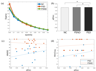 |
10 | Surface-based Cortical Network Analysis in Post-Stroke Dementia with Subcortical lesions Video Permission Withheld
Wenwen Li1, Huaying Cai2, Linhui Ni2, Guocan Han2, Zhiyong Zhao1, and Dan Wu1
1Key Laboratory for Biomedical Engineering of Ministry of Education, Department of Biomedical Engineering, College of Biomedical Engineering & Instrument Science, Zhejiang University, Hangzhou, China, 2Department of radiology, Sir Run Run Shaw Hospital, School of Medicine, Zhejiang University, Hangzhou, China
This study aimed to investigate alterations of cortical network in post-stroke dementia (PSD) patients with subcortical lesions using a surface-based morphometry analysis. We calculated nine cortical morphometric metrics and constructed the structural covariate network for each participant. Comparisons between groups showed that PSD and post-stroke nondemented (PSND) groups both displayed an increased local efficiency and a similar global efficiency compared with the normal controls group, and the cortical network in three groups all remained a small-world property. Moreover, we found PSD-specific decreases in gray matter volume and surface area in lateral occipital cortex and middle temporal gyrus.
|
||
2947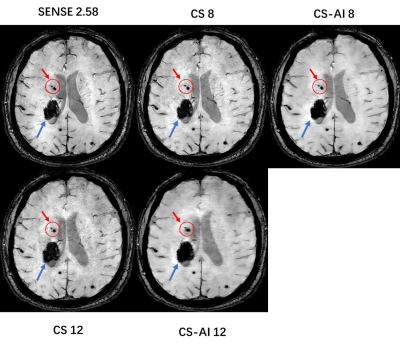 |
11 | Clinical Feasibility of Compressed SENSE Artificial Intelligence Accelerated SWI in Cerebral Hemorrhage Imaging
Zhenzhen Shi1, Cong Ning1, Chang Zhai1, Dan Tong1, Yi Zhu2, and Ke Jiang2
1Radiology, The First Hospital Of Jilin University, Changchun, China, 2Philips Healthcare, Beijing, China
Susceptibility weighted imaging (SWI) is more sensitive than T2* gradient echo MRI in detecting intracranial hemorrhage, but its acquisition time is too long. The purpose of this study is to prospectively assess the clinical feasibility of Compressed SENSE Artificial Intelligence (CS-AI) SWI by comparing it with SENSE SWI and Compressed SENSE (CS) SWI. The results suggested that the CS-AI SWI with an acceleration factor 8 was able to reduce the acquisition time of SWI by 74.58% while maintaining the image quality and accuracy of the diagnosis of intracerebral hemorrhage, except for the diagnosis of cerebral microbleeds.
|
||
2948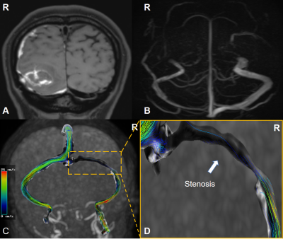 |
12 | A preliminary study of hemodynamic assessments of cerebral venous sinus stenosis using 4D flow MRI Video Not Available
hongwei zhou1, kexin zhao1, derui kong1, chong zheng1, yueluan jiang2, and xiaoye wang3
1the First Hospital of Jilin University, Chang Chun, China, 2MR Scientific Marketing, Siemens Healthineers, Bei Jing, China, 3MR Clinical Marketing, Siemens Healthineers Ltd, Bei Jing, China
4D flow MRI provides detailed insight into the alterations of hemodynamic,including quantitative assessment of flow parameters for the venous system. This technique overcomes the missing hemodynamic information of standard magnetic resonance imaging and the invasive disadvantage of digital subtraction angiography (DSA). Through data visualization and quantification, the particle tracking map is obtained and the peak velocities, wall shear stress (WSS) is calculated. The dynamics of venous sinus stenosis will change especially in severe area.
|
||
2949 |
13 | Effect of Dobutamine Stress on Ocular Blood Flow Measurements Using 3D Pseudocontinuous Arterial Spin Labeling Video Not Available
linkun cai1, haijun niu1, pengling ren2, yawen liu1, tingting zhang2, jing li2, hao wang2, wei zheng3, hongxia yin2, and zhenchang wang1
1Beijing Advanced Innovation Center for Biomedical Engineering, School of Biological Science and Medical Engineering, Beihang University, Beijing, China, 2Beijing Friendship Hospital, Beijing, China, 3Chinese Academy of Sciences, Beijing, China
Dobutamine stress testing is a very widely used tool in cardiological investigation. However, little is known about the effect of dobutamine on adult ocular blood flow with clinical dosage. In this study, we infused dobutamine in human subjects and examined the effects of dobutamine on ocular blood flow using 3D pseudocontinuous arterial spin labeling(3D-pcASL). Dobutamine infusion significantly increased the ocular blood flow. And the heart rate and diastolic blood pressure were the main factors affecting the ocular blood flow.
|
||
2950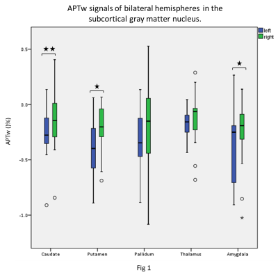 |
14 | Subcortical Gray Matter Nucleus Lateralization of Amide Proton Transfer Weighted Signals in Young Healthy Subjects Video Permission Withheld
Yuhan Jiang1, Yangyingqiu Liu2, Bingbing Gao2, Qingwei Song2, and Yanwei Miao2
1Radiology, the First Affiliated Hospital of Dalian Medical University, Dalian, China, 2the First Affiliated Hospital of Dalian Medical University, Dalian, China
Amide proton transfer weighted (APTw) imaging is a novel molecular imaging technique to acquire the proton exchanged signals from the amide in proteins or peptides to water. Previous studies discovered lateralization in the brain structure and function, including the subcortical gray matter nucleus. However, it is not clear whether APTw also has lateral advantage. In this study, we applied the automatic brain segmentation method to quantify the APTw signal values of subcortical gray matter nucleus in young healthy subjects. The increased APTw signals were present in the right side of the subcortical gray matter nucleus,which may suppose related with right-handedness.
|
||
 Back to Meeting Home
 Back to the Program-at-a-Glance
The International Society for Magnetic Resonance in Medicine is accredited by the Accreditation Council for Continuing Medical Education to provide continuing medical education for physicians.
 Back to Meeting Home
 Back to the Program-at-a-Glance
 View the Presentation