Online Gather.town Pitches
Neurodegeneration: Aging, AD & MS
Joint Annual Meeting ISMRM-ESMRMB & ISMRT 31st Annual Meeting • 07-12 May 2022 • London, UK

Online Gather.town Pitches
Neurodegeneration: Aging, AD & MS
| Booth # | ||||
|---|---|---|---|---|
3954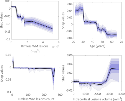 |
1 | A modern machine-learning approach to reveal the role of multiple sclerosis lesions in inducing cortical tissue loss Video Permission Withheld
Constantina Treaba1, Allegra Conti2, Ambica Mehndiratta1, Valeria Barletta1,3, Caterina Mainero1,3, and Nicola Toschi1,2
1Radiology, Massachusetts General Hospital; A. Martinos Center for Biomedical Imaging, Boston, MA, United States, 2Department of Biomedicine and Prevention, University of Rome “Tor Vergata", Rome, Italy, 3Harvard Medical School, Boston, MA, United States
In multiple sclerosis (MS), cortical atrophy could result from extensive axonal transection from distant white matter lesion and/or local cortical demyelination. The destructive potential of MS white matter lesions, however, seem to differ across lesion types, being higher for chronic active lesions detectable on susceptibility weighted images by their characteristic paramagnetic rim. Using 7T MRI that has shown increased sensitivity to cortical lesions and modern machine learning algorithms, we demonstrate that both white matter and cortical lesions are main determinants of cortical thinning. Despite their destructive capacity, chronic active lesions are not major contributors of cortical tissue loss.
|
||
3955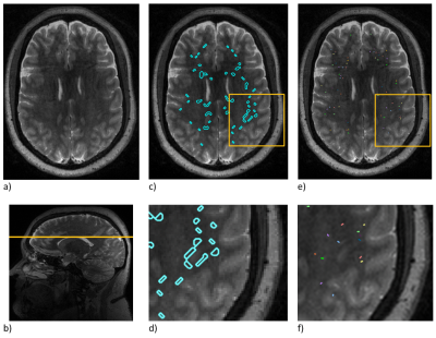 |
2 | Evaluation of automatic segmentation of perivascular spaces in multiple sclerosis patients and healthy controls on 3T MRI
Joan Brewer1, Ilena George2, Gale Edison1, Joy Zhang2, Sarah King2, James Sumowski2, Priti Balchandani2, Gaurav Verma2, Rebecca Feldman1, and Sam Horng2
1University of British Columbia Okanagan, Kelowna, BC, Canada, 2Icahn School of Medicine at Mount Sinai, New York, NY, United States Multiple Sclerosis (MS) is an autoimmune disease; disease can be measured on MRI by the appearance or enlargement of lesions. PVS measurements on MRI may represent a more predictive biomarker of disease. Because manual measurement of PVSs is time intensive, our collaborators developed a tool to automatically segment PVSs. Here, we investigated the utility of the automatic segmentations. We correlated the automated PVS counts with manual segmentations (0.66), as well as the PVS areas (0.55). Differences between healthy control and MS groups in this preliminary analysis were not statistically significant. Future work will expand from 45 to 300 patients. |
||
3956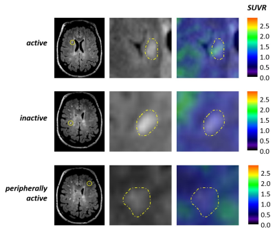 |
3 | White matter inflammation and its relation to myelin content in vivo in patients at different stages of multiple sclerosis.
Valeria Barletta1, Elena Herranz1, Constantina Andrada Treaba1, Ambica Mehndiratta2, Russell Ouellette3, Tobias Granberg4, Eric Klawiter5, Carolina Ionete6, Jacob Sloane7, and Caterina Mainero1
1Department of Radiology, Massachusetts General Hospital, Harvard Medical School, Athinoula A. Martinos Center for Biomedical Imaging, Boston, MA, United States, 2Department of Radiology, Massachusetts General Hospital, Athinoula A. Martinos Center for Biomedical Imaging, Boston, MA, United States, 3Karolinska Institutet, Department of Clinical Neuroscience, Stockholm, Sweden, 4Karolinska University Hospital, Department of Neuroradiology, Stockholm, Sweden, 5Department of Neurology, Massachusetts General Hospital, Boston, MA, United States, 6Umass Memorial Medical Center, Worcester, MA, United States, 7Department of Neurology, Beth Israel Deaconess Medical Center, Boston, MA, United States
We combined 11C-PBR28 magnetic resonance-positron emission tomography, marking activated microglia, with synthetic MRI to measure myelin content, on a group of 33 patients affected by multiple sclerosis, to quantify neuroinflammation in the brain white matter and assess its relation to myelin content and disease burden. Microglia activation was higher in the white matter of multiple sclerosis patients compared to 16 healthy volunteers. Microglia activation within perilesional WM correlated the most with worse disease outcomes. Peripherally active lesions were related to higher disability and progressive disease. Perilesional myelin content was lower for higher inflammation and correlated with disease burden.
|
||
3957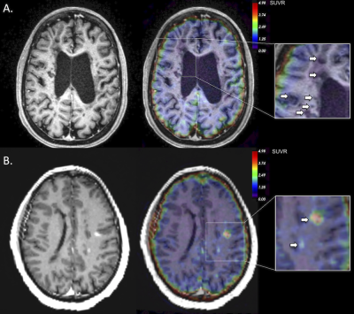 |
4 | Initial in vivo evidence of fibrin deposition in multiple sclerosis patients using fibrin-targeted 64Cu-FBP8 positron emission tomography Video Permission Withheld
Constantina Treaba1, Ciprian Catana1,2, Eric Klawiter2,3, Susie Huang1,2, Grae Arabasz1, Valeria Barletta1,2, Elena Herranz1,2, Jacob Sloane4, Peter Caravan1,2, and Caterina Mainero1,2
1Radiology, Massachusetts General Hospital; A. Martinos Center for Biomedical Imaging, Boston, MA, United States, 2Harvard Medical School, Boston, MA, United States, 3Neurology, Massachusetts General Hospital, Boston, MA, United States, 4Neurology, Beth Israel Deaconess Medical Center, Boston, MA, United States
In multiple sclerosis, experimental and histopathological studies point to fibrinogen as a link between a damaged brain blood barrier and the initiation of the inflammatory demyelination and neurodegeneration in both cortex and white matter. Nevertheless, although the widespread accumulation of fibrinogen as fibrin deposits within multiple sclerosis lesions is well documented on postmortem brain tissue, in vivo evidence is still lacking. Using a novel fibrin-specific positron emission tomography probe, 64Cu-FBP8, we were able to demonstrate in vivo fibrin deposition not only in active white matter multiple sclerosis lesions but also in the cortex of a few multiple sclerosis patients.
|
||
3958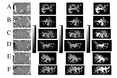 |
5 | Quantitative T2* and T1 mapping of post-mortem MS tissue at 7T for discrimination of normal-appearing and dirty-appearing white matter pathology
Risavarshni Thevakumaran 1,2, Adam Groh 3,4, Jo Anne Stratton3,4, and David Rudko 1,2,3
1Biological and Biomedical Engineering, McGill University, Montreal, QC, Canada, 2McConnell Brain Imaging Centre, Montreal, QC, Canada, 3Neurology and Neurosurgery, McGill University, Montreal, QC, Canada, 4Montreal Neurological Institute-Hospital, Montreal, QC, Canada
Quantitative T2* and T1 relaxometry metrics calculated at 7T have high sensitivity to myelin and can be jointly used to identify dirty-appearing white matter (DAWM) and normal-appearing white matter (NAWM) pathology in post-mortem, multiple sclerosis (MS) brain tissue. Relaxometry mapping was performed on fixed, cerebral brain samples from MS patients and healthy donors. T2* and T1 distributions in WM from MS tissue exhibited bimodality and were shifted to higher relaxation time values compared to healthy tissue. Using k-means clustering applied to 2D T2* and T1 MS tissue data, regions of DAWM were detected in periventricular WM for MS tissue samples.
|
||
3959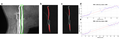 |
6 | Evaluation of the robustness of carotid pulse wave velocity measurement using a single-slice oblique-sagittal phase-contrast MRI
Jianing Tang1, Soroush Heidari Pahlavian1, Helena Chui2, and Lirong Yan1,2
1USC Mark and Mary Stevens Neuroimaging and Informatics Institute, Keck School of Medicine, University of Southern California, Los Angeles, CA, United States, 2Department of Neurology, Keck School of Medicine, University of Southern California, Los Angeles, CA, United States
Characterization of cerebral vascular compliance (VC) could offer more insights into the vascular contribution to cognitive impairment. Cerebral VC can be measured using ASL methods but with a long scan time. A recent study introduced a fast MRI technique to assess global cerebral VC by measuring carotid pulse wave velocity (cPWV) using a single-slice oblique-sagittal PC-MRI. In this study, the robustness of cPWV measurement to the variations of ROI selection has been demonstrated. The results from a pilot study on an aged cohort suggests elevated cPWV is associated with cognitive decline and could be a potential marker for cognitive impairment.
|
||
 |
3960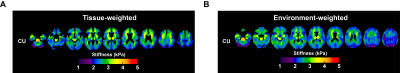 |
7 | Differential Effect of Dementia Etiology on Cortical Stiffness as Assessed by MR Elastography
KowsalyaDevi Pavuluri1, Jonathan M. Scott2, John Huston III1, Richard L. Ehman1, Armando Manduca1,3, Clifford R. Jack1, Rodolfo Savica4, Bradley F Boeve5, Kejal Kantarci1, Ronald C. Petersen4, and Matthew C. Murphy1
1Department of Radiology, Mayo Clinic, Rochester, MN, United States, 2Mayo Clinic Medical Scientist Training Program, Mayo Clinic, Rochester, MN, United States, 3Department of Physiology and Biomedical Engineering, Mayo Clinic, Rochester, MN, United States, 4Department of Neurology, Mayo Clinic, Rochester, MN, United States, 5Division of Pulmonary and Critical Care Medicine, Mayo Clinic, Rochester, MN, United States
Dementia is a progressive neurodegenerative syndrome characterized by impairment in memory and activities of daily living, altered behavior, personality, and other cognitive dysfunctions. Around 50 million people have dementia worldwide, and there are nearly 10 million new cases every year. MR elastography is a non-invasive imaging technique to measure the mechanical properties of tissues and has demonstrated sensitivity to neurodegenerative processes. In this study we utilized two neural network inversions to investigate the viscoelastic property changes in cortical regions with both tissue- and environment-weighting in various etiologies of dementia including Alzheimer’s, frontotemporal dementia, and dementia with Lewy bodies.
|
|
3961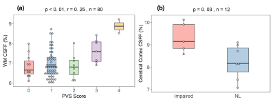 |
8 | MRI-based parenchyma CSF fraction (CSFF) mapping is a potential biomarker of brain drainage function: a multimodal imaging study
Liangdong Zhou1, Thanh Nguyen1, and Yi Li1
1Radiology, Weill Cornell Medicine, New York, NY, United States
We demonstrated that, our proposed T2 relaxometry-based free water mapping, CSFF, could be an imaging biomarker of dilated PVS and CSF clearance function by correlating it with multiple brain clearance-related measures including PVS score, PC-MRI based aqueduct CSF flow, dynamic PET based ventricle CSF clearance, amyloid beta and tau deposits. Our results show that CSFF is positively associated with PVS score, aging, amyloid beta and tau deposits. And it negatively correlates with net aqueduct CSF flow, ventricle CSF flow, and cognitive score.
|
||
3962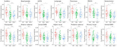 |
9 | Resting-state functional networks in normal aging and asymptomatic Alzheimer's disease
Junjie Wu1, Qixiang Lin2, Benjamin B. Risk3, Aditya S. Bisht2, David W. Loring2, Felicia C. Goldstein2, Allan I. Levey2, James J. Lah2, and Deqiang Qiu1,4
1Department of Radiology and Imaging Sciences, Emory University School of Medicine, Atlanta, GA, United States, 2Department of Neurology, Emory University School of Medicine, Atlanta, GA, United States, 3Department of Biostatistics and Bioinformatics, Emory University Rollins School of Public Health, Atlanta, GA, United States, 4Joint Department of Biomedical Engineering, Emory University and Georgia Institute of Technology, Atlanta, GA, United States
We systematically studied alterations in 14 resting-state functional networks due to normal aging and AD pathology in the asymptomatic phase, as well as their relationship with CSF biomarkers for AD and neuropsychological assessments. We found disrupted functional connectivity in most of the functional networks with normal aging and compromised left executive control network (LECN) due to asymptomatic AD pathology. Furthermore, LECN is associated with overall cognition. Tau closely correlates with functional networks in asymptomatic AD.
|
||
3963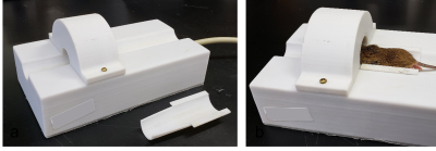 |
10 | Studying cerebrospinal fluid bulk flow in mice using high-resolution dynamic macromolecular gadolinium enhanced MRI on a whole-body 3T system
Wingchi Edmund Kwok1, Molly Brady2, Akib Rahman3, Alexander Solorzano3, Ronald Wood3, and Rashid Deane3
1Imaging Sciences, University of Rochester Medical Center, Rochester, NY, United States, 2Tufts University, Boston, MA, United States, 3University of Rochester, Rochester, NY, United States
Study of CSF flow can be used to assess CSF clearance pathways for the better understanding of neurological diseases, such as Alzheimer's disease. We developed a technique for macromolecular gadolinium enhanced dynamic MRI of CSF in mice on a whole-body 3T system. A specially designed 4-channel RF receive coil was developed, and a high-resolution 3D spoiled gradient-echo T1 sequence was used. Gadolinium-albumin contrast was injected into the cisterna magna. Contrast enhancement was detected in various locations including the olfactory lobe, nasal cavity, lymphatic system and spine cord. Lower contrast elimination rates were observed in old mice than in young mice.
|
||
3964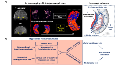 |
11 | In vivo mapping of hippocampal venous vasculature and oxygen saturation using dual-echo SWI/QSM on 7T: a potential marker for neurodegeneration
Chenyang Li1, Li Jiang1, Marco Muccio1, Sagar Buch2, Hanzhang Lu3, E.Mark Haacke2, and Yulin Ge1
1Department of Radiology, NYU Grossman School of Medicine, New York, NY, United States, 2Department of Radiology, Wayne State University School of Medicine, Detroit, MI, United States, 3Department of Radiology and Radiological Science, Johns Hopkins University School of Medicine, Baltimore, MD, United States
In this study, we reconstructed the venous drainage course of Basal vein of Rosenthal (BVR) and its tributaries using dual-echo SWI/QSM. Consistent with existing knowledge of venous anatomy, our results show that BVR drains blood from multiple tissue structures such as insula, amygdala and hippocampus. Inferior ventricular vein (IVV) and medial atrial vein (MAV), two tributaries of BVR, are anatomically relevant to the venous drainage in hippocampus. With the proposed technique, we extracted the venous blood susceptibility value in BVR and IVV/MAV to examine its feasibility in characterizing changes of venous oxygenation level related to hippocampal neurodegeneration.
|
||
 Back to Meeting Home
 Back to the Program-at-a-Glance
The International Society for Magnetic Resonance in Medicine is accredited by the Accreditation Council for Continuing Medical Education to provide continuing medical education for physicians.
 Back to Meeting Home
 Back to the Program-at-a-Glance
 View the Presentation