Online Gather.town Pitches
Multiple Sclerosis, Alzheimer's & Dementia I
Joint Annual Meeting ISMRM-ESMRMB & ISMRT 31st Annual Meeting • 07-12 May 2022 • London, UK

Online Gather.town Pitches
Multiple Sclerosis, Alzheimer's & Dementia I
| Booth # | ||||
|---|---|---|---|---|
3252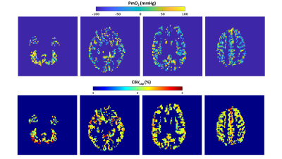 |
1 | Evidence from dual-calibrated fMRI for raised mitochondrial oxygen tension in the MS brain
Antonio Maria Chiarelli1, Eleonora Patitucci2, Hannah Chandler2, Valentina Tomassini1,3,4,5, Michael Germuska2,6, and Richard Wise1,2
1Department of Neuroscience, Imaging and Clinical Sciences, University G. D'Annunzio of Chieti Pescara, Chieti Scalo, Italy, 2CUBRIC, School of Psychology, Cardiff University, Cardiff, United Kingdom, 3Helen Durham Centre for Neuroinflammation, University Hospital of Wales, Cardiff, United Kingdom, 4MS Centre, Neurology Unit, “SS. Annunziata” University Hospital, Chieti, Italy, 5Division of Psychological Medicine and Clinical Neurosciences, University Hospital of Wales, Cardiff, United Kingdom, 6School of Physics and Astronomy, Cardiff University, Cardiff, United Kingdom
Dysfunction of energy supply or usage may be present in Multiple Sclerosis. We investigated the use of a simple oxygen diffusion model to infer mitochondrial oxygen tension from dual-calibrated fMRI (dc-fMRI) data. We observed a significant reduction of grey matter CBF and CMRO2 in people with MS but no significant difference in OEF or BOLD-sensitive blood volume. Assuming no substantial tissue or vascular remodelling in MS, these results imply, within a simple flow-diffusion model of oxygen from capillaries into the tissue, an elevated partial pressure of oxygen at the mitochondria which may indicate mitochondrial dysfunction.
|
||
3253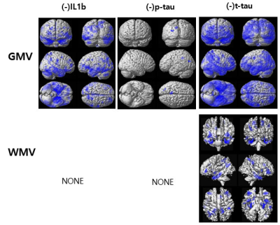 |
2 | Association of Blood Biomarkers and Brain Tissue Volume Loss in Patients of Mild Cognitive Impairment and Alzheimer’s Disease
Geon-Ho Jahng1, Kyung Mi Lee2, Sang Tae Kim3, Soonchan Park1, Sue Min Jung4, Hak Young Rhee5, Chang-Woo Ryu1, and Eui Jong Kim2
1Radiology, Kyung Hee University Hospital at Gangdong, Seoul, Korea, Republic of, 2Radiology, Kyung Hee University Hospital, Seoul, Korea, Republic of, 3Surgery & Neurology, Bundang Hospital, Seongnam city, Korea, Republic of, 4Biomedical Engineering, Kyung Hee University, Yongin-si, Korea, Republic of, 5Neurology, Kyung Hee University Hospital at Gangdong, Seoul, Korea, Republic of
To report the values of a battery of blood inflammatory biomarkers from cognitive normal (CN) to Alzheimer’s disease (AD) in our reasonably sized patient cohort and to evaluate the association between gray matter volume (GMV) loss in AD and plasma levels of blood biomarkers, we included 38 elderly CN elderly, 40 mild cognitive impairment (MCI), and 33 AD subjects and obtained blood biomarkers and GMV. We found that GMV was negatively associated with the levels of IL1b, P-tau, and T-tau. There were no significant associations between brain tissue volumes and the levels of mAβ, NLRP3, miR155, oAβ, and Nogo-A.
|
||
3254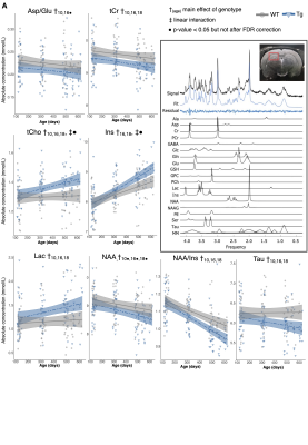 |
3 | Neurochemical and cognitive changes precede structural abnormalities in the TgF344-AD rat model of Alzheimer’s disease
Caitlin F Fowler1,2, Dana Goerzen2, Gabriel A Devenyi2,3, Dan Madularu4, M Mallar F Chakravarty1,2,3, and Jamie Near2,5
1Biological and Biomedical Engineering, McGill University, Montreal, QC, Canada, 2Cerebral Imaging Centre, Douglas Mental Health University Institute, Verdun, QC, Canada, 3Psychiatry, McGill University, Montreal, QC, Canada, 4Center for Translational Neuroimaging, Northeastern University, Boston, MA, United States, 5Physical Studies Research Platform, Sunnybrook Research Institute, Toronto, ON, Canada
Alzheimer’s disease is a progressive neurodegenerative disorder with a decades-long pre-symptomatic phase, substantiating the need for prodromal biomarker development and early intervention. We employed longitudinal magnetic resonance imaging and spectroscopy in conjunction with behavioural testing to characterize the chronological order of appearance and progression of multiple facets of disease pathology in the TgF344-AD rat model. The TgF344-AD rat demonstrated impaired spatial reference memory by 4 months of age, followed by neurochemical abnormalities by 10 months and major structural changes by 16 months, most of which recapitulated documented changes in brain structure and tissue chemistry in human Alzheimer’s disease patients.
|
||
3255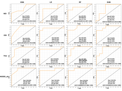 |
4 | Machine learning to detect microstructural brain changes in patients with amnestic mild cognitive impairment based on NODDI
Xiuwei Fu1, Yu Zhang2, Tongtong Li2, Yuanyuan Chen3, Xianchang Zhang4, and Hongyan Ni5
1Department of Radiology, Tianjin Medical University General Hospital, Tianjin, China, 2Department of Radiology, First Central Clinical institution, Tianjin Medical University, Tianjin, China, 3Institute of Medical Engineering and Translational Medicine, Tianjin University, Tianjin, China, 4MR Collaboration, Siemens Healthineers Ltd., Beijing, China, 5Department of Radiology, Tianjin First Central Hospital, Tianjin, China This study investigated the changes in brain microstructure in patients with amnestic mild cognitive impairment (aMCI) using neurite orientation dispersion and density imaging (NODDI) combined with machine learning. Neurite density index (NDI) was significantly decreased in white matter, orientation dispersion index (ODI) was significantly decreased in gray matter, and volume fraction of isotropic water molecules (Viso) was significantly increased in the aMCI group. Further correlation and receiver operating characteristic (ROC) curve analyses showed NODDI may reflect the clinical cognitive status of aMCI. NODDI combined with a machine learning algorithm could be a promising alternative for early diagnosis of MCI. |
||
3256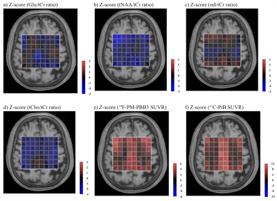 |
5 | Glutamatergic dysfunction associated with tau depositions in Alzheimer’s disease
Kiwamu Matsuoka1,2, Kosei Hirata1, Naomi Kokubo1, Kenji Tagai1, Hironobu Endo1, Keisuke Takahata1, Hitoshi Shinotoh1, Maiko Ono1, Chie Seki1, Kazunori Kawamura3, Ming-Rong Zhang3, Hitoshi Shimada1,4, Yuhei Takado1, and Makoto Higuchi1
1Department of Functional Brain Imaging, Institute for Quantum Medical Science, Quantum Life and Medical Science Directorate, National Institutes for Quantum Science and Technology, Chiba-shi, Japan, 2Department of Psychiatry, Nara Medical University, Kashihara-shi, Japan, 3Department of Advanced Nuclear Medicine Sciences, Institute for Quantum Medical Science, Quantum Life and Medical Science Directorate, National Institutes for Quantum Science and Technology, Chiba-shi, Japan, 4Department of Functional Neurology & Neurosurgery, Center for Integrated Human Brain Science, Brain Research Institute, Niigata University, Niigata-shi, Japan
Glutamatergic neurons and cingulate cortices have crucial roles in the cognitive dysfunction of Alzheimer's disease (AD). This study aimed to evaluate regional vulnerabilities of the glutamatergic system in AD at the level of the cingulate gyrus in relation to tau and amyloid-β (Aβ) depositions. Combining MRSI and PET, we found that the glutamatergic system in the posterior cingulate cortex (PCC) is vulnerable to tau deposits but not Aβ from the early stage of AD, and glutamate in the PCC region may be a marker of disease progression in AD.
|
||
3257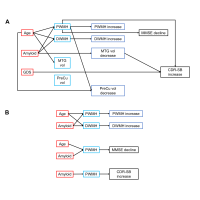 |
6 | Brain structure, amyloid and behavioural features for predicting clinical progression in subjective cognitive decline
Siwei Liu1, Xiao Luo2, Yeerfan Jiaerken2, Joanna Su Xian Chong1, Hee Youn Shim1, Minming Zhang2, and Juan Helen Zhou1
1National University of Singapore, Singapore, Singapore, 2Second Affiliated Hospital, Zhejiang University School of Medicine, Hangzhou, China Subjective cognitive decline (SCD) is a risk factor for dementia. However, multiple pathologies, including amyloid deposition, cerebrovascular pathology, and depression, contribute to the heterogeneity in SCD. We included 170 non-demented elderly with 2-years follow-up to examine brain structural abnormalities in SCD. We found progressive and stable SCD individuals differed in deep white matter hyperintensities and temporoparietal grey matter atrophy. We used multi-model brain and behavioural factors to predict cognitive impairment and dementia progression. Periventricular white matter hyperintensities mediated the effect of amyloid accumulation on cognitive decline and disease severity, while depressive symptoms directly predicted disease severity. |
||
3258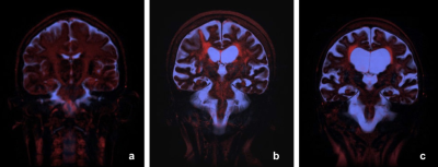 |
7 | Observing Clearance Pathway in Alzheimer Patients Using T2 Component Analysis
Koichi Oshio1, Masao Yui2, Seiko Shimizu2, Shinya Yamada3,4, Madoka Nakajima4, Chihiro Akiba5, Hideki Bandai5, and Masakazu Miyajima6
1Department of Radiology, Juntendo University, Tokyo, Japan, 2Canon Medical Systems Corporation, Otawara, Japan, 3Neurosurgery, Kugayama Hospital, Tokyo, Japan, 4Department of Neurosurgery, Juntendo University, Tokyo, Japan, 5Department of Neurosurgery, Juntendo Tokyo Koto Geriatric Medical Center, Tokyo, Japan, 6Department of Neurosurgery, Junterndo Tokyo Koto Geriatric Medical Center, Tokyo, Japan
A method to visualize possible waste clearance pathway in the brain was proposed earlier. The method is based on an assumption that there exist water containing macromolecules along the clearance pathway, and this water can be visualized using T2 component analysis. Preliminary results of this method applied for Alzheimer patients, as well as healthy volunteers, are presented. Distinct differences between the patients and healthy volunteers were observed. It is expected that this method can be a powerful tool for understanding the patho-physiology of the Alzheimer disease.
|
||
3259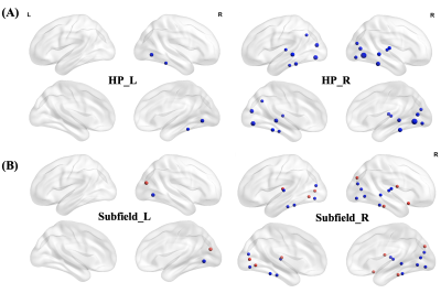 |
8 | Hippocampal subfield-specific alterations in post-stroke dementia with subcortical lesion Video Not Available
Yihan Wu1,2, Huaying Cai3, Linhui Ni3, Guocan Han4, Zhiyong Zhao1, and Dan Wu1
1Key Laboratory for Biomedical Engineering of Ministry of Education, Department of Biomedical Engineering, College of Biomedical Engineering & Instrument Science, Zhejiang University, Hangzhou, China, Hangzhou, China, 2College of Biosystems Engineering and Food Science, Zhejiang University, Hangzhou, China, Hangzhou, China, 3Department of Neurology, Neuroscience Center, Sir Run Run Shaw Hospital, Zhejiang University, Hangzhou, China,, Hangzhou, China, 4Department of Radiology, Neuroscience Center, Sir Run Run Shaw Hospital, Zhejiang University, Hangzhou, China, Hangzhou, China The present study aimed to explore functional and structural alterations in hippocampus and its subfields in post-stroke dementia (PSD). We collected resting-state fMRI and DTI data from 24 PSD patients, 36 post-stroke nondemented (PSND) patients and 21 normal controls (NC). Comparisons between groups found that PSD patients showed subfield-specific changes in the microstructures of hippocampus compared with the other two groups. Moreover, we not only found more PSD-related changes of functional connectivity with cortical regions in the subfields than entire hippocampus, but also revealed the different contributions of subfields on the connectivity changes of entire hippocampus. |
||
3260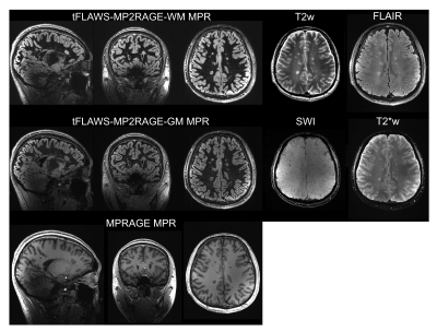 |
9 | Improved Multiple Sclerosis Lesion Visualization using Tailored FLAWS-MP2RAGE at 7T
Zhe Zhang1,2, Chenyang Gao3, Wanlin Zhu1,2, Yingkui Zhang1,2, Qingle Kong4, Decai Tian3, Jing Jing1,2, and Yongjun Wang1,2,3
1Tiantan Neuroimaging Center of Excellence, Beijing Tiantan Hospital, Capital Medical University, Beijing, China, 2China National Clinical Research Center for Neurological Diseases, Beijing, China, 3Department of Neurology, Beijing Tiantan Hospital, Capital Medical University, Beijing, China, 4MR Collaboration, Siemens Healthineers Ltd., Beijing, China
Accurate visualization and evaluation of the MS lesions is an essential step for clinical practice. FLAWS-MP2RAGE has been proposed and applied at ultra-high field to better visualize both white matter and gray matter lesions. This work proposed the optimized method, tailored FLAWS(tFLAWS)-MP2RAGE, using a new image combination method for better lesion contrast. Results of MS patients showed higher lesion CNR of tFLAWS-MP2RAGE compared with conventional FLAWS-MP2RAGE and MPRAGE methods. This method might be a potential tool for lesion detection and evaluation at UHF for MS and other brain diseases.
|
||
3261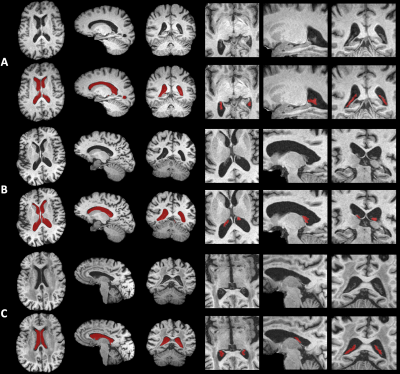 |
10 | The Contribution of Increased Choroid Plexus Volume in the Alzheimer’s Disease
Jiaxin Li1, Yueqin Hu2, Xue Feng 3, Weiying Dai4, Craig H. Meyer3, David C. Alsop5, and Li Zhao1
1College of Biomedical Engineering & Instrument, Zhejiang University, Hangzhou, China, 2Psychology, Beijing Normal University, Beijing, China, 3Biomedical Engineering, University of Virginia, Charlottesville, VA, United States, 4Department of Computer Science, State University of New York at Binghamton, Binghamton, NY, United States, 5Radiology, Beth Israel Deaconess Medical Center and Harvard Medical School, Boston, MA, United States
Alzheimer’s disease (AD) may be caused by the dysfunction of glymphatic system, in which anatomic changes of the choroid plexus may be associated with reduced CSF production. Our previous work showed significantly increased choroid plexus volume in AD patients compared to healthy controls. However, contributions of the lateral ventricle volume were not investigated. Here, the lateral ventricle and choroid plexus were segmented using two cascaded deep learning models and the volumes were measured on 733 subjects. Increased lateral ventricle volume was found in AD patients, similar to previous studies. More importantly, choroid plexus volume showed a unique contribution to AD.
|
||
3262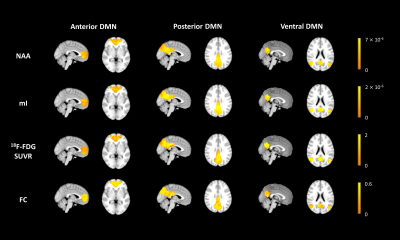 |
11 | Coupled Metabolic and Functional Changes in Default Mode Network Subsystems of Alzheimer's Disease: A Hybrid 3D 1H-MRSI/FDG-PET/fMRI Study
Wenli Li1, Miao Zhang2, Rong Guo3,4, Yudu Li3,4, Yibo Zhao3,4, Jialin Hu1, Yaoyu Zhang1, Zhi-Pei Liang3,4, and Yao Li1
1School of Biomedical Engineering, Shanghai Jiao Tong University, Shanghai, China, 2Department of Nuclear Medicine, Ruijin Hospital, Shanghai Jiao Tong University School of Medicine, Shanghai, China, 3Beckman Institute for Advanced Science and Technology, University of Illinois at Urbana-Champaign, Urbana, IL, United States, 4Department of Electrical and Computer Engineering, University of Illinois at Urbana-Champaign, Urbana, IL, United States
The default mode network (DMN) is the first large-scale system disrupted in Alzheimer’s disease (AD). However, the functional and metabolic changes in the DMN subsystems of AD patients are not yet fully understood. This study investigated the coupled functional and metabolic changes of the DMN subsystems by combining high-resolution 3D MRSI, resting-state fMRI, and 18F-FDG PET on a hybrid PET/MR scanner. Our results showed that the DMN subsystems have distinct functional and metabolic associations and contributions to cognitive decline in AD patients. Our findings may provide useful insights into the system-level pathophysiology of AD.
|
||
3263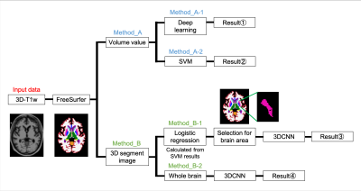 |
12 | Determination of Alzheimer's disease based on morphology and atrophy using machine learning combined with automated segmentation Video Permission Withheld
Natsuki Ikemitsu1, Yuki Kanazawa2, Akihiro Haga2, Hiroaki Hayashi3, Yuki Matsumoto2, and Masafumi Harada2
1Graduate school of Health Science, Tokushima University, Tokushima, Japan, 2Institute of Biomedical Sciences, Tokushima University Graduate School, Tokushima, Japan, 3Faculty of Health Sciences, Institute of Medical, Pharmaceutical and Health Sciences, Kanazawa University, Kanazawa, Japan To classify healthy subjects or patients with Alzheimer's disease (AD) using three-dimensional T1w data, we developed a machine learning system which can capture morphology features and determine atrophy of brain tissue in early-stage AD. Deep learning, a support vector machine (SVM), and 3D convolutional neural networks (3DCNN) were performed. The accuracies of SVM and deep learning based on volume values were comparable and greatly exceeded the accuracy of 3DCNN. It was found that atrophic features were more considerable than morphological features in early-stage AD. |
||
 |
3264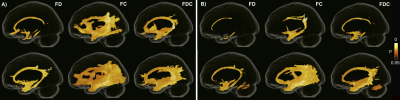 |
13 | Tau accumulation is associated with fiber-specific white matter degeneration in Alzheimer's disease
Khazar Ahmadi1, Joana B. Pereira1,2, Danielle van Westen1,3, Markus Nilsson4, Nicola Spotorno1, and Oskar Hansson1,5
1Clinical Memory Research Unit, Department of Clinical Sciences, Lund University, Lund, Sweden, 2Department of Neurobiology, Care Sciences and Society, Karolinska Institute, Stockholm, Sweden, 3Diagnostic Radiology, Lund University, Lund, Sweden, 4Department of Medical Radiation Physics, Lund University, Lund, Sweden, 5Memory Clinic, Skåne University Hospital, Malmö, Sweden
Fixel-based analysis (FBA) enables investigation of micro- and macrostructural damage in white matter (WM) fiber bundles. However, the link between microscopic changes in fiber density (FD) and macroscopic morphological alterations in fiber cross-section (FC) with the pathological hallmarks of Alzheimer’s disease (AD) including regional tau and amyloid-β (Aβ) accumulation has not been explored yet. Here, we show that reduction in FC in the AD continuum is closely connected with tau tangles in contrast to Aβ pathology.
|
|
3265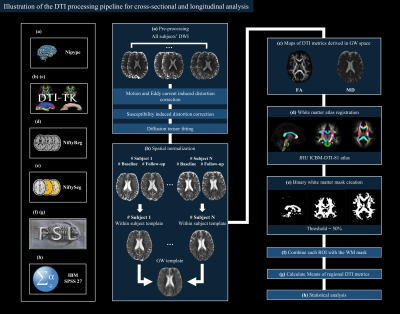 |
14 | What to do for an accurate cross-sectional and longitudinal processing of diffusion tensor imaging: application to frontotemporal dementia
Fan Huang1, Jonathan D. Rohrer2, Hui Zhang3, and Martina Bocchetta2
1Department of Medical Physics and Biomedical Engineering, University College London, London, United Kingdom, 2Dementia Research Centre, Department of Neurodegenerative Disease, UCL Queen Square Institute of Neurology, University College London, London, United Kingdom, 3Centre for Medical Image Computing (CMIC), Department of Computer Science, University College London, London, United Kingdom
We considered the steps needed to analyse Diffusion Tensor Imaging (DTI) cross-sectionally and longitudinally, following a state-of-the-art approach which takes into account image quality, acquisition consistency, artifact correction, spatial correspondence. We applied this method to a small cohort of frontotemporal dementia (FTD) patients and controls to replicate the findings in the existing literature.
|
||
3266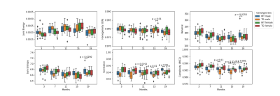 |
15 | Correlation between MRI texture analysis and histo-pathology of Alzheimer’s disease: evaluation in the hippocampus of the Tgf344-AD rat model.
Emma Muñoz-Moreno1, Raul Tudela2,3, Xavier López-Gil1, and Guadalupe Soria4,5
1Magnetic Resonance Imaging Core Facility, IDIBAPS, Barcelona, Spain, 2CIBER-BBN, Barcelona, Spain, 3Universitat de Barcelona, Barcelona, Spain, 4Laboratory of Surgical Neuroanatomy, Universitat de Barcelona, Barcelona, Spain, 5Institute of Neurosciences, Universitat de Barcelona, Barcelona, Spain
β-amyloid plaques and neuroinflammation, two of the pathological processes associated to Alzehimer's disease (AD) are not visible by conventional MRI. However, its presence may induce image changes that can be evaluated using texture analysis. We evaluated the potential of texture analysis of conventional MRI to characterize these changes. T2-weighted MRI of a transgenic rat model of AD were acquired and texture measures in hippocampus were correlated with inmunohistochemical quantification of plaques and microglia, showing significant correlation. Coherently, significant differences in the texture measures were found between transgenic and control animals, pointing to the potential of texture analysis as AD biomarker.
|
||
 Back to Meeting Home
 Back to the Program-at-a-Glance
The International Society for Magnetic Resonance in Medicine is accredited by the Accreditation Council for Continuing Medical Education to provide continuing medical education for physicians.
 Back to Meeting Home
 Back to the Program-at-a-Glance
 View the Presentation