Online Gather.town Pitches
Multiple Sclerosis
Joint Annual Meeting ISMRM-ESMRMB & ISMRT 31st Annual Meeting • 07-12 May 2022 • London, UK

| Booth # | ||||
|---|---|---|---|---|
4512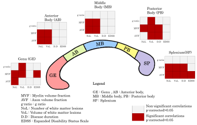 |
1 | Microstructure changes of the normal-appearing white matter in the Corpus Callosum in Relapsing-Remitting Multiple Sclerosis
Lester Melie-Garcia1,2,3, Muhamed Barakovic1,2,3, Matthias Weigel1,2,3,4, Reza Rahmanzadeh1,2,3, Riccardo Galbusera1,2,4, Po-Jui Lu1,2,3, Alessandro Cagol1,2,3, Antoine Lutti5, Sabine Schaedelin6, Pascal Benkert6, Alexandra Todea1,2,3,7,8, Esther Ruberte1,9, Ernst-Wilhelm Radue1, Ludwig Kappos1,2,3, Jens Kuhle3, and Cristina Granziera1,2,3
1Translational Imaging in Neurology (ThINK), Biomedical Engineering Department, University of Basel, Basel, Switzerland, 2Department of Medicine, University Hospital Basel, Basel, Switzerland, 3Neurologic Clinic and Policlinic, MS Center and Research Center for Clinical Neuroimmunology and Neuroscience Basel (RC2NB), University Hospital Basel, Basel, Switzerland, 4Division of Radiological Physics, Department of Radiology, University Hospital Basel, Basel, Switzerland, 5Laboratory for Research in Neuroimaging (LREN), Department of Clinical Neurosciences, Lausanne University Hospital (CHUV) and University of Lausanne, Lausanne, Switzerland, 6Clinical Trial Unit, Department of Clinical Research, University Hospital Basel, Basel, Switzerland, 7Division of Neuroradiology, Department of Radiology, University Hospital Basel, Basel, Switzerland, 8Translational Imaging in Neurology (ThINK) Basel, Biomedical Engineering Department, University of Basel, Basel, Switzerland, 9Medical Image Analysis Center (MIAC), Basel, Switzerland
A scarce body of literature studies the Corpus Callosum (CC) normal-appearing white matter (NAWM) in Relapsing-Remitting Multiple Sclerosis (RRMS). This work aimed to characterize the degree of the alterations in different CC segments by assessing the extent of damage to myelin and axon, as measured with myelin volume fraction, axonal volume fraction, and g-ratio. These microstructural measures correlated with disease duration, EDSS, number, and volume of the lesions in other white matter regions. In sum, this work shows that CC pathology in RRMS patients is related to focal damage and/or diffuse neurodegeneration, which is clinically relevant.
|
||
4513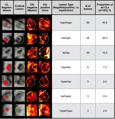 |
2 | Iron and Myelin Content in Cortical Lesions in Multiple Sclerosis using Magnetic Susceptibility Source Separation
Jannis Müller1,2, Po-Jui Lu1,2, Hyeong-Geol Shin3, Reza Rahmanzadeh1,2, Charidimos Tsagkas1,2, Muhamed Barakovic1,2, Riccardo Galbusera1,2, Özgür Yaldizli1,2, Matthias Weigel1,4, Yi Wang5, Thanh Nguyen5, Jens Kuhle2, Jongho Lee3, and Cristina Granziera1,2
1Department of Biomedical Engineering, Faculty of Medicine, University Hospital Basel and University of Basel, Translational Imaging in Neurology (ThINk) Basel, Basel, Switzerland, 2University Hospital Basel and University of Basel, Neurologic Clinic and Policlinic, MS Center and Research Center for Clinical Neuroimmunology and Neuroscience Basel (RC2NB), Basel, Switzerland, 3Department of Electrical and Computer Engineering, Seoul National University, Laboratory for Imaging Science and Technology, Seoul, Korea, Republic of, 4Division of Radiological Physics, Department of Radiology, University Hospital Basel and University of Basel, Basel, Switzerland, 5Department of Radiology, Weill Medical College of Cornell University, New York, NY, United States The visualization and characterization of cortical MS lesions is challenging on conventional MRI. In the present study, we used a susceptibility source separation algorithm to divide the positive and negative susceptibility of quantitative susceptibility mapping (QSM) MRI, so that we could disentangle signal alterations due to myelin loss or iron accumulation. In 19 MS patient with 123 cortical lesions, we found that the major part of QSM susceptibility of cortical lesions is driven by myelin and iron loss, while only a small proportion of lesions (12/123, 9.8%) showed an increment of susceptibility that is caused by iron accumulation. |
||
4514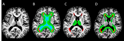 |
3 | Periventricular tissue damage in multiple sclerosis assessed by quantitative magnetization transfer
Lukas Pirpamer1, Andrej Vovk2, Anna Damulina3, Michael Khalil3, Reinhold Schmidt1, Christian Enzinger4,5, and Stefan Ropele3
1Department of Neurology, Division of Neurogeriatrics, Medical University of Graz, Graz, Austria, 2Medicinska fakulteta, Univerza v Ljubljani, Ljubljana, Slovenia, 3Department of Neurology, Division of General Neurology, Medical University of Graz, Graz, Austria, 4Neurology, Medical University of Graz, Graz, Austria, 5Department of Radiology, Division of Neuroradiology, Medical University of Graz, Graz, Austria
Periventricular tissue damage in multiple sclerosis has been suggested to be mediated by toxic soluble factors in the cerebrospinal-fluid. Magnetization transfer imaging has shown great potential to reveal microstructural tissue changes and has been used to study the association of periventricular normal-appearing white matter (NAWM) changes to the distance from the ventricle. We assessed high-resolution magnetization transfer saturation imaging (MTsat) in equidistant bands around the ventricle of MS patients and compared the resulting MTsat gradient of the thalamus and NAWM. While PV-MTsat values in NAWM are consistent to the literature, MTsat values in the outer thalamic bands were higher than controls, likely caused by increased iron levels.
|
||
4515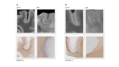 |
4 | Disentangling characteristics of subpial lesions in multiple sclerosis using multiparametric postmortem MRI Video Permission Withheld
Riccardo Galbusera1,2,3, Erik Bahn4, Matthias Weigel1,2,3, Po-Jui Lu1,2,3, Jonas Franz4, Muhamed Barakovic1,2,3, Sabine Schaedelin5, Lester Melie-Garcia1,2,3, Reza Rahmanzadeh1,2,3, Peter Dechent6, Antoine Lutti7, Govind Bhagavatheeshwaran8, Wolfgang Brück4, Ludwig Kappos2,3, Christine Stadelmann4, and Cristina Granziera1,2
1Neurology Clinic and Policlinic, Departments of Medicine, Clinical Research and Biomedical Engineering, University Hospital Basel and University of Basel, Basel, Switzerland, 2Translational Imaging in Neurology (ThINk) Basel, Department of Biomedical Engineering, University Hospital Basel and University of Basel, Basel, Switzerland, 3Research Center for Clinical Neuroimmunology and Neuroscience (RC2NB) Basel, University Hospital Basel and University of Basel, Basel, Switzerland, 4Institute of Neuropathology, University Medical Center Göttingen, Göttingen, Germany, 5Clinical Trial Unit, Department of Clinical Research, University Hospital Basel, University of Basel, Basel, Switzerland, 6Department of Cognitive Neurology, MR-Research in Neurosciences, University Medical Center Göttingen, Göttingen, Germany, 7Centre for Research in Neuroscience - Department of Clinical Neurosciences, Laboratoire de recherche en neuroimagerie (LREN) University Hospital and University of Lausanne, Lausanne, Switzerland, 8National Institute of Neurological Disorders and Stroke, Bethesda, MD, Bethesda, MD, United States
We have characterized the imaging correlates of subpial demyelination in the cerebral cortex of MS patients by exploiting multiparametric postmortem qMRI and histopathology. MTsat, qT1 and AD were the measures that best captured subpial lesions pathology. Additionally, we found that some subpial lesions exhibit a juxta-cortical rim of increased susceptibility and show lower MWF than the ones without rim.
|
||
4516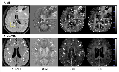 |
5 | Hypo-diamagnetism on χ-separation: a potential marker for the differential diagnosis between MS and NMOSD.
Jinhee Jang1, Hyeong-Geol Shin2, Hyebin Lee1, Dohoon Park1, Junghwa Kang3, Yoonho Nam3, Jongho Lee2, and Woojun Kim4
1Radiology, Seoul St. Mary's Hospital, Seoul, Korea, Republic of, 2Department of Electrical and Computer Engineering, Seoul National University, Seoul, Korea, Republic of, 3Biomedical Engineering, Hankuk University of Foreign Studies, Yongin, Korea, Republic of, 4Neurology, Seoul St. Mary's Hospital, Seoul, Korea, Republic of Using χ-separation, detailed characterization of MS and NMOSD lesions were possible. Majority (490/611, 80%) of MS lesions were hypo-diamagnetic on χneg maps, suggesting loss of diamagnetic myelin. On the contrary, only 14.2% (32/225) of NMOSD lesions were hypo-diamagnetic. On χpos maps, some MS and a few NMOSD lesions showed paramagnetic rim round the lesion, which was identical to paramagnetic rim sign on SWI or QSM. The proportion of hypo-diamagnetic lesions showed excellent diagnostic performances (AUROC 0.961, 95%CI 0.907-1), as compared to central vein sign and paramagnetic rim lesion sign. |
||
 |
4517 | 6 | Demyelination and remyelination in cuprizone mouse model detected by CEST MRI at 3T
Zilin CHEN1, Jianpan HUANG1, Joseph H.C. LAI1, Kai-Hei TSE2, and Kannie W.Y. CHAN1,3,4,5
1Department of Biomedical Engineering, City University of Hong Kong, Hong Kong, China, 2Department of Health Technology and Informatics, The Hong Kong Polytechnic University, Hong Kong, China, 3Russell H. Morgan Department of Radiology and Radiological Science, The Johns Hopkins University School of Medicine, Baltimore, MD, United States, 4City University of Hong Kong Shenzhen Research Institute, Shenzhen, China, 5Hong Kong Centre for Cerebro-Cardiovascular Health Engineering (COCHE), Hong Kong, China Multiple sclerosis (MS) is a common demyelinating disease that heavily relies on differential diagnosis. Specific CEST contrast is known to be sensitive to alterations in proteins and lipids, the major components of myelin. This includes amide protons at 3.5 ppm and relayed nuclear Overhauser effect (rNOE) at -1.6~-3.5 ppm. Here, we study these CEST contrasts and their uniqueness towards myelin changes in a cuprizone model, which recapitulates remyelination and demyelination in MS. We observed substantial changes of rNOE and amide, during demyelination (P<0.05) and remyelination, indicating great potential of CEST MRI in monitoring myelin change and MS identification at 3T. |
|
 |
4518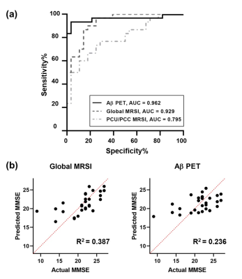 |
7 | Study of Neurometabolic Signature in Alzheimer’s Disease using High-Resolution 3D 1H-MRSI
Yaoyu Zhang1, Jialin Hu1, Miao Zhang2, Rong Guo3,4, Yudu Li3,4, Yibo Zhao3,4, Ziyu Meng1, Danni Wang1, Wenli Li1, Biao Li2, Jun Liu5, Binyin Li5, Zhi-Pei Liang3,4, and Yao Li1
1School of Biomedical Engineering, Shanghai Jiao Tong University, Shanghai, China, 2Department of Nuclear Medicine, Ruijin Hospital, Shanghai Jiao Tong University School of Medicine, Shanghai, China, 3Beckman Institute for Advanced Science and Technology, University of Illinois at Urbana-Champaign, Urbana, IL, United States, 4Department of Electrical and Computer Engineering, University of Illinois at Urbana-Champaign, Urbana, IL, United States, 5Department of Neurology and Institute of Neurology, Ruijin Hospital, Shanghai Jiao Tong University School of Medicine, Shanghai, China
Early and accurate diagnosis of AD is clinically important. Neurometabolic signals measured noninvasively by MRSI showed potential. Previous MRSI studies, limited to single voxel/slice techniques, could only examine neurometabolite concentration from limited brain regions. Using a high-resolution 3D MRSI technique, we assessed neurometabolic signature in AD by integrating neurometabolite concentrations from multiple brain regions. The discriminative power of global neurometabolic signature was evaluated in comparison with that of Aβ PET for both AD detection and predicting cognitive decline, showing promising results. The study provides a good foundation for further investigation using neurometabolic signature for early and accurate diagnosis of AD.
|
|
4519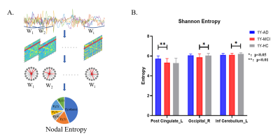 |
8 | Instability and hyper-connected pattern of dynamic functional network in MCI converters Video Permission Withheld
Zhihan Chen1,2, Keliang Chen2, Yuxin Li1,2, Qianhua Zhao1,2, and Liqin Yang1,2
1Fudan University, Shanghai, China, 2Huashan Hospital, Shanghai, China In this study, based on rs-fMRI, we analyzed the dynamic characteristics of 3 groups: MCI convert to AD within one-year follow-up (1Y_AD), stable in MCI within one-year follow-up (1Y_MCI) and revision from MCI within one-year follow-up (1Y_HC), including nodal entropy and measurements of dynamic brain functional network. We found that with the disease progression, the brain functional network was reconfigured into a highly-integrated/segregated pattern, and dwell time of highly-integrated pattern is associated with executive function in 1Y_AD. These findings provide the evidence for developing dynamic measurements as potential biomarkers for tracking mechanism of AD. |
||
4520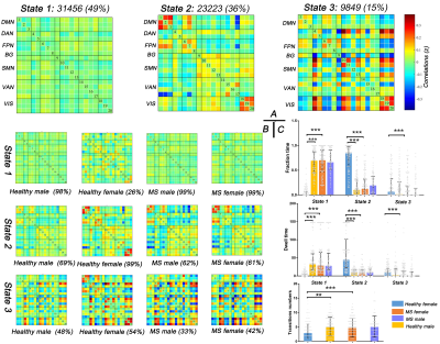 |
9 | Gender-related differences in static and dynamic functional connectivity in multiple sclerosis: a multicenter study
Yao Wang1, Fuqing Zhou1, Yaou Liu2, and Qian Long 3
1Department of Radiology, The First Affiliated Hospital of Nanchang University, Nanchang, China, 2Department of Radiology, Beijing Tiantan Hospital, Beijing, China, 3MR Research, GE Healthcare, Beijing, China
The study investigated gender-related differences in patients with multiple sclerosis by assessing static and dynamic functional connectivity within large scale functional networks. Our results observed that MS could eliminate the effect of gender on static and dynamic functional connectivity in healthy controls, and affect static connectivity in male patients and dynamic connectivty in female patients. Correlation analyses suggested these characteristics of static and dynamic connectivity were related to disability and gray matter volume. Thus, our findings underline the importance of gender in founctional impairment and reorganization in MS
|
||
4521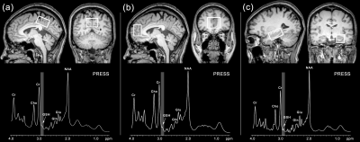 |
10 | Hippocampal glutathione-glutamate couplings predict cognitive impairment in patients with relapsing-remitting multiple sclerosis Video Permission Withheld
Fuyan Li1, Wei Zong1, Fuxin Ren1, Ning Li1, Xiao Li1, Zongrui Dai2, Weibo Chen3, Muwei Li4, and Fei Gao1
1Department of Radiology, Shandong Provincial Hospital, Cheeloo College of Medicine, Shandong University, Jinan, China, 2Westa College, Southwest University, Chongqing, China, 3Philips Healthcare, Shanghai, China, 4Vanderbilt University Institute of Imaging Science, Nashville, TN, United States Cognitive impairment is a common symptom of MS. GSH and Glu, keeping a homeostasis in the stable stage, were considered as key players in oxidative stress defending and synaptic plasticity, respectively. We aim to explore the changes of GSH and Glu levels and GSH-Glu couplings in RRMS and their association with cognitive impairment. Our findings indicate that oxidative stress and glutamatergic dysfunction may contribute to cognitive impairment of MS in a regional specificity manner. Hippocampal GSH-Glu decoupling may offer a crucial noninvasive measure of early cognitive impairment and provide a new strategy for the treatment of MS patients. |
||
4522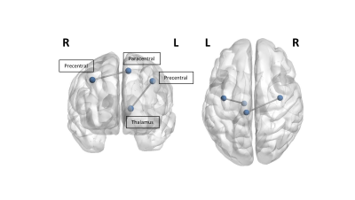 |
11 | Selective Disruption of the Motor Connectome in Non-Familial Amyotrophic Lateral Sclerosis
Sicong Tu1, Arkiev D'Souza1,2, Chenyu Wang1,2, Christina Maher1, Colin Mahoney1, William Huynh1, Michael Barnett1,2,3, and Matthew Kiernan1,3
1Brain and Mind Centre; The University of Sydney, Sydney, Australia, 2Sydney Neuroimaging and Analysis Centre, Sydney, Australia, 3Department of Neurology, Royal Prince Alfred Hospital, Sydney, Australia
Amyotrophic lateral sclerosis (ALS) is a rapidly progressing neurodegenerative disorder with a widespread cortical disease signature. ALS patients demonstrate global network alterations in the white matter connectome in the early stage of disease. Selective disruption of cortical motor associated nodes with the thalamus and contralateral motor cortices is present at disease onset. Disease duration is associated with reduced structural interhemispheric cortical motor connectivity. Focal motor abnormalities present in the white matter connectome may be a sensitive marker in the earliest stages of ALS prior to functional decline.
|
||
4523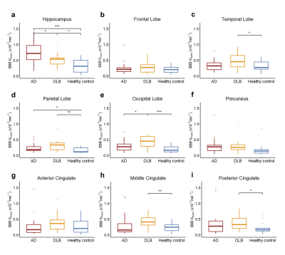 |
12 | Blood-Brain Barrier Leakage in Patients with Alzheimer’s Disease and Dementia with Lewy Bodies
Ziming Xu1, Zhichao Chen2, Lingyun Ma2, Yajie Wang1, Hao Wu3, Hao Lu4, Yong Ji2, and Huijun Chen1
1Center for Biomedical Imaging Research, School of Medicine, Tsinghua University, Beijing, China, 2Department of Neurology, Beijing Tiantan Hospital, Capital Medical University, China National Clinical Research Center for Neurological Diseases, Beijing, China, 3Department of Neurology, Tianjin Huanhu Hospital, Tianjin, China, 4Department of Radiology, Tianjin Huanhu Hospital, Tianjin, China
Previous in vivo studies have shown that dysfunction of blood-brain barrier is highly associated with the process of Alzheimer’s disease (AD), but similar studies in dementia with Lewy bodies (DLB) have rarely been reported. In this study, for the first time, differences of BBB leakage among patients with AD, DLB and age-matched healthy control in different brain regions had been demonstrated, which may serve as one potential tool for diagnosis of different neurodegenerative diseases and contribute to our understanding of their pathophysiological basis.
|
||
4524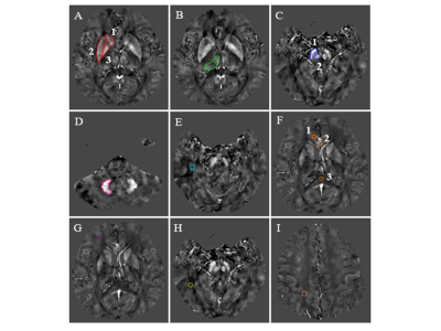 |
13 | Monitoring mild cognitive impairment of workers exposed to occupational aluminum based on quantitative susceptibility mapping
Zhiyi Zhang1, Haoru Jiang1, Xiangru Sun1, Xiaochun Wang1, Bin Wang1, Qiao Niu1, Huaxing Meng1, Jiangfeng Du1, Guoqiang Yang1, Bo Liu1, Hui Zhang1, and Yan Tan1
1Shanxi Medical University, Taiyuan, China
In this work, we presented a study which investigated the diagnostic value of quantitative susceptibility mapping (QSM) in mild cognitive impairment (MCI) of occupational aluminum (Al) workers, we found that QSM might be a reliable neuroimaging marker for the diagnosis of MCI.
|
||
4525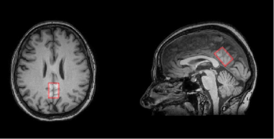 |
14 | Predicting conversion from normal aging to mild cognitive impairment using 1H-MRS of posterior cingulate cortex
Anna Kiryanova1, Andrei Manzhurtsev1,2,3, Olga Bozhko2,4, Yana Fedorova4, and Natalia Semenova 1,2,3,5
1Lomonosov Moscow State University, Moscow, Russian Federation, 2Clinical and Research Institute of Emergency Pediatric Surgery and Trauma, Moscow, Russian Federation, 3Emanuel Institute of Biochemical Physics of RAS, Moscow, Russian Federation, 4Mental Health Research Center, Moscow, Russian Federation, 5Semenov Federal Research Center of Chemical Physics of the Russian Academy of Sciences., Moscow, Russian Federation
This study aims to identify possible biomarkers of minimal cognitive impairment (MCI) in the posterior cingulate cortex using MR spectroscopy. Absolute concentrations of metabolites such as Cr, NAA, Cho, Glx, Ins were determined in subjects with MCI and in subjects from the normal group. Our study revealed a statistically significant increase in the absolute concentration of choline-containing compounds, as well as the Cho/Cr value in PCC in the MCI group, what may indicate the destruction of cell membranes. The results expand our knowledge in understanding the causes of MCI and are promising for the study of Cho as biomarkers.
|
||
4526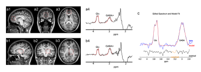 |
15 | Reduced GABA levels in the medial prefrontal cortex are associated with cognitive impairment in patients with NMOSD
Yang Yang1, Qiuyu Yu1, Peng Wu2, Hui Dai1, Xiaojuan Wu1, Shuting Han1, and Yonggang Li1
1The First Affiliated Hospital of Soochow University, Suzhou, China, 2Philips Healthcare, Shanghai, China
Cognitive impairment is a symptom present in part of patients with neuromyelitis optica spectrum disorder (NMOSD). We demonstrated decreased GABA levels in the medial prefrontal cortex (mPFC) in NMOSD participants compared to healthy controls. The mPFC GABA levels showed significant associations with cognitive performance in patients with NMOSD. This study suggested that the changes in regional GABA levels might be a potential metabolic feature of cognitive decline in patients with NMOSD.
|
||
 Back to Meeting Home
 Back to the Program-at-a-Glance
The International Society for Magnetic Resonance in Medicine is accredited by the Accreditation Council for Continuing Medical Education to provide continuing medical education for physicians.
 Back to Meeting Home
 Back to the Program-at-a-Glance
 View the Presentation