Online Gather.town Pitches
Imaging in Parkinson's Disease
Joint Annual Meeting ISMRM-ESMRMB & ISMRT 31st Annual Meeting • 07-12 May 2022 • London, UK

Online Gather.town Pitches
Imaging in Parkinson's Disease
| Booth # | ||||
|---|---|---|---|---|
2951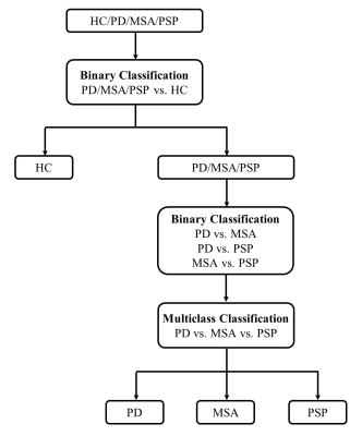 |
1 | Stratified Parkinsonism classification based on multi-modality MRI Video Permission Withheld
Yijun Leng1,2, Xueling Liu2, Daoying Geng1,2,3, Fengtao Liu4, Pu-Yeh Wu5, Yuxin Li2,3, and Liqin Yang2,3
1Academy for Engineering & Technology, Fudan University, Shanghai, China, 2Department of Radiology, Huashan Hospital, Shanghai, China, 3Institute of Functional and Molecular Medical Imaging, Fudan University, Shanghai, China, 4Department of Neurology, Huashan Hospital, Shanghai, China, 5GE Healthcare, Beijing, China
For differential diagnosis between PD and atypical parkinsonisms, we designed a stratified automated method based on SVM and multi-modality MRI, including T1WI, QSM and DTI. 163 patients (96 PD, 27 MSA, 40 PSP) and 65 healthy controls were recruited. Features including volume, cortical thickness, magnetic susceptibility, FA and MD of 124 ROIs were calculated for SVM classification. The result showed that SVM classification based on susceptibility enabled accurate differentiation of patients with parkinsonian disorders and controls, and classification of PD, MSA and PSP was allowed by using T1 and DTI.
|
||
2952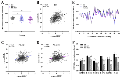 |
2 | Region-specific neurovascular decoupling associated with cognitive decline in Parkinson’s disease Video Not Available
Song'an Shang1, Weiqiang Dou2, and Jingtao Wu3
1Nanjing First Hospital, Nanjing Medical University, Nanjing, China, 2GE Healthcare, MR Research China, Beijing, China, 3Clinical Medical College, Yangzhou University, Yangzhou, China
Cognitive deficits are prominent non-motor symptoms in Parkinson’s disease (PD) and have been involved with neurovascular unit. However, sufficient neuroimaging studies are lacked investigating the associated modulating mechanisms. This study aimed to identify the contribution of neurovascular decoupling to the pathogenesis of cognitive decline in PD by integrating informative data obtained from the blood oxygen level-dependent and arterial-spin-labeling approaches. The involvement of neurovascular decoupling in cognitive impairment in PD is regionally specific and most prominent in the visual-spatial cortices, which could potentially provide a complementary understanding for the pathophysiological mechanisms underlying cognitive deficits in PD.
|
||
2953 |
3 | Structural and functional changes of the cerebellum in patients with Parkinson's disease: a VBM and rs-fMRI study
xiu Cheng1, jun Wang1, pengfei Zhang1, jing Zhang1, kai Ai2, and wanjun Hu1
1Lanzhou University Second Hospital, Lanzhou, China, 2Philips Healthcare, Xi'an, China
To investigate the structural and functional alterations of cerebellum in patients with Parkinson's disease (PD) using voxel-based morphometry and resting-state functional MRI. We analyzed alterations gray matter volume (GMV), regional homogeneity (ReHo), amplitude of low-frequency fluctuation (ALFF) in the cerebellum of PD patients. Furthermore, the correlation analysis between GMV, ALFF, ReHo values and clinical scale scores was performed. Our results showed that the structure and function of the cerebellum of PD patients are significantly different from healthy controls (HC), and these changes are significantly correlated with clinical scales scores, which suggested that the cerebellum plays an important role in PD.
|
||
2954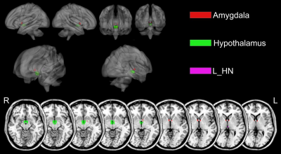 |
4 | Gray matter myelin quantification in Parkinson’s disease based on MP2RAGE T1 mapping Video Permission Withheld
Yu Shen1, Yan Bai1, Xianchang Zhang2, Yaping Wu1, Yu Luo1, Zhun Huang1, Xipeng Yue1, Menghuan Zhang1, and Meiyun Wang1
1Henan Provincial People's Hospital, Zhengzhou, China, 2MR Collaboration, Siemens Healthineers Ltd, Beijing, China
Quantitative R1(1/T1) imaging has been proposed as surrogate imaging biomarkers for myelin content in cortical gray matter. This study, using high resolution T1 maps generated by MP2RAGE sequence, quantified gray matter myelin in Parkinson’s disease (PD) patients and compared the values with those in healthy controls. The results revealed that myelin (represented by R1) was significantly decreased in PD in several cortical and subcortical brain areas, which may be anatomic evidence for the spectrum of symptoms in PD, such as deficits of memory, mood, and cognition. Myelin quantification based on MP2RAGE-T1 mapping may be a valuable biomarker for characterizing PD.
|
||
2955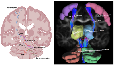 |
5 | Distinct patterns of tremor network microstructure for essential tremor and Parkinson’s disease
Thomas Welton1,2, Amadis Aliya Ong3, Septian Hartono1,2, Yao-Chia Shih4, Amanda Lee3, Eng-King Tan1,5, and Ling Ling Chan1,3
1National Neuroscience Institute, Singapore, Singapore, 2Duke-NUS Graduate Medical School, Singapore, Singapore, 3Diagnostic Radiology, Singapore General Hospital, Singapore, Singapore, 4Graduate Institute of Medicine, Yuan Ze University, Taiwan, Taiwan, 5Neurology, Singapore General Hospital, Singapore, Singapore
Differential diagnosis of essential tremor and Parkinson’s disease can be challenging due to overlapping clinical presentations, but these diseases differ in neuropathological features, which may be detected using diffusion spectrum imaging. We identified parts of a tremor-related cerebello-thalamo-cortical network based on diffusion spectrum imaging that differed among a cohort comprising essential tremor and Parkinson’s patients, and healthy-controls. The patient groups had minimally-overlapping areas of abnormal microstructure in the tremor-network, and these were significantly correlated with clinical and kinetic measures of tremor severity. A future DSI-based marker of tremor-network microstructure may aid in differential diagnosis of essential tremor and Parkinson’s disease.
|
||
2956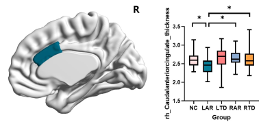 |
6 | Gray and white matter alterations in different predominant side and type of motor symptom in Parkinson’ disease
Jingwen Chen1, Cheng Zhou1, Jingjing Wu1, Tao Guo1, Xueqin Bai1, Haoting Wu1, Xiaocao Liu1, Jiaqi Wen1, Zhengye Cao1, Yong Zhang2, Xiaojun Guan1, Xiaojun Xu1, and Minming Zhang1
1Radiology, The second affiliated hospital, Zhejiang University school of medicine, Hangzhou, China, 2GE Healthcare, Shanghai, China
It has been overlooked in clinical that there exists a complex relationship between laterality and motor symptoms in Parkinson’s disease (PD) and these factors may influence their motor and nonmotor symptoms in together. Here we found PD with left-side akinetic/rigid (L-AR) signs performed worst in clinical manifestations. And the underlying brain structure alterations was revealed severer damage in anterior cingulate and its connected fibers of right hemisphere by a multimodal imaging approach including cortical thickness and diffusion-weighted MRI measures.
|
||
2957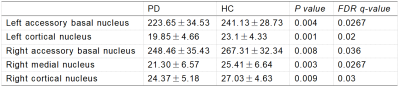 |
7 | Automated volumetry of amygdala subregions in Parkinson’s disease patients
mingrui qu1, bingbing gao1, yuhan jiang1, yangyingqiu liu1, yuan li1, qingwei song1, and yanwei miao1
1the First Affiliated Hospital of Dalian Medical University, Dalian, China
Cognitive impairment is very common in patients with Parkinson’s disease (PD). The amygdala in the limbic system of midbrain has been proved to play a unique role in cognitive function. However, there are almost no studies on changes in amygdala structure, especially the volume of amygdala subregions, in PD patients. In this study, automated volumetry of amygdala subregions in PD patients was performed and compared with healthy controls, and found that PD patients have amygdala subregions atrophy, especially in bilateral accessory basal nucleus, bilateral cortical nucleus and right medial nucleus.
|
||
2958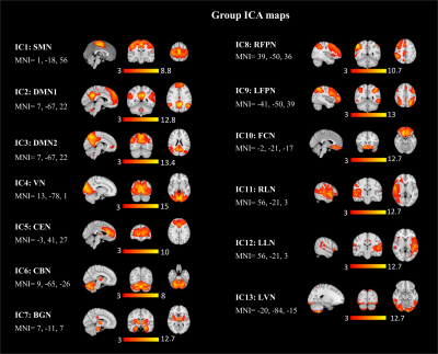 |
8 | Altered Visual Network and Global Fluctuation in the Resting-state fMRI of Patients with Parkinson’s Disease
Destaw Bayabil Mekbib1, Weiying Dai2, Miao Cai3, Xiaoli Liu3, and Li Zhao1
1Key Laboratory for Biomedical Engineering of Ministry of Education, Zhejiang University, Hangzhou, China, 2Computer Science, Binghamton Univeristy, State University of New York, Binghamton, NY, United States, 3Neurology, Zhejiang Hospital, Hangzhou, China
Resting-state fMRI plays an increasing role in understanding the neural mechanisms of Parkinson's disease (PD). However, neural network alterations, regional and global signal associations are not fully understood in PD patients. Here, we studied the whole-brain neural networks including the inter-network and their correlations with the global fluctuation in 37 PD patients and 20 healthy controls. Our findings revealed that the visual cortex had reduced neural network connectivity in PD patients and altered connections from default mode network to other networks. Weaker correlations were observed between the global signal and regional networks in the PD patients than healthy controls.
|
||
2959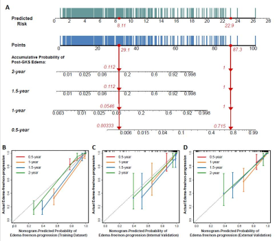 |
9 | Predicting Peritumoral Edema Development after Gamma Knife Radiosurgery of Meningiomas Using Radiomics: A Multicenter Study Video Permission Withheld
Xuanxuan Li1, Yiping Lu1, Pu-Yeh Wu2, Tonggang Yu3, and Bo Yin1
1Huashan Hospital, Fudan University, Shanghai, China, 2GE Healthcare, Beijing, China, 3Shanghai Gamma Hospital, Huashan Hospital, Fudan University, Shanghai, China
The aim of this study is to adopt machine learning and deep learning methods to predict the risk of post-GKS edema for meningiomas. 595 multicenter cases were included to train and validate 38 random survival forest (RSF) and DeepSurv models. The RSF model incorporating clinical, semantic, and ADC radiomic features achieved the best performance with a C-index of 0.861 in internal validation, and 0.780 in external validation. The derived nomogram had excellent discrimination and calibration. The proposed RSF model with a nomogram represents a non-invasive and cost-effective tool to predict post-GKS edema risks, thus facilitates personalized decision-making in meningioma treatment.
|
||
2960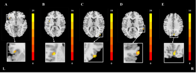 |
10 | Parkinson’s disease state modulates the effect of nigral iron deposition on the large-scale brain networks.
Jiaqi Wen1, Xiaojun Guan1, Tao Guo1, Jingjing Wu1, Xueqin Bai1, Cheng Zhou1, Haoting Wu1, Xiaocao Liu1, Jingwen Chen1, Zhengye Cao1, Yong Zhang2, Luyan Gu3, Jiali Pu3, Baorong Zhang3, Minming Zhang1, and Xiaojun Xu1
1Department of Radiology, The Second Affiliated Hospital, Zhejiang University School of Medicine, Hangzhou, China, 2GE Healthcare, Shanghai, China, Shanghai, China, 3Department of Neurology, The Second Affiliated Hospital, Zhejiang University School of Medicine, Hangzhou, China
In Parkinson’s disease (PD), nigral iron deposition exacerbates a-synuclein aggregation. In our study, we divided PD and normal controls (NC) into high iron group and normal iron group according to the median of nigral magnetic susceptibility of NC, respectively. ICA method was applied to separate brain large-scale networks. The PD*iron interaction effect on brain functional networks were investigated by mixed effect analysis. We found that PD disease status specifically moderates the effect of nigral iron deposition on the function of some regions in BGN and VN, which partially mediates the relationship between nigral iron deposition and disease severity.
|
||
2961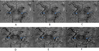 |
11 | Optimization of TE and Postprocessing for Nigrosome-1 Imaging using Wave-CAIPI Susceptibility Weighted Imaging at 3T
Caixia Fu1, Youmin Zhang2, Qing Li3, Wei Liu1, Naying He2, Yu Liu2, Daniel Polak4, E. Mark Haacke 5, and Fuhua Yan2
1MR Application Development, Siemens Shenzhen Magnetic Resonance Ltd., Shenzhen, China, 2Department of Radiology, Ruijin Hospital, Shanghai Jiao Tong University School of Medicine, Shanghai, China, 3MR Collaborations, Siemens Healthineers Ltd., Shanghai, China, 4MR Application Predevelopment, Siemens Healthcare GmbH, Erlangen, Germany, 5Department of Radiology, Wayne State University, Detroit, MI, United States
In this work, Wave-CAIPI accelerated 3D SWI has been used to achieve whole brain imaging in about 3min 30s. Both short-TE and long-TE protocols were compared for the detection of the Nigrosome-1(N1) sign. The phase-mask formula and number of phase mask multiplications were adjusted to explorer their influence on the contrast-to-noise (CNR) of the N1 sign relative to adjacent tissues. The result showed that long-TE protocol had better CNR and made it easier to recognize the N1 sign. The adjusted phase-mask formula with an increased number of phase mask multiplications also helped to improve the CNR of the N1 sign.
|
||
2962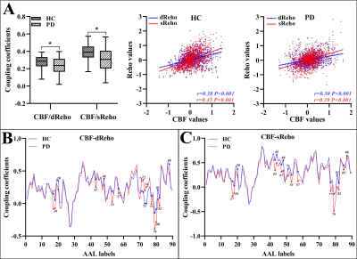 |
12 | Early disturbance of dynamic synchronization and neurovascular coupling in cognitively normal Parkinson’s disease: an integrated MRI study Video Not Available
Song'an Shang1, Weiqiang Dou2, and Jingtao Wu3
1Nanjing First Hospital, Nanjing Medical University, Nanjing, China, 2GE Healthcare, MR Research China, Beijing, China, 3Clinical Medical College, Yangzhou University, Yangzhou, China
For Parkinson’s disease (PD), the time-varying properties of functional coherence and their coupling to regional perfusion are rarely elucidated. We aimed to investigate early disruption of dynamic regional homogeneity (dReho) pattern and neurovascular coupling (NC) in PD patients before onset of cognitive impairment and their classification performance. PD patients at early stage exhibited an impaired dynamic pattern of neuronal synchronization and disrupted NC. The features of CBF/dReho provided robust performance in differentiating PD from healthy controls. With this findings, the insights into early pathophysiological mechanism underlying PD from regional dynamic pattern and NC were reinforced.
|
||
2963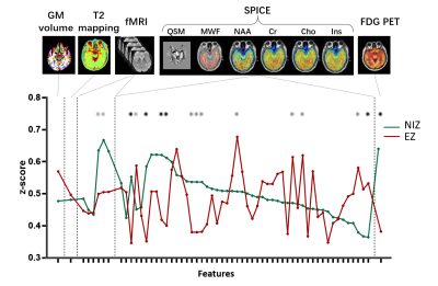 |
13 | Multimodal imaging signatures of the epileptogenic zone
Hui Huang1, Siyu Yuan1, Miao Zhang2, Wei Liu3, Yibo Zhao4,5, Rong Guo4,5, Yudu Li4,5, Lihong Tang1, Zhi-Pei Liang4,5, Yao Li1, and Jie Luo1
1School of Biomedical Engineering, Shanghai Jiao Tong University, Shanghai, China, 2Department of Nuclear Medicine, Ruijin Hospital, Shanghai Jiao Tong University School of Medicine, Shanghai, China, 3Department of Neurosurgery, Ruijin Hospital, Shanghai Jiao Tong University School of Medicine, Shanghai, China, 4Department of Electrical and Computer Engineering, University of Illinois at Urbana Champaign, Urbana, IL, United States, 5Beckman Institute for Advanced Sciences and Technology, University of Illinois at Urbana Champaign, Urbana, IL, United States
Accurate detection of the epileptogenic zone for surgical planning often relies on stereo-electroencephalography, which is limited by low spatial sampling. Development of noninvasive brain high resolution imaging to better identify epileptogenic zone is of great value. This study aimed to investigate the imaging signature for detection of epileptogenic zones among features extracted from structural, functional, and metabolic imaging using hybrid PET/MR scanner. Our results highlighted the value of combination between metabolic and functional imaging features.
|
||
 |
2964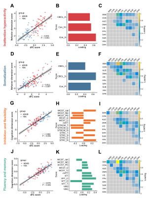 |
14 | Brain Dynamic Functional Connectivity are linked with Attention-Deficit/Hyperactivity Disorder Related Cognitive and Behavioral Dimensions
Lekai Luo1, Lizhou Chen1, Qian Li1, Ning He2, Yuanyuan Li2, Wanfang You1, Yuxia Wang1, Yaxuan Wang1, John A. Sweeney1,3, Lanting Guo2, Qiyong Gong1, and Fei Li1
1Huaxi MR Research Center (HMRRC), Department of Radiology, West China Hospital of Sichuan University, Chengdu, China, 2Department of Psychiatry, West China Hospital of Sichuan University, Chengdu, China, 3Department of Psychiatry and Behavioral Neuroscience, University of Cincinnati, Cincinnati, OH, United States
Sparse canonical correlation analysis (sCCA) was used to delineate multivariate relationship between dynamic functional connectivity (dFC) and behavior or cognition scores in a cohort of children with and without Attention-Deficit/Hyperactivity Disorder (ADHD). We identified four distinct patterns of dFC, each corresponded to a specific dimension of behavior (inattention/hyperactivity, somatization) or cognitive function (inhibition and flexibility, fluency and memory). Altered dFC within the default mode network (DMN) and between DMN and sensorimotor network (SMN) were common to all dimensions.
|
|
 Back to Meeting Home
 Back to the Program-at-a-Glance
The International Society for Magnetic Resonance in Medicine is accredited by the Accreditation Council for Continuing Medical Education to provide continuing medical education for physicians.
 Back to Meeting Home
 Back to the Program-at-a-Glance
 View the Presentation