Online Gather.town Pitches
Imaging White Matter & Spinal Cord
Joint Annual Meeting ISMRM-ESMRMB & ISMRT 31st Annual Meeting • 07-12 May 2022 • London, UK

Online Gather.town Pitches
Imaging White Matter & Spinal Cord
| Booth # | ||||
|---|---|---|---|---|
2965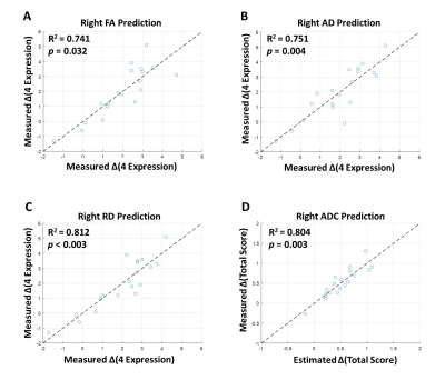 |
1 | WM integrity of right AF in predicting language recovery of chronic poststroke aphasia after LF-rTMS treatment
Bing-Fong Lin1, Fei Hon1, Po-Yi Tsai2,3, and Chia-Feng Lu1
1Department of Biomedical Imaging and Radiological Sciences, National Yang Ming Chiao Tung University, Taipei, Taiwan, Taipei, Taiwan, 2Department of Physical Medicine and Rehabilitation, Taipei Veterans General Hospital, Taipei, Taiwan, Taipei, Taiwan, 3School of Medicine, National Yang Ming Chiao Tung University, Taipei, Taiwan, Taipei, Taiwan
Low-frequency repetitive transcranial magnetic stimulation (LF-rTMS) provided promising results to facilitate the language recovery in stroke patients with non-fluent aphasia 1. The contralesional inhibitory LF-rTMS treatment can induce the functional reorganization within language networks and recovery of language function in chronic aphasic stroke 2, 3. This study evaluated the right microstructural integrity of the arcuate fasciculus before the intervention and its association with the language improvement after the LF-rTMS treatment.
|
||
2966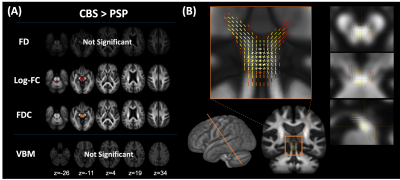 |
2 | Fixel-Based Analysis of White Matter Degeneration in Patients with Progressive Supranuclear Palsy and Corticobasal Syndrome
Wataru Uchida1, Koji Kamagata1, Christina Andica1, Yuya Saito1, Kaito Takabayashi1, Akifumi Hagiwara1, Shohei Fujita1,2, Toshiaki Akashi1, Akihiko Wada1, Masaaki Hori3, and Shigeki Aoki1
1Department of Radiology, Juntendo University Graduate School of Medicine, Tokyo, Japan, 2Department of Radiology, The University of Tokyo, Tokyo, Japan, 3Department of Radiology, Toho University Omori Medical Center, Tokyo, Japan
Progressive supranuclear palsy (PSP) and corticobasal syndrome (CBS) are neurodegenerative disorders associated with corticobasal degeneration pathology. A previous study evaluated white matter (WM) alteration using diffusion tensor imaging; however, the details are unclear due to the crossing fiber. This study evaluated cross-sectional and longitudinal WM changes in PSP and CBS using fixel-based analysis. This study also demonstrated consistent cross-sectional and longitudinal WM degeneration with previous histopathological studies and higher sensitivity than classical magnetic resonance imaging findings in differentiating PSP and CBS. Our results suggest that FBA can be a biomarker to classify PSP and CBS and estimate disease progression.
|
||
2967 |
3 | White matter microstructure alterations using Generalized Q-Sampling Imaging in Moderate and Severe Obstructive Sleep Apnea
Peng Wang1, Xinchun Li1, Qi Wan1, Wenjin Zou2, Yihao Guo3, Mengzhu Wang4, Jianfeng Hu1, Yu Peng1, Xiaoying Xia1, Xiaobin Xie1, and Jieqiong Liu1
1Department of Radiology, The First Affiliated Hospital of Guangzhou Medical University, Guangzhou, China, 2The Affiliated Brain Hospital of Guangzhou Medical University (Guangzhou Huiai Hospital), Guangzhou, China, 3MR Collaboration, Siemens Healthineers Ltd., Guangzhou, China, 4MR Scientific Marketing, Siemens Healthineers, Guangzhou, China
In the current study, we assessed possible alterations of brain white matter (WM) microstructure in patients with obstructive sleep apnea (OSA) using generalized q-sampling imaging (GQI). We found higher normalized quantitative anisotropy values of the bilateral superior longitudinal fasciculus, uncinate fasciculus, fornix, and corpus callosum bundle in the OSA group vs controls, which were related to multiple clinical indices of OSA.
|
||
2968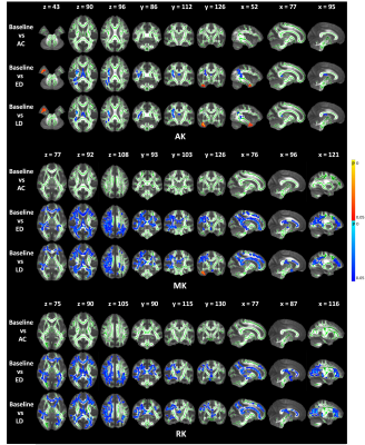 |
4 | Radiation-induced White Matter Abnormalities in Nasopharyngeal Carcinoma: Evaluation by DKI and NODDI Video Permission Withheld
Yuhao Lin1, Xiaofei Lv2, Jiahui Liang2, Yanqiu Feng1, and Xinyuan Zhang1
1School of Biomedical Engineering, Southern Medical University, Guangzhou, China, 2Department of Medical Imaging, Sun Yat-sen University Cancer Center, State Key Laboratory of Oncology in South China, Collaborative Innovation Center for Cancer Medicine, Guangdong Key Laboratory of Nasopharyngeal Carcinoma Diagnosis and Therapy, Guangzhou, China
Based on the multi-shell diffusion MRI data acquisition and tract-based spatial statistics (TBSS), we conducted a longitudinal study to explore the changing patterns of DKI and NODDI metrics after radiotherapy for nasopharyngeal carcinoma (NPC) patients. We found most of the significant regions showed a “self-recovery” mechanism but the temporal lobe regions show a progressive changing pattern over time after RT. Besides, we also found that the change of DKI metrics has a stronger dose-correlation than DTI and NODDI metrics. Our study indicates the DKI and NODDI metrics can provide more comprehensive brain microstructure changes compared with conventional DTI.
|
||
2969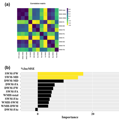 |
5 | Role of the superficial white matter in processing speed decline in cerebral small vessel disease
Shuyue Wang1, Fan Zhang2, Peiyu Huang1, Hui Hong1, Yeerfan Jiaerken1, Xinfeng Yu1, Ruiting zhang1, Qingze Zeng1, Yao Zhang1, Ron Kikinis2, Yogesh Rathi2, Nikos Makris2, Ofer Pasternak2, Minming Zhang1, and Lauren J. O’Donnell2
1The Second Affiliated Hospital of Zhejiang University School of Medicine, Hangzhou, China, 2Harvard Medical School, Boston, MA, United States
We assess microstructural alterations in superficial white matter (SWM) in cerebral small vessel disease (CSVD) and evaluate their contributions to the decline in processing speed, which is the main dysfunction in CSVD. We identify that the significant decline in processing speed may relate to the involvement of WMH in the SWM under high burden of disease. The increased extracellular free water may be the main SWM microstructural change under low burden of disease. These observations suggest that the SWM may serve as a potential target for monitoring pathophysiological processes in CSVD. This study extends the current understanding of CSVD-related dysfunction.
|
||
2970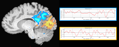 |
6 | Structure-function relationships in the human hippocampus: new insights using track-weighted dynamic functional connectivity Video Permission Withheld
Marshall A Dalton1, Jinglei Lv1, Arkiev D'Souza1, and Fernando Calamante1
1The University of Sydney, Sydney, Australia
The hippocampus is a brain structure central to a broad range of cognitive functions including episodic memory. In recent years, we have developed a greater understanding of the structural and functional connectivity of the human hippocampus. Despite these advances, we lack a detailed understanding of structure-function relationships of cortico-hippocampal connectivity. We addressed this gap by combining high-quality data from the Human Connectome Project with cutting-edge fibre-tracking and track-weighted dynamic functional connectivity methods to quantitatively characterise the relationship between anatomical and functional connectivity of the human hippocampus. Our results contribute to ongoing efforts to characterise structure-function relationships of the hippocampus.
|
||
2971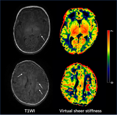 |
7 | Quantitative assessment of neonatal white matter injury stiffness by virtual magnetic resonance elastography
Miaomiao Wang1, Congcong Liu1, Xianjun Li1, and Jian Yang1
1the first affiliated hospital of Xi'an Jiaotong university, Xi'an, China
Punctate white matter lesion (PWML) is the most common injury in neonates. Due to newborns are a vulnerable population, the limited imaging protocols are not sufficient to fully understand the injury. Le Bihan et.al recently proposed a virtual MR elastography (vMRE) method based on multiple b-values diffusion sequences, which is attractive for evaluation of brain development and injury. This study aims to quantitatively assess the stiffness of PWML using vMRE. Compared with white matter regions, a significant increased virtual sheer stiffness is observed in lesions, and it may be a feasible clinical evaluation of the pathophysiological state of brain tissue.
|
||
2972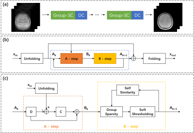 |
8 | Accelerating Myelin Water Content Quantification using Deep Non-Local Sparse Model
Bowen Li1, Huajun She1, Quan Chen2, Zhijun Wang1, and Yiping P. Du1
1School of Biomedical Engineering, Shanghai Jiao Tong University, Shanghai, China, 2Department of Radiology, Stanford University, Stanford, CA, United States
A grouping sparse coding (Group-SC) model is used in this study for the acceleration of myelin water content quantification. Images with improved quality are obtained using the Group-SC algorithm, in the aspect of minimal artefacts and good data consistency with fully-sampled labels. Myelin water fraction (MWF) maps reconstructed using Group-SC demonstrate more natural spatial distribution of myelin water in the brain. The proposed Group-SC algorithm has demonstrated its potential for the acceleration of the myelin water content quantification at R = 6.
|
||
2973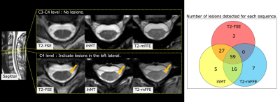 |
9 | Inhomogeneous Magnetization Transfer (ihMT) Helps to Detect Multiple Sclerosis Plaques in The Spinal Cord.
Ryuna Kurosawa1, Hajime Yokota2, Takafumi Yoda1, Takayuki Sada1, Koji Matsumoto1, Takashi Namiki3, Masami Yoneyama3, Guillaume Gilbert4, Yoshitada Masuda1, and Takashi Uno2
1Department of Radiology, Chiba Univercity Hospital, Chiba, Japan, 2Diagnostic Radiology and Radiation Oncology, Graduate School of Medicine, Chiba University, Chiba, Japan, 3Philips Japan, Tokyo, Japan, 4Philips Canada, Mississauga, ON, Canada
Demyelination assessment with MRI for multiple sclerosis (MS) is a reliable biomarker for monitoring disease progression. Recently, inhomogeneous magnetization transfer (ihMT) combined sequencing has been developed for myelin-specific imaging. The purpose of this study was to evaluate ihMT in delineating MS lesions in the spinal cord by comparing the conventional imaging methods. The lesion-to-white matter contrast-to-noise ratio (CNR) on ihMT was significantly higher than the other methods. The qualitative analysis revealed clarity of lesion detection, but false positives were a concern. Our results in MS patients suggest that ihMT may be a helpful sequence for lesion detection.
|
||
2974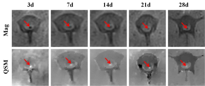 |
10 | Application of MRI in tracking and evaluating of SPIONS labeled stromal stem cells transplanted in traumatic spinal cord injury of beagle Video Permission Withheld
Xiaoli Mai1, Jilei Zhang2, Junting Zou1, Yuanyuan Xie3, Bin Wang3, and Bing Zhang1
1Radiology Department, Nanjing Drum Tower Hospital, the affiliated hospital of Nanjing University Medical School, Nanjing, China, 2Philips Healthcare, Shanghai, China, 3Nanjing Drum Tower Hospital, Nanjing, China
Stromal cell transplantation plays an important role in the treatment of traumatic spinal cord injury (SCI). The multi-parameter MRI, including DTI, T2 , T2* mapping and QSM , of all beagles were performed before and after operation. We found that the transplanted stromal stem cells with Ruicun-labeled in the injured spinal cord could be quantified by QSM within 28 days after transplantation. FA value can be used to quantitatively evaluate the integrity of spinal fibers and the recovery of spinal nerve function. Thus, MRI can used to track the dynamic changes of SPIONS labeled stromal stem cells transplanted.
|
||
2975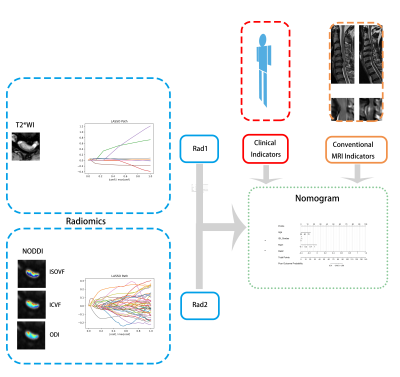 |
11 | Radiomic approach for predicting short-term postoperative recovery of cervical spondylotic myelopathy based on NODDI and T2*WI Video Permission Withheld
Meng-Ze Zhang1, Han-Qiang Ou-Yang1,2,3, Chun-Jie Wang1, Jian-Fang Liu1, Dan Jin1, Xian-Chang Zhang4, Qiang Zhao1, Xiao-Guang Liu1,2,3, Zhong-Jun Liu1,2,3, Ning Lang1, Xing-Wen Sun1, Liang Jiang1,2,3, and Hui-Shu Yuan1
1Peking University Third Hospital, Beijing, China, 2Engineering Research Center of Bone and Joint Precision Medicine, Beijing, China, 3Beijing Key Laboratory of Spinal Disease Research, Beijing, China, 4MR Collaboration, Siemens Healthineers Ltd, Beijing, China
This study used radiomics based on T2*-weighted imaging (T2*WI) and neurite orientation dispersion and density imaging (NODDI) to predict the short-term recovery of patients with cervical spondylotic myelopathy (CSM). By classifying patients into good and poor outcomes based on the 3-month recovery rate, we found both T2*WI- and NODDI-based radiomic features to have good prognostic power. Furthermore, radiomic score based on NODDI was an independent predictor, whereas the other features were not. These findings suggest that radiomics based on NODDI has good prognostic power for CSM.
|
||
2976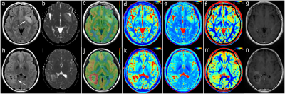 |
12 | The Diagnostic Value of Quantitative Analysis of Synthetic MRI, ASL and DWI in Grading Gliomas as Compared to Conventional MRI: A Preliminary Study
Xin Ge1,2, Xueying Huang2, Aijun Wang2, Kai Zhu2, Xiaocheng Wei3, Min Li4, Ying Shen1,2, Wenxiao Liu1,2, Peng Yong1,2, Ruirui Lv1,2, Xuhong Yang1,2, and Xiaodong Wang2
1Ningxia Medical University, Yinchuan, China, 2General Hospital of Ningxia Medical University, Yinchuan, China, 3GE Healthcare, MR Research, Beijing, China, Beijing, China, 4GE Healthcare, MR Enhancement Application, Beijing, China, Beijing, China
This work sought to investigate the performance of synthetic MRI, ASL and DWI in differentiating LGGs from HGGs as compared to conventional MRI. It was concluded that the T1 and PD from synthetic MRI can be used as novel quantitative imaging biomarkers for grading gliomas. Combining T1, PD, CBF and ADC may explore as an effective strategy to improve the ability for discriminating gliomas grade, and outperformed conventional approaches.
|
||
2977 |
13 | Can MAP-MRI model be used for advance prediction of Grade II/III diffuse glioma genetics?
Peng Wang1, Yang Gao1, Qiong Wu1, Jinlong He1, Shenghui Xie1, Shaoyu Wang2, and Huapeng Zhang2
1Radiology, Affiliated Hospital of Inner Mongolia Medical University, Hohhot, China, 2Siemens Healthineers, Shanghai, China
Although surgery is still the most effective treatment for diffuse glioma, patients with unresectable primary, recurrent, or multicentric illness have few options. In some cases, genotyping predictions may be useful for targeted therapy. We performed pretreatment MRI scans and genetic sequencing of tumor tissue, as well as correlation analysis, to see if essential molecular genetic events linked with diffuse glioma may be predicted in advance and noninvasively. The findings revealed that the MAP-MRI model can reliably predict tumor genotyping with high robustness and diagnostic validity, implying that it could be useful in the development of more standardized treatment.
|
||
2978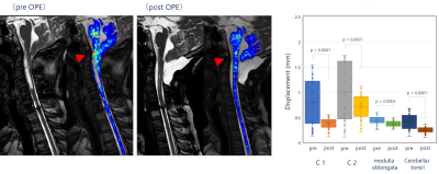 |
14 | Quantitative Evaluation of Spinal Cord Cine-MRI with Optical Flow Analysis
Tomohiko Horie1, Natsuo Konta1, Masateru Kawakubo2, Hiroshi Hamano3, Han Soo Chang4, Tetsu Niwa5, Kagayaki Kuroda6, and Mitsunori Matsumae4
1Department of Radiology, Tokai University Hospital, Isehara, Japan, 2Department of Health Sciences, Faculty of Medical Sciences, Kyushu University, Fukuoka, Japan, 3Philips Japan, Shinagawa, Japan, 4Department of Neurosurgery, Tokai University School of Medicine, Isehara, Japan, 5Department of Radiology, Tokai University School of Medicine, Isehara, Japan, 66) Department of Human and Information Science, School of Information Science and Technology, Tokai University, Hiratsuka, Japan
There are still many unclear issues in the motions of the spinal cord. This study is the first report to use optical flow analysis for quantitative evaluation of spinal cord motions. In the study of healthy volunteers, we found large displacement of the upper thoracic spinal cord. The study of clinical cases revealed that the spinal cord displacement changed before and after surgery. Therefore, these results suggested that quantitative evaluation of spinal cord motions is feasible using optical flow analysis.
|
||
| 2979 | 15 | Feasibility of 3D-MENSA sequence for qualitative assessment of lumbosacral plexus nerve root in comparison with 3D-MERGE MRI Video Permission Withheld
Shuang Hu1, BoWen Hou1, YiTong Li1, Yao Zhang1, Weiyin Vivian Liu2, and XiaoMing Li1
1Tongji Hospital,Tongji Medical College,Huazhong University of Science and Technology, Wuhan,China, China, 2GE Healthcare, Beijing, China., China
Lumbar radiculopathy is a worldwide cause of disability. Mechanical compression of nerve root may lead to nerve inflammatory changes. Peripheral blood vessels overlapping with inflammatory changes can make radiologists confused and result misdiagnosis. To differ lesions from vessels in lumbosacral plexus nerve imaging, three-dimensional motion-sensitized driven equilibrium prepared rapid gradient echo (3D-MERGE) and three-dimensional multiecho in the steady-state acquisition (3D-MENSA) were investigated. Our study showed 3D-MENSA images showed greater nerve-to-vein CNR and CR than 3D-MERGE and the measurement repeatability of the same observer was good. Overall, 3D-MENSA sequence provided images with superior vascular suppression and offered better conspicuity of LSP.
|
||
 Back to Meeting Home
 Back to the Program-at-a-Glance
The International Society for Magnetic Resonance in Medicine is accredited by the Accreditation Council for Continuing Medical Education to provide continuing medical education for physicians.
 Back to Meeting Home
 Back to the Program-at-a-Glance
 View the Presentation