Weekday Course
Physics for Clinicians I
Joint Annual Meeting ISMRM-ESMRMB & ISMRT 31st Annual Meeting • 07-12 May 2022 • London, UK

| Physics for Clinicians I.A | |||
| 14:45 | Where Does the Signal Come From?
Robert Thomen
Where exactly does the signal of an NMR experiment come from? How is tissue contrasted in an MR image? What can be done to improve signal? What are T1, T2, and Boltzmann Magnetization? In this lecture we will examine the foundations of the NMR experiment and build an expression for the detected signal which determines the voxel brightness in MRI. We will take a conceptual approach to understanding NMR beginning with the NMR experiment, precession, nuclear magnetization, and relaxation.
|
||
| 15:15 | Overview of MR Hardware & Its Function
Rebecca Feldman
Magnetic Resonance Imaging is a powerful tool use to non-invasively and non-destructively image the human body. This is done by carefully timing and manipulating the magnetic fields. The system that makes this happen is generally composed of at three different types of electromagnets, as well as systems for control and maintenance. The physics underlying the function of these systems will be discussed, without mathematical detail.
|
||
| Physics for Clinicians I.B | |||
| 15:45 | Anatomy of a Pulse Sequence
Michael Schär
In MRI, the previously described electromagnetic fields are applied sequentially to localize the signal within the body. The timings and strengths of these fields are described by standard parameters and visualized with pulse sequence diagrams. In this talk, the most basic MRI parameters and sequences will be introduced, and how changing them affects the image contrast.
|
||
| 16:15 | 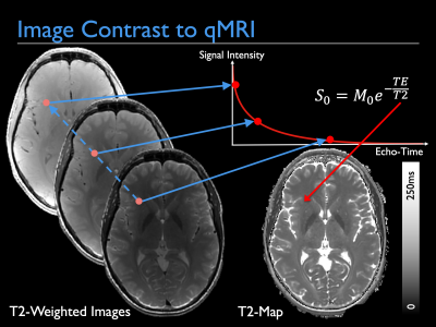 |
From Image Contrast to Quantitative Imaging
Tobias Wood
This talk will provide a grounding in how to turn standard MR contrast images into quantitative maps. Such maps represent meaningful physical and biological parameters with known units. This provides new information on a voxel-wise level with which to base clinical diagnoses. This development can be seen as part of the continuing development of MRI as a field, turning the scanner from a fancy camera into a precise scientific measuring instrument. |
|
 Back to Meeting Home
 Back to the Program-at-a-Glance
The International Society for Magnetic Resonance in Medicine is accredited by the Accreditation Council for Continuing Medical Education to provide continuing medical education for physicians.
 Back to Meeting Home
 Back to the Program-at-a-Glance
 View the Presentation