Weekday Course
The Heart-Brain Connection: Linking Cardiovascular to Cerebral Function
Joint Annual Meeting ISMRM-ESMRMB & ISMRT 31st Annual Meeting • 07-12 May 2022 • London, UK

Weekday Course
The Heart-Brain Connection: Linking Cardiovascular to Cerebral Function
| Basic Science | |||
| 17:00 | Heart & Brain: Physiology, Cerebrovascular Reactivity & Perfusion
Claudine Gauthier
|
||
| 17:20 | The Central Autonomic Network in Health
Nicola Toschi
|
||
| 17:40 | 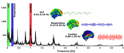 |
Functional Connectivity & Brain Perfusion: Cardiorespiratory brain pulsations
Vesa Kiviniemi
A century after the detection of three pulsation mechanisms in the human brain during neurosurgical procedures, ultrafast fMRI enables exact depiction of the vasomotor waves, respiratory and cardiac pulsations non-invasively. Arterial blood impulses and respiration induced venous pulsations form continuosly propagating CSF waves that flush brain paravascular spaces and drive the glymphatic solute transport. Sleep increases all these pulsations in areas of increased brain interstiatial flushing. Brain diseases such as Alzheimer's disease, epilepsy, and PCNSL invading the perivascular space, significantly alter the pulsation dynamics of the brain, which enables direct visualization of individual pathology with ultrafast BOLD scans.
|
|
| Applications | |||
| 18:00 | Brain MRI Signal Changes Due to Cardiac Disease in Aging
Fang Yu
The heart and the brain are intimately linked. Herein, we review the changes in the brain as we age that can be detected using clinical MRI, and how they relate to cardiovascular disease. Additionally, we will review imaging techniques including diffusion imaging for tissue microstructure and quantitative susceptibility mapping for iron accumulation as it relates to these neurologic changes.
|
||
| 18:20 | Role of MRI to Examine Brain Structural, Metabolic, Hemodynamic, and Functional Deficits in Adults After Heart Failure
Rajesh Kumar
Novel MRI procedures and analytical methods offer a unique opportunity to assess the brain structural, metabolic, hemodynamic, blood brain barrier, resting-state functional connectivity, and functional responses to autonomic challenges status in heart failure (HF) subjects. In a series of experiments in patients with HF, we characterized brain injury, abnormal metabolites, hemodynamics, resting-state functional connectivity, and abnormal functional responses to autonomic challenges, in autonomic, mood, and cognitive control sites, functions that are deficient in HF. Also, potential pathological mechanisms, including compromised CBF and BBB function, contributing to brain damages in HF will be discussed.
|
||
| 18:40 | Brain MRI Signal Changes in Autonomic Dysfunction (Multiple System Atrophy, Hypotension, Dysautonomia)
Stephen Jones
The first step of understanding the neuroimaging of autonomic function is to focus on normal subjects with normal brains, and then investigate the structural and functional correlates of the autonomic system. The next step is to investigate diseases affecting the autonomic system, namely those causing dysautonomia. This step may be even more revealing than normal subjects, since often the understanding of disease helps to better define normal function. This presentation will provide multiple examples of diseases associate with either primary or secondary dysautonomia. Examples include multiple systems atrophy (MSA), multiple sclerosis, tumors, stroke, and epilepsy.
|
||
 Back to Meeting Home
 Back to the Program-at-a-Glance
The International Society for Magnetic Resonance in Medicine is accredited by the Accreditation Council for Continuing Medical Education to provide continuing medical education for physicians.
 Back to Meeting Home
 Back to the Program-at-a-Glance
 View the Presentation