Oral Session
Data Analysis Outside the Head
Joint Annual Meeting ISMRM-ESMRMB & ISMRT 31st Annual Meeting • 07-12 May 2022 • London, UK

| 09:15 | 0615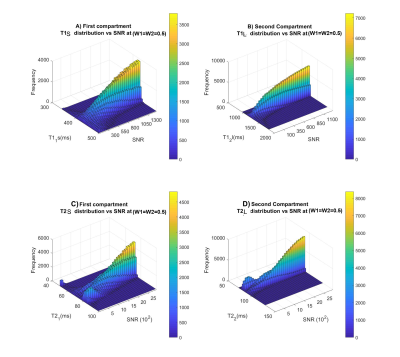 |
Evaluation of precision of multi-compartment model on relaxation properties in breast cancer on 3T using numerical simulation
Roshni Senthilkumar1,2, Sai Man Cheung2, Kwok-Shing Chan3, and Jiabao He2
1Radiology Physics, University Hospital Coventry and Warwickshire, Coventry, United Kingdom, 2Institute of Medical Sciences, School of Medicine, Medical Sciences and Nutrition, University of Aberdeen, Fosterhill, Aberdeen, Scotland, 3Donders Institute for Brain, Cognition and Behaviour, AJ Nijmegen, Germany
MR relaxation properties of T1 and T2 are well known to alter in cancer, reflecting diseased tissue micro-environment. Multiple compartment models have been developed to better approximate tissue classes within an imaging voxel, allowing more accurate estimation of disease load for more precise treatment planning and monitoring. However, multiple compartment models is more sensitive to noise and suffers from potential overfitting, due to from significantly increased number of variables for fitting exponential functions and consequently more complex cost function. We therefore conducted numerical simulation to establish, the first in the literature, applicability condition of multiple compartment model in breast cancer.
|
|
| 09:27 | 0616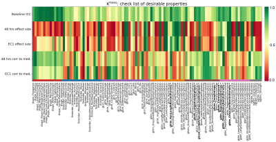 |
A principled approach to select DCE-MRI derived radiomics features for evaluation in phase I/II trials
Michael Berks1, Damien J McHugh2, Nuria Porta3, Ross A Little1, Susan Cheung1, Gordon C Jayson4,5, Geoff J M Parker6,7, and James P B O'Connor1,8,9
1Quantitative Biomedical Imaging Laboratory, Division of Cancer Sciences, University of Manchester, Manchester, United Kingdom, 2Medical Physics, The Christie Hospital NHS Trust, Manchester, United Kingdom, 3Clinical Trials and Statistics Unit, The Institute of Cancer Research, London, United Kingdom, 4Division of Cancer Sciences, University of Manchester, Manchester, United Kingdom, 5Department of Medical Oncology, The Christie Hospital NHS Trust, Manchester, United Kingdom, 6Centre for Medical Image Computing, Department of Medical Physics and Biomedical Engineering, University College London, London, United Kingdom, 7Bioxydyn Ltd, Manchester, United Kingdom, 8Department of Radiology, The Christie Hospital NHS Trust, Manchester, United Kingdom, 9Division of Radiotherapy and Imaging, The Institute of Cancer Research, London, United Kingdom
DCE-MRI biomarkers such as change in median Ktrans have a proven role in drug development in phase I/II trials. There is current interest in using approaches such as radiomics to extract additional information relating to spatial heterogeneity from images and one emerging application is to apply these analyses to clinical trial data where imaging is used to monitor pharmacodynamic change in the tumour microenvironment. Here, we explore the properties of radiomics features extracted from maps of Ktrans and aim to identify features that are repeatable at baseline, show consistent treatment effect and provide additional, independent information to the median Ktrans.
|
|
| 09:39 | 0617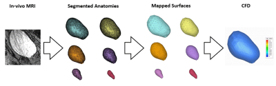 |
Predictive Bladder Urodynamics Using Real-Time Magnetic Resonance Imaging: A Pilot Study
Labib Shahid1, Juan Pablo Gonzalez-Pereira1, Cody Johnson1, Yanheng Li2, David Rowinski2, and Alejandro Roldán-Alzate1
1University of Wisconsin-Madison, Madison, WI, United States, 2Convergent Science, Inc., Madison, WI, United States
Multi-channel urodynamics is an invasive diagnostic method used to assess bladder biomechanics. MRI-based CFD has shown high potential as a clinical tool primarily focused on cardiovascular flows. In this pilot study, we demonstrate MRI-based CFD as a tool to study urodynamics. Real-time MRI on one subject provided bladder wall surfaces at multiple time points during voiding. We developed a surface mapping algorithm that processes the bladder geometries before inputting them into a CFD simulation. Coupling MRI with CFD successfully visualized and quantified urine flow dynamics. This provides a non-invasive tool to investigate urodynamics in common urological conditions such as BPH/LUTS.
|
|
| 09:51 | 0618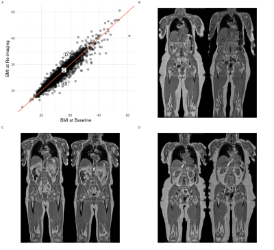 |
Changes in Abdominal Organs from the UK Biobank Longitudinal Imaging Study Video Permission Withheld
Brandon Whitcher1, Marjola Thanaj1, Madeleine Cule2, Nicolas Basty1, Elena P. Sorokin2, Jimmy D. Bell1, and E. Louise Thomas1
1Research Centre for Optimal Health, University of Westminster, London, United Kingdom, 2Calico Life Sciences LLC, South San Francisco, CA, United States
We investigated changes in body composition in 3,088 free-living participants of the UK Biobank imaging study. Statistical models utilised organ and tissue segmentations from the neck-to-knee Dixon acquisitions and multiecho sequences of the liver/pancreas. A significant decrease in grip strength was observed, and small, but statistically significant, decreases in all skeletal muscle measurements. Significant increases in visceral adipose tissue and intermuscular fat in the thighs were also found in the absence of changes in BMI, waist circumference and ectopic-fat deposition. We have shown that even after a relatively short period of time significant changes in body composition are observable.
|
|
| 10:03 | 0619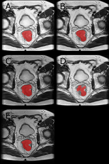 |
Comparison of Image Normalization Techniques for Rectal Cancer Segmentation in Multi-Center Data: Initial results
Steffen Albert1, Barbara D. Wichtmann2, Wenzhao Zhao3, Jürgen Hesser3, Ulrike I. Attenberger2, Lothar R. Schad1, and Frank G. Zöllner1
1Computer Assisted Clinical Medicine, Mannheim Institute for Intelligent Systems in Medicine, Medical Faculty Mannheim, Heidelberg University, Mannheim, Germany, 2Department of Diagnostic and Interventional Radiology, University Hospital Bonn, Bonn, Germany, 3Data Analysis and Modeling in Medicine, Mannheim Institute for Intelligent Systems in Medicine, Medical Faculty Mannheim, Heidelberg University, Mannheim, Germany We evaluated the influence of normalization (setting mean and standard deviation, histogram matching and percentiles) on the segmentation of rectal cancer on multimodal images when operating on multicenter data as part of a Radiomics pipeline. We used two different networks for segmentation. When training and evaluating on all data or data from a single center, normalization did not play a significant role. In contrast, when training on one center and evaluating on all others, it did play a major role. Best results are obtained by normalization using percentiles. Fixing the mean and standard deviation did not work well. |
|
| 10:15 | 0620 |
DCE-MRI with free-breathing compressed sensing VIBE for predicting chemotherapy response and patient outcome in pancreatic adenocarcinoma Video Not Available
Yoshihiko Fukukura1, Fumitaka Ejima1, Takuro Ayukawa1, Kiyohisa Kamimura1, Masanori Nakajo1, Hiroaki Nagano1, Koji Takumi1, Marcel Dominik Nickel2, Hiroshi Imai3, and Takashi Yoshiura1
1Kagoshima University Graduate School of Medical and Dental Sciences, Kagoshima, Japan, 2MR Application Predevelopment, Siemens Healthcare GmbH, Erlangen, Germany, 3Siemens Healthcare K.K., Tokyo, Japan
This study focused on the feasibility of dynamic contrast-enhanced MRI (DCE-MRI) with motion-resolved compressed sensing T1-weighted volumetric interpolated breath-hold examination (CS-VIBE) for prediction of treatment response to chemotherapy and patient outcome in pancreatic ductal adenocarcinoma (PDAC). Our results showed tumor ve was significantly higher in the response group than in the non-response group and was an independent predictor of progression-free survival in patients with PDAC treated with chemotherapy. These results suggest that DCE-MRI obtained with CS-VIBE may be useful for predicting treatment response to chemotherapy and patient outcome in PDAC.
|
 Back to Meeting Home
 Back to the Program-at-a-Glance
The International Society for Magnetic Resonance in Medicine is accredited by the Accreditation Council for Continuing Medical Education to provide continuing medical education for physicians.
 Back to Meeting Home
 Back to the Program-at-a-Glance
 View the Presentation