Oral Session
Signal Representations
Joint Annual Meeting ISMRM-ESMRMB & ISMRT 31st Annual Meeting • 07-12 May 2022 • London, UK

| 17:00 | 0748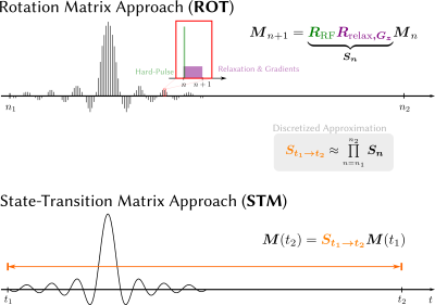 |
Efficient Bloch Simulation Based on Precomputed State-Transition Matrices
Nick Scholand1,2,3, Christina Graf1, and Martin Uecker1,2,3
1Institute of Medical Engineering, Graz University of Technology, Graz, Austria, 2Department of Interventional and Diagnostic Radiology, University Medical Center Göttingen, Göttingen, Germany, 3German Centre for Cardiovascular Research, Göttingen, Germany The Bloch equations describe the effects of relaxation and external fields on the magnetization. Its coefficients are time-dependent, requiring computationally expensive techniques for its solution in the generic case. Currently, most techniques used in MRI are based on temporal discretization, but this requires very small time steps to achieve sufficiently small approximation errors. |
|
| 17:12 | 0749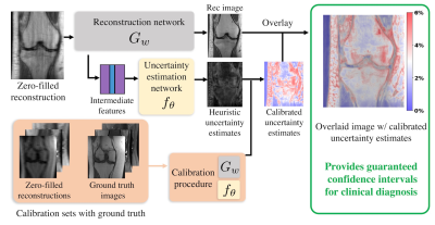 |
Rigorous Uncertainty Estimation for MRI Reconstruction
Ke Wang1,2, Anastasios Angelopoulos1, Alfredo De Goyeneche1, Amit Kohli1, Efrat Shimron1, Stella Yu1,2, Jitendra Malik1, and Michael Lustig1
1Electrical Engineering and Computer Sciences, University of California, Berkeley, Berkeley, CA, United States, 2International Computer Science Institute, University of California, Berkeley, Berkeley, CA, United States
Deep-learning (DL)-based MRI reconstructions have shown great potential to reduce scan time while maintaining diagnostic image quality. However, their adoption has been plagued with fears that the models will hallucinate or eliminate important anatomical features. To address this issue, we develop a framework to identify when and where a reconstruction model is producing potentially misleading results. Specifically, our framework produces confidence intervals at each pixel of a reconstruction image such that 95% of these intervals contain the true pixel value with high probability. In-vivo 2D knee and brain reconstruction results demonstrate the effectiveness of our proposed uncertainty estimation framework.
|
|
| 17:24 | 0750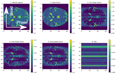 |
Phase distribution graphs for differentiable and efficient simulations of arbitrary MRI sequences
Jonathan Endres1, Hoai Nam Dang1, Felix Glang2, Alexander Loktyushin2, Simon Weinmüller1, and Moritz Zaiss1,2
1Department of Neuroradiology, Universitätsklinik Erlangen, Erlangen, Germany, 2Magnetic Resonance Center, Max-Planck-Institute for Biological Cybernetics, Tübingen, Germany
We propose an alternate method for simulating arbitrary MRI sequences, forming complete Bloch simulations. It is closely related to Extended Phase Graphs but imposes no restrictions on the alignment of timing or gradients, extends it to support T2' relaxation and 3D gradient encoding, is fully differentiable, provides additional insight into the composition of the measured signal while outperforming isochromat-based Bloch simulations in execution speed by using an analytical description of the signal instead of relying on a Monte-Carlo simulation.
|
|
| 17:36 | 0751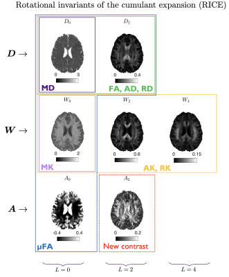 |
Rotational invariants of the cumulant expansion (RICE)
Santiago Coelho1, Els Fieremans1, and Dmitry S. Novikov1
1Center for Advanced Imaging Innovation and Research (CAI2R), Department of Radiology, New York University, School of Medicine, New York, NY, United States
We studied the symmetries of the diffusion and covariance tensors, both accessible through a general b-tensor acquisition. We showed that there is an unexplored contrast in the non-symmetric part of the covariance tensor and use it to compute the size-shape covariance of compartmental diffusion tensors. We measure all rotational invariants of the cumulant tensors in a normal volunteer, explore the novel size-shape contrast, and provide the relations between these invariants and typical diffusion contrasts.
|
|
| 17:48 | 0752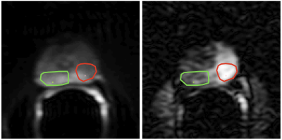 |
Bayesian Estimation of Diffusivity Spectra: Application to Prostate Diffusion MRI
William M Wells1, Stephan E Maier1, and Carl-Fredrik Westin1
1Brigham and Women's Hospital, Harvard Medical School, Boston, MA, United States
There is great interest to quantify the spectrum of diffusivities that underlie the observed diffusion signal decay; separate tissue compartments can be identified by their spectral peaks. This spectrum is defined by the inverse Laplace transform, but unfortunately this transform is very sensitive to noise omnipresent in diffusion MRI. We present a Bayesian method of inverse Laplace transform that uses Gibbs sampling to provide spectra along with an estimate of the noise related uncertainty in the spectra; this uncertainty information is valuable in interpreting the results. This is applied to multi-b high SNR endorectal coil diffusion data of the prostate.
|
|
| 18:00 | 0753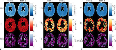 |
Voxel-wise compartmental modelling of blood-brain barrier water exchange measurements using FEXI
Elizabeth Powell1, Yolanda Ohene2,3, Marco Battiston4, Laura M Parkes2,3, and Geoff JM Parker5,6
1Centre for Medical Image Computing, Department of Computer Science, University College London, London, United Kingdom, 2Division of Neuroscience and Experimental Psychology, University of Manchester, Manchester, United Kingdom, 3Geoffrey Jefferson Brain Research Centre, University of Manchester, Manchester, United Kingdom, 4NMR Research Unit, Queen Square Multiple Sclerosis Centre, Department of Neuroinflammation, UCL Queen Square Institute of Neurology, University College London, London, United Kingdom, 5Centre for Medical Image Computing, Department of Medical Physics and Biomedical Engineering, University College London, London, United Kingdom, 6Bioxydyn Limited, Manchester, United Kingdom
We propose compartmental modelling of blood-brain barrier (BBB) water exchange measurements using diffusion-filtered exchange imaging (FEXI), with the aim of providing greater biophysical insight into BBB function than is possible using the apparent exchange rate (AXR) approach. As relaxation time differences between blood and extravascular tissue have not yet been accounted for in FEXI-based BBB permeability measurements, we use simulations to quantify potential biases in exchange rates from both the AXR and compartmental approaches. Finally, we evaluate the repeatability of the AXR and compartmental models in a cohort of healthy subjects.
|
|
| 18:12 | 0754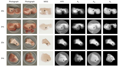 |
Mapping the myelin bilayer with short-T2 MRI: Validation studies
Emily Louise Baadsvik1, Markus Weiger1, Romain Froidevaux1, Wolfgang Faigle2, Benjamin Victor Ineichen3, and Klaas Paul Pruessmann1
1Institute for Biomedical Engineering, ETH Zurich and University of Zurich, Zurich, Switzerland, 2Neuroimmunology and MS Research Section, Neurology Clinic, University of Zurich, University Hospital Zurich, Zurich, Switzerland, 3Department of Neuroradiology, Clinical Neuroscience Center, University Hospital Zurich, University of Zurich, Zurich, Switzerland
Various techniques for myelin mapping based on signal from the lipid-protein bilayer have been proposed, and a common way of validating these techniques is using ex-vivo tissue samples. However, it is still unclear to what extent experimental factors such as tissue storage conditions and processing affect MR signal. In this work, we investigate how long-term deep-frozen storage impacts tissue signal, and evaluate whether signal component mapping is feasible in non-D2O-exchanged samples. We also determine whether animal tissue can act as a substitute for human tissue and investigate signal differences between white and grey matter.
|
 Back to Meeting Home
 Back to the Program-at-a-Glance
The International Society for Magnetic Resonance in Medicine is accredited by the Accreditation Council for Continuing Medical Education to provide continuing medical education for physicians.
 Back to Meeting Home
 Back to the Program-at-a-Glance
 View the Presentation