Oral Session
Advances in Image Acquisition for Diffusion & Perfusion
Joint Annual Meeting ISMRM-ESMRMB & ISMRT 31st Annual Meeting • 07-12 May 2022 • London, UK

Oral Session
Advances in Image Acquisition for Diffusion & Perfusion
| 09:15 | 0042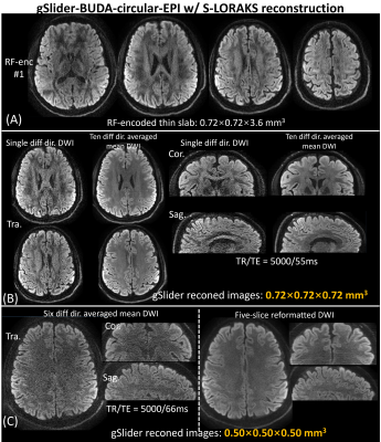 |
High-fidelity submillimeter-isotropic-resolution diffusion MRI through gSlider-BUDA and circular EPI with S-LORAKS reconstruction
Congyu Liao1,2, Uten Yarach3, Xiaozhi Cao1,2, Siddharth Srinivasan Iyer4, Nan Wang1,2, Tae Hyung Kim5,6, Berkin Bilgic5,6, Adam Kerr1,7, and Kawin Setsompop1,2
1Department of Radiology, Stanford University, Stanford, CA, United States, 2Department of Electrical Engineering, Stanford University, Stanford, CA, United States, 3Radiologic Technology Department, Associated Medical Sciences, Chiang Mai University, Chiang Mai, Thailand, 4Department of Electrical Engineering and Computer Science, Massachusetts Institute of Technology, Cambridge, MA, United States, 5Athinoula A. Martinos Center for Biomedical Imaging, Massachusetts General Hospital, Charlestown, MA, United States, 6Department of Radiology, Harvard Medical School, Boston, MA, United States, 7Stanford Center for Cognitive and Neurobiological Imaging, Stanford University, Stanford, CA, United States
BUDA-cEPI combines a circular-EPI (cEPI) trajectory with Blip-Up and Down Acquisition (BUDA), to achieve high-fidelity diffusion imaging. It employs full-ramp-sampling to efficiently cover a circular k-space to shorten the readout-train and mitigate T2*-blurring. For further readout-shortening, a combined phase-encoding(PE) & readout(RO) partial-Fourier (pF) is employed, where PE-pF is resolved via opposing the blip-up and down EPI-shots, and S-LORAKS reconstruction is used to effectively fill-out the remaining k-space. This resulted in an acquisition with markedly-reduced TE and ~40% reduced echo-train-length and T2*-blurring compared to standard-EPI at the same Rinplane and PE-pF. To achieve high-SNR, BUDA-cEPI is also combined with a gSlider-multi-slab acquisition.
|
|
| 09:27 | 0043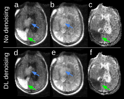 |
Diffusion-Weighted Imaging at 0.064 T
Rafael O'Halloran1, Hadrien Dyvorne1, Laura Sacolick1, Jo Schlemper1, Michal Sofka1, Sadegh Salehi1, Samantha By1, Riana Schleicher2, Edmond Knopp1, Kevin Sheth3, and W. Taylor Kimberly2
1Hyperfine, Inc, Guilford, CT, United States, 2Neurology, Massachusetts General Hospital, Boston, MA, United States, 3Yale New Haven Hospital, New Haven, CT, United States
A sequence is described for DWI on a portable, 0.064T scanner. Images in a healthy subject and a patient with pathology are shown.
|
|
09:39 |
0044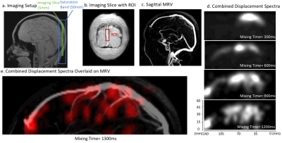 |
“Reverse Perfusion” Imaging of the Cerebral Venous System with Displacement Spectrum Imaging (DiSpect)
Ekin Karasan1, Zhiyong Zhang2, and Michael Lustig1
1University of California, Berkeley, Berkeley, CA, United States, 2Biomedical Engineering, Shanghai Jiao Tong University, Shanghai, China
Displacement Spectrum Imaging (DiSpect) is a Fourier encoding variant of Displacement Encoding with Stimulated Echoes (DENSE). DiSpect can resolve a multi-dimensional spectrum of displacements that spins exhibit between tagging and imaging. We previously demonstrated ASL-like retrospective vessel-selective perfusion imaging with DiSpect. Here, we demonstrate the versatility of DiSpect by doing the reverse; tracing how blood drains from the capillary bed through the cerebral venous system. We image two locations in the superior sagittal sinus and trace how the superior superficial veins drain into the superior sagittal sinus, in a sense measuring reverse perfusion.
|
|
| 09:51 | 0045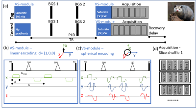 |
Perfusion Observations using Spherical Slice-shuffled Ultra-high-field MRI (POSSUM)
Jana Hutter1,2, Raphael Tomi-Tricot3, Thomas A Wilkinson1,2, Philippa A Bridgen1,2, Enrico DeVita2, Shaihan A Malik1,2, and Joseph V Hajnal1,2
1London Collaborative Ultra high field System (LoCUS), London, United Kingdom, 2Centre for Medical Engineering, King's College London, London, United Kingdom, 3Siemens Healthcare Limited, Frimley, United Kingdom
Spin tagging perfusion MR imaging allows the movement of blood in human tissue to be visualized and quantified non-invasively and contrast agent-free. Velicity-selection Arterial Spin Labeling (VSASL) relies on the velocity instead of the location for tagging. It is less dependent on the anatomy and coil coverage but includes directional dependence and can suffer from reduced SNR. POSSUM expands VSASL to spherical, directional-independant encoding and slice shuffling to enable more robust perfusion imaging at 7T.
|
|
| 10:03 | 0046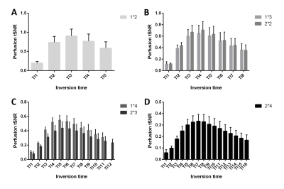 |
Optimization of perfusion measurements at 7 T using pulsed ASL with simultaneous multi-slice EPI readouts at multiple post-labelling delays
Dimo Ivanov1, Sriranga Kashyap1,2, Laurentius Huber1, Josef Pfeuffer3, Kâmil Uludağ4, and Benedikt A Poser1
1Department of Cognitive Neuroscience, Maastricht University, Maastricht, Netherlands, 2Techna Institute, University Health Network, Toronto, ON, Canada, 3Siemens Healthineers, Erlangen, Germany, 4Techna Institute & Koerner Scientist in MR Imaging, Toronto, ON, Canada
Arterial spin labeling (ASL) at 7T is beneficial due to the higher signal-to-noise ratio (SNR) and the longer T1 of blood and tissues. A whole-brain 7T pulsed ASL approach with simultaneous multi-slice (SMS) echo planar imaging readouts at multiple inversion times is presented. The interplay between the number of inversion times acquired, the total acceleration factor employed, and the temporal SNR was investigated. In summary, a protocol with in-plane acceleration factor 2 and SMS factor 2 offers the best compromise between perfusion tSNR and number of inversion times and can be used in clinical applications investigating perfusion parameters beyond CBF.
|
|
| 10:15 | 0047 |
The Open Science Initiative for Perfusion Imaging (OSIPI): Early Results from the DCE-MRI Challenge
Eve Shalom1, Harrison Kim2, Rianne A. van der Heijden3, Zaki Ahmed4, Reyna Patel5, David A. Hormuth6, Julie C. DiCarlo7, Nicholas J. Sisco8, Richard D. Dortch8, Ashley M. Stokes8, Marianna Inglese9,10, Matthew Grech-Sollars11,12, Nicola Toschi13,14, Prativa Sahoo15, Anup Singh16, Sanjay K. Verma17, Divya K. Rathore18, Ali Nabavizadeh19, Hamidreza Saligheh Rad20,21, Leland S. Hu22,
Laura C. Bell23, Steven Sourbron24, and Anahita Fathi Kazerooni25
1School of Physics and Astronomy, University of Leeds, Leeds, United Kingdom, 2The University of Alabama at Birmingham, Birmingham, AL, United States, 3Department of Radiology & Nuclear Medicine, Erasmus MC University Medical Center, Rotterdam, Netherlands, 4Mayo Clinic, Rochester, MN, United States, 5Department of Radiology, Neuroradiology Division, Mayo Clinic, Scottsdale, AZ, United States, 6Oden Institute for Computational Engineering and Sciences, The University of Texas at Austin, Austin, TX, United States, 7University of Texas at Austin, Austin, TX, United States, 8Neurological Imaging, Barrow Neurological Innovation Center, Phoenix, AZ, United States, 9Department of Surgery and Cancer, Imperial College London, London, United Kingdom, 10Department of Biomedicine and Prevention, University of Rome “Tor Vergata”, Roma, Italy, 11Department of Surgery & Cancer, Imperial College London, London, United Kingdom, 12Department of Medical Physics, Royal Surrey NHS Foundation Trust, Guildford, United Kingdom, 13Department of Biomedicine and Prevention, University of Rome "Tor Vergata", Rome, Italy, 14Athinoula A. Martinos Center for Biomedical Imaging, Harvard Medical School, Boston, MA, United States, 15University Medicine Göttingen, Göttingen, Germany, 16Indian Institute of Technology Delhi, New Delhi, India, 17Singapore Bioimaging Consortium (SBIC), Singapore, Singapore, 18GPU.IO, Pune, India, 19Department of Radiology, Perelman School of Medicine, University of Pennsylvania, Philadelphia, PA, United States, 20Quantitative MR Imaging and Spectroscopy Group, Research Center for Molecular and Cellular Imaging, Tehran University of Medical Sciences, Tehran, Iran (Islamic Republic of), 21Centre for Computational Imaging & Simulation Technologies in Biomedicine, School of Computing / School of Medicine, University of Leeds, Leeds, United Kingdom, 22Neuroradiology Division, Department of Radiology, Mayo Clinic, Phoenix, AZ, United States, 23Genentech, Inc., South San Francisco, CA, United States, 24Department of Infection, Immunity and Cardiovascular Disease, University of Sheffield, Sheffield, United Kingdom, 25Department of Radiology, University of Pennsylvania, Philadelphia, PA, United States
While there is growing evidence that DCE-MRI may provide insights about the response of patients to therapies, there is a lack of standardized software quantification tools, resulting in variability in reported Ktrans values across different studies and limiting its utility in clinical applications. We have designed and launched the Open Science Initiative for Perfusion Imaging (OSIPI)-DCE challenge to provide recommended and benchmarked analysis tools for Ktrans estimation in the brain, by evaluating and comparing DCE software tools in terms of accuracy, repeatability, and reproducibility. Here, we report on the preliminary results of this challenge.
|
 Back to Meeting Home
 Back to the Program-at-a-Glance
The International Society for Magnetic Resonance in Medicine is accredited by the Accreditation Council for Continuing Medical Education to provide continuing medical education for physicians.
 Back to Meeting Home
 Back to the Program-at-a-Glance
 View the Presentation