Oral Session
Advances in Acquisition for Spectroscopy & X-Nuclei Imaging
Joint Annual Meeting ISMRM-ESMRMB & ISMRT 31st Annual Meeting • 07-12 May 2022 • London, UK

Oral Session
Advances in Acquisition for Spectroscopy & X-Nuclei Imaging
| 14:30 | 0291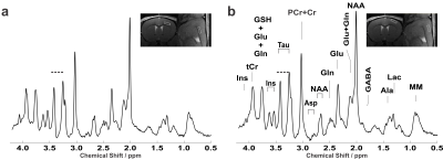 |
Cortical layer-specific neurochemical profiles in the in-vivo mouse brain Video Permission Withheld
Alireza Abaei1, Dinesh K Deelchand 2, Francesco Roselli 3, and Volker Rasche 1
1Core Facility Small Animal Imaging (CF-SANI), University of Ulm, Ulm, Germany, 2Center for Magnetic Resonance Research, University of Minnesota, Minneapolis, MN, United States, 3German Center for Neurodegenerative Diseases (DZNE), Ulm, Germany
The goal of this study was to demonstrate the feasibility to detect neurochemical differences in sub-cortical domains of the murine brain, namely upper (I-IIII) and deep (V-VI) layers. To achieve sub-microliter voxel prescription in these layers, an ultra-high field of 11.7T was used along with the single-voxel LASER sequence. Results show significantly higher taurine (13%) and phosphatidylethanolamine (36%) concentrations in the superficial cortex region compared to the deep layer, in good agreement with cellular composition of these two regions.
|
|
| 14:42 | 0292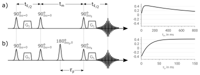 |
An Alternative to WEX: T1-Independent Exchange Rate Quantification using Phase Sensitive Water Exchange Spectroscopy
Nils Marc Joel Plaehn1, Simon Mayer1,2, Petra Albertová1, Peter Michael Jakob1, and Fabian Tobias Gutjahr1
1Experimental Physics 5, University of Würzburg, Würzburg, Germany, 2Leibniz Institute of Plant Genetics and Crop Plant Research (IPK), Gatersleben, Germany
Water Exchange Spectroscopy (WEX) is one of the most widely used approaches to assess the exchange rate of labile protons from a solute to water. In this work, we propose as an versatile alternative “Phase Sensitive WEX” (PS-WEX), which uses an additional RF-pulse and a fixed mixing-time delay. By exploiting the phase sensitivity of the PS-WEX pathway the dynamic range can be increased and the T1-dependence of the signal is completely reduced. This is demonstrated in simulations and phantom measurements. The fixed mixing-time reduces the influence of the T1-decay on the signal curve leading to improved fit quality.
|
|
| 14:54 | 0293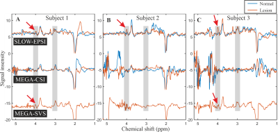 |
Clinical Applicability of SLOW-editing compared to MEGA-editing for the Evaluation of the IDH-mutation Status in Glioma Patients at 7T
Guodong Weng1,2, Johannes Slotboom1,2, and Piotr Radojewski1,2
1Institute for Diagnostic and Interventional Neuroradiology, Support Center for Advanced Neuroimaging (SCAN), University of Bern, Bern, Switzerland, 2Translational Imaging Center, sitem-insel AG, Bern, Switzerland Purpose: comparison of SLOW-editing and MEGA-editing for detection of 2HG in glioma-suspected patients at 7T. Methods: EPSI-based B0/B0+ robust SLOW-editing and semiLASER-SVS/CSI-based MEGA-editing were applied in 10 patients. The edited 2HG signal at 4.01 ppm aimed to be detected with TE = 68 – 75 ms. Results: 8 of 9 patients were identified successfully using SLOW-editing which has a higher spectral quality and success rate compared to MEGA-semiLASER. semiLASER-SVS/CSI-based MEGA-editing showed strong ghosting artifacts around 4.01ppm making 2HG-identification impossible. Conclusions: in vivo 2HG-editing can be performed using SLOW-EPSI within 10 minutes measurement time and is the preferrable method at UHF. |
|
| 15:06 | 0294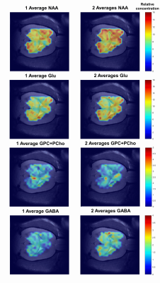 |
Fast high-resolution metabolite mapping on a preclinical 14.1T scanner using 1H-FID-MRSI
Dunja Simicic1,2,3, Brayan Alves1,2, Jessie Julie Mosso1,2,3, Thanh Phong Lê3,4, Ruud B. van Heeswijk5, Jana Starcukova6, Antoine Klauser1,7, Bernhard Strasser8, Wolfgang Bogner8, and Cristina Cudalbu1,2
1CIBM Center for Biomedical Imaging, Lausanne, Switzerland, 2Animal Imaging and Technology, EPFL, Lausanne, Switzerland, 3Laboratory of Functional and Metabolic Imaging, EPFL, Lausanne, Switzerland, 4HES-SO University of Applied Sciences and Arts Western Switzerland, Geneva School of Health Sciences, Geneva, Switzerland, 5Department of Diagnostic and Interventional Radiology, Lausanne University Hospital and University of Lausanne, Lausanne, Switzerland, 6Institute of Scientific Instruments, Czech Academy of Sciences, Brno, Czech Republic, 7Department of radiology and medical informatics, University of Geneva, Geneva, Switzerland, 8Department of Radiology, Medical University Vienna, MR Center of Excellence, Vienna, Austria
1H-MRSI enables a simultaneous acquisition of MR-spectra from multiple spatial locations inside the brain. While 1H-MRSI is increasingly used in the human brain, its implementation in preclinical setting is limited because of the smaller size of rodent brain. At UHF for humans, 1H-FID-MRSI acquisitions are increasingly used (T2 and J-evolution minimization, increased SNR). We present the first implementation of fast 1H-FID-MRSI in the rat brain at 14.1T and exploit its potential for an increased brain coverage, reliable and accurate quantification results and metabolic maps. Our results set the grounds for a wider application of 1H-FID-MRSI in the preclinical setting.
|
|
| 15:18 | 0295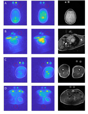 |
Clinically feasible B1 field correction for multi-organ sodium imaging at 3T
Michael Vaeggemose1,2, Rolf F. Schulte3, and Christoffer Laustsen2
1GE Healtcare, Broendby, Denmark, 2MR Research Centre, Department of Clinical Medicine, Aarhus University, Aarhus N, Denmark, 3GE Healtcare, Munich, Germany
Sodium (23Na) MRI allows non-invasive examinations of intra-organ sodium concentrations in vivo. B1 field corrections are important determinants of the sodium signal level. However, low signal-to-noise ratio (SNR) in sodium MRI makes accurate B1 mapping in reasonable scan times challenging. The aim of this study is to evaluate Bloch-Siegert off-resonance B1 field correction of sodium images in thigh muscle, heart, kidney, and brain with the use of MRI in healthy human subjects using a 3D FLORET readout trajectory.
|
|
| 15:30 | 0296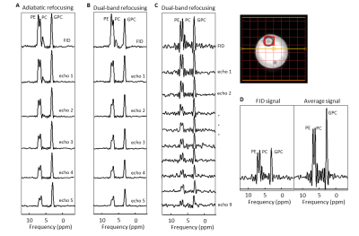 |
31P multi-echo imaging with low B1+ dual-band refocusing pulses
Zahra Shams1, Wybe J.M. van der Kemp1, Evita C. Wiegers1, Jacobus J.M. Zwanenburg1, Jannie P. Wijnen1, and Dennis W.J. Klomp1
1Department of Radiology, University Medical Center Utrecht, Utrecht, Netherlands
We developed a multi-echo sequence to detect phosphomonoesters (PME) and phosphodiesters (PDE), aiming for high signal-to-noise and T2-contrast to noise ratio per unit of time, with the constraint of a maximum available B1 of ~15μT. In line with MRI, the candidates were multi-echo MRSI with 180° pulses (full refocusing) and fast spin echo (FSE) with modulated variable flip angles. Multi-echo MRSI resulted in higher SNR at the same SAR level compared to a FSE with modulated refocusing flip angles. Using 9 dual-band echo pulses improved the SNR for PDE and PME more than two-fold compared to the FID signal alone.
|
|
| 15:42 | 0297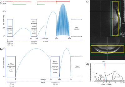 |
Super-fast and accurate estimation of relaxation and chemical exchange rate using 31P-MT-MR fingerprinting at 7T in the human brain
Mark Stephan Widmaier1,2, Song-I Lim1,2, and Lijing Xin1,3
1CIBM Center for Biomedical Imaging, Lausanne, Switzerland, 2Laboratory for Functional and Metabolic Imaging, EPFL, Lausanne, Switzerland, 3Animal Imaging and Technology, EPFL, Lausanne, Switzerland In this abstract, we introduce 31P-MT-MRF, using a SAR efficient magnetization transfer (MT) approach. We extended the magnet resonance fingerprinting (MRF) framework to overcome obstacles of in vivo human brain 31P measurements. This abstract reports the reproducibility and robustness of 31P-MT-MRF estimations of the relaxation parameters of y-ATP and PCr as well as the chemical exchange rate kCK on healthy volunteers in vivo in human brain at 7T. We were able to demonstrate that our method is 3 times faster as state-of-the-art MT methods [6] and estimates are bias free enabling ultra-fast kCK measurement in 2:15 min scan time. |
 Back to Meeting Home
 Back to the Program-at-a-Glance
The International Society for Magnetic Resonance in Medicine is accredited by the Accreditation Council for Continuing Medical Education to provide continuing medical education for physicians.
 Back to Meeting Home
 Back to the Program-at-a-Glance
 View the Presentation