Oral Session
Field & Eddy Current Mapping & Compensation
Joint Annual Meeting ISMRM-ESMRMB & ISMRT 31st Annual Meeting • 07-12 May 2022 • London, UK

Oral Session
Field & Eddy Current Mapping & Compensation
| 09:15 | 0232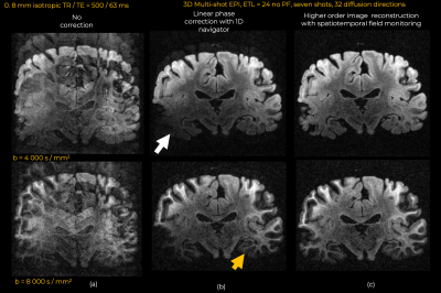 |
Distortion- and ghosting-free high b-value ex vivo human brain diffusion MRI achieved with spatiotemporal magnetic field monitoring
Gabriel Ramos-Llorden1, Daniel Park1, Christian Mirkes2, Cameron M. Cushing3, Paul Weavers3, Hong-Hsi Lee1, Qiyuan Tian1, Alina Scholz4, Boris Keil4, Berkin Bilgic1,5, Anastasia Yendiki1, Thomas Witzel6, and Susie Y. Huang1,5
1Athinoula A. Martinos Center for Biomedical Imaging, Massachusetts General Hospital, Harvard Medical School, Charlestown, MA, United States, 2Skope Magnetic Resonance Technologies AG, Zurich, Switzerland, 3Skope Magnetic Resonance Inc, Lake Mills, WI, United States, 4Institute of Medical Physics and Radiation Protection, Giessen, Germany, 5Harvard-MIT Division of Health Sciences and Technology, Cambridge, MA, United States, 6Q Bio Inc,, San Carlos, MA, United States
Whole brain ex vivo diffusion MRI needs high b-value encoding to compensate for the reduced diffusivity, requiring human brain scanners with ultra-strong diffusion gradients. At that regime, eddy currents become too severe, to the point image distortions and ghosting artifacts cannot be enough mitigated with conventional image reconstruction and post-processing approaches. On a 3T Connectom scanner , we demonstrate the importance of spatiotemporal field monitoring to measure and model eddy currents. When Fourier reconstruction is informed with the unwanted spatiotemporal evolution of the phase, distortion- and ghosting-free high-resolution DW images can be achieved.
|
|
09:27 |
0233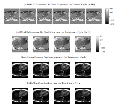 |
Field-Map Combination Method for Phase-Cycled bSSFP using Inherent Bo Mapping
Anjali Datta1, Dwight Nishimura1, and Corey Baron2
1Stanford University, Stanford, CA, United States, 2Western University, London, ON, Canada
Phase-cycled bSSFP enables off-resonance-robust bSSFP imaging. Conventional phase-cycle combination methods weight brighter phase-cycles more, assuming that passband signal is brighter than off-resonant signal. However, near-band signal can be hyperintense, most notably for flowing spins, so traditional methods fail to mitigate these artifacts. Incorporating knowledge of Bo enables inclusion of only passband signal, and exclusion of dark bands and near-band hyperintense artifacts. Using a golden-angle radial trajectory for a free-breathing, phase-cycled acquisition enables reconstruction of a Bo-map time series without any additional scans. This facilitates field-map-based combination throughout the respiratory and cardiac cycles, resulting in substantially reduced hyperintense artifacts than root-sum-of-squares.
|
|
| 09:39 | 0234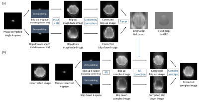 |
Top-up algorithm for single k-space from single-shot EPI
Seon-Ha Hwang1, Hyun-Soo Lee2, Seung Hong Choi3, and Sung-Hong Park1
1Department of Bio and Brain Engineering, Korea Advanced Institute of Science and Technology, Daejeon, Korea, Republic of, 2Siemens Healthineers, Seoul, Korea, Republic of, 3Department of Radiology, Seoul National University College of Medicine, Seoul, Korea, Republic of
Top-up algorithm requires two EPI images with opposite gradient polarities to make one corrected image. Here, top-up algorithm with a single k-space is proposed for single-shot pseudo-centric EPI, where a k-space can be separated into two halves with opposite distortions. A field map could be estimated by applying top-up algorithm to the two halves. The estimated map compensated the distortions well while preserving inner brain structures. The proposed approach provided 1.8 times higher perfusion SNR than conventional linear EPI. The approach is promising for magnetization-prepared imaging that requires high SNR, high temporal resolution, and minimal distortion with no additional scan.
|
|
| 09:51 | 0235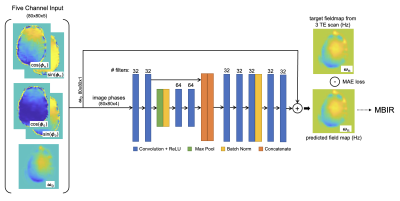 |
FieldMapNet MRI: Learning-based mapping from single echo time BOLD fMRI data to fieldmaps with model-based image reconstruction
Melissa W. Haskell1, Anish Lahiri1, Jon-Fredrik Nielsen2, Jeffrey A. Fessler1, and Douglas C. Noll2
1Electrical Engineering and Computer Science, University of Michigan, Ann Arbor, MI, United States, 2Biomedical Engineering, University of Michigan, Ann Arbor, MI, United States
Artifacts due to B0 field off-resonance result in image distortions and blurring in non-Cartesian acquisitions. FieldMapNet is a learning-based method to map from each image in a spiral-in BOLD fMRI acquisition to a corresponding B0 fieldmap at that timepoint. We train FieldMapNet in a supervised fashion using custom data acquired at three echo times to generate ground truth dynamic fieldmaps. We then perform a tailored B0 correction at each fMRI timepoint using a model-based image reconstruction (MBIR). We show improved image space RMSE using FieldMapNet vs. a separately acquired GRE fieldmap, and a reduction in distortion artifacts and blurring.
|
|
| 10:03 | 0236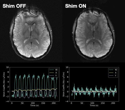 |
Real-time shimming using FID navigators
Tess E Wallace1,2, Tobias Kober3,4,5, Jason P Stockmann1,6, Jonathan R Polimeni1,6, Simon K Warfield1,2, and Onur Afacan1,2
1Radiology, Harvard Medical School, Boston, MA, United States, 2Computational Radiology Laboratory, Boston Children's Hospital, Boston, MA, United States, 3Advanced Clinical Imaging Technology Group, Siemens Healthcare, Lausanne, Switzerland, 4Radiology, Lausanne University Hospital and University of Lausanne, Lausanne, Switzerland, 5LTS5, École Polytechnique Fédérale de Lausanne, Lausanne, Switzerland, 6Athinoula A. Martinos Center for Biomedical Imaging, Massachusetts General Hospital, Boston, MA, United States
Real-time B0 shimming enables improved compensation for the effects of dynamic field perturbations in MRI compared to retrospective approaches, which are computationally intensive and cannot compensate for inherent losses of information. FID navigators can rapidly characterize spatiotemporal field changes using a forward model that predicts the effect of B0 shim changes on the measured FID signal, making them ideally suited for real-time applications. In this work, we implement real-time field control using FID navigators to compensate for spatially linear field changes and demonstrate substantially improved T2*-weighted image quality in the presence of field perturbations in three volunteers scanned at 3T.
|
|
| 10:15 | 0237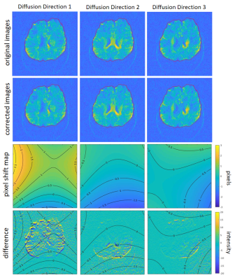 |
Receive Array Coil-driven Eddy Current Correction of Diffusion Weighted MRI Data
Negin Yaghmaie1,2, Warda T. Syeda3,4, Yasmin Blunck1,2, Rebecca Glarin1,5, Daniel Staeb6, Kieran O'Brien6, Scott Kolbe7,8,9, and Leigh A. Johnston1,2
1Melbourne Brain Centre Imaging Unit, The University of Melbourne, Melbourne, Australia, 2Department of Biomedical Engineering, The University of Melbourne, Melbourne, Australia, 3Melbourne Neuropsychiatry Centre, The University of Melbourne, Melbourne, Australia, 4Department of Medicine, The University of Melbourne, Melbourne, Australia, 5Department of Radiology, Royal Melbourne Hospital, Melbourne, Australia, 6MR Research Collaborations, Siemens Healthcare Pty Ltd, Australia, 7Department of Neuroscience, Central Clinical School, Monash University, Melbourne, Australia, 8Department of Radiology, Alfred Hospital, Melbourne, Australia, 9Department of Medicine and Radiology, The University of Melbourne, Melbourne, Australia
An eddy current correction algorithm is proposed that exploits the spatially distributed nature of the receive coil array with signal acquisition immediately preceding image readout in PGSE-EPI diffusion-weighted imaging. The array coil phase for each diffusion gradient direction is expanded using spherical harmonics to yield an estimate of the eddy current-induced field shift in the FOV, and the resultant eddy current-induced pixel shift maps. Distortion corrected diffusion-weighted images are subsequently produced using the estimated pixel shift maps for each diffusion direction, with the method demonstrated in phantom and in-vivo 7T experimental data.
|
|
| 10:27 | 0238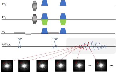 |
Imaging eddy currents induced by crusher gradients using SPEEDI with sub-millisecond temporal resolution Video Permission Withheld
Alessandro M Scotti1, Zheng Zhong2, and Xiaohong Joe Zhou1,3,4,5
1Center for MR Research, University of Illinois at Chicago, Chicago, IL, United States, 2Department of Radiology, Stanford University, Stanford, CA, United States, 3Department of Radiology, University of Illinois at Chicago, Chicago, IL, United States, 4Department of Neurosurgery, University of Illinois at Chicago, Chicago, IL, United States, 5Department of Biomedical Engineering, University of Illinois at Chicago, Chicago, IL, United States
Crusher gradients, typically having very high amplitude, can induce a substantial amount of eddy currents, causing various artifacts. In order to fully characterize the spatiotemporal characteristics of the currents, we have developed a spin echo version of the previously reported SPEEDI technique. The method consists of a series of spin echo acquisitions where each echo point is assigned to a different k-space, reaching a temporal resolution equal to the dwell time. The measured phase difference between sequences with positive and negative crushers revealed B0 and linear eddy currents with time constants equal to the ones measured by the manufacturer’s tool.
|
|
| 10:39 | 0239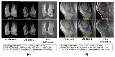 |
Ultrashort Echo Time Double Echo Steady State (UTE-DESS) Imaging with Eddy Current Compensation
Hyungseok Jang1, Jiyo S Athertya1, Yajun Ma1, Alecio F Lombardi1, Saeed Jerban1, Christine B Chung1,2, Eric Y Chang1,2, and Jiang Du1
1Radiology, University of California, San Diego, San Diego, CA, United States, 2Radiology Service, Veterans Affairs San Diego Healthcare System, San Diego, CA, United States
The double echo steady state (DESS) sequence has been investigated extensively due to its short scan time and flexible image contrast achieved from a combination of a free induction decay (FID)-like S+ signal and simulated echo-based S- signal. Ultrashort echo time-based DESS (UTE-DESS) has recently been proposed for imaging of short T2 tissues in the human knee joint, but the effect of eddy currents in this technique has not yet been investigated. In this study, we demonstrate the effects of B0 and linear eddy current and elucidate the importance of eddy current compensation (ECC) for reliable UTE-DESS imaging.
|
 Back to Meeting Home
 Back to the Program-at-a-Glance
The International Society for Magnetic Resonance in Medicine is accredited by the Accreditation Council for Continuing Medical Education to provide continuing medical education for physicians.
 Back to Meeting Home
 Back to the Program-at-a-Glance
 View the Presentation