Oral Session
Novel Encoding Methods & Trajectories
Joint Annual Meeting ISMRM-ESMRMB & ISMRT 31st Annual Meeting • 07-12 May 2022 • London, UK

Oral Session
Novel Encoding Methods & Trajectories
14:30 |
0503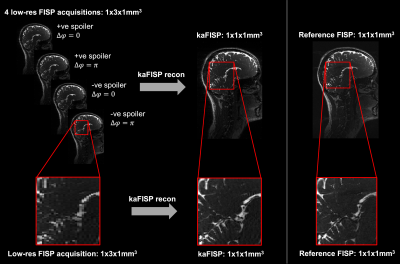 |
SNR-efficient SSFP with k-space aliasing
Peter J Lally1,2, Ben Statton3, Paul M Matthews1,4, Mark Chiew5, Karla L Miller5, and Neal K Bangerter6
1Department of Brain Sciences, Imperial College London, London, United Kingdom, 2UK Dementia Research Institute Centre for Care Research and Technology, London, United Kingdom, 3MRC London Institute of Medical Sciences, Imperial College London, London, United Kingdom, 4UK Dementia Research Institute Centre at Imperial College London, London, United Kingdom, 5Wellcome Centre for Integrative Neuroimaging, Nuffield Department of Clinical Neurosciences, University of Oxford, Oxford, United Kingdom, 6Department of Bioengineering, Imperial College London, London, United Kingdom
Steady state free precession (SSFP) imaging with unbalanced gradients (e.g. FISP, PSIF, DESS) causes aliasing in k-space which is often overlooked, or at best, minimised with large spoilers. Here we exploit this phenomenon and use the aliased signals to increase the SNR-efficiency of SSFP imaging.
|
|
| 14:42 | 0504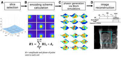 |
Nonlinear encoding scheme design for gradient-free projection imaging in an inhomogeneous Bo at low-field
Kartiga Selvaganesan1, Yonghyun Ha2, Gigi Galiana2, and Todd Constable2
1Biomedical Engineering, Yale University, New Haven, CT, United States, 2Radiology and Biomedical Imaging, Yale University, New Haven, CT, United States
Gradient-free imaging at low-fields can significantly reduce the cost of and increase access to MRI devices. Here we propose to exploit the Bloch-Siegert shift effect to perform gradient-free, RF spatial encoding at 24mT. We have developed a simulation algorithm that designs various nonlinear encoding schemes and evaluates their performance through image reconstruction. Our results indicate that this technique is tolerant to noise and B0 inhomogeneities; this is an important step towards demonstrating the feasibility of performing low-field imaging with nonlinear RF spatial encoding using the Bloch-Siegert shift.
|
|
| 14:54 | 0505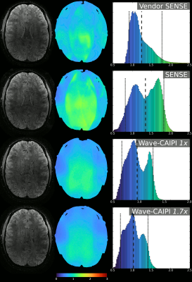 |
Ultra-high field done ultra-fast: Enhancing Wave-CAIPI using an single-axis insert head gradient
Thomas Roos1,2,3, Edwin Versteeg1, and Jeroen Siero1
1Highfield research group, University Medical Center Utrecht, Utrecht, Netherlands, 2Delft University of Technology, Delft, Netherlands, 3Spinoza Centre for Neuroimaging, Amsterdam, Netherlands Acceleration techniques like SENSE have enabled significant decreases in scan time, but the $$$G$$$-factor penalty on SNR limits attainable accelerations. Simulations and 7T phantom & in-vivo acquisitions show significant improvements in $$$G$$$-factor when using Wave-CAIPI, especially when utilizing the higher performance of the insert gradient.
|
|
| 15:06 | 0506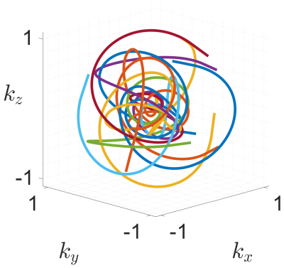 |
Design and Analysis of Field-of-View Independent k-space Trajectories for Magnetic Resonance Imaging
Tobias Speidel1, Patrick Metze1, Kilian Stumpf1, Thomas Hüfken1, and Volker Rasche1
1Internal Medicine II, Ulm University Medical Center, Ulm, Germany
The calculation of k-space trajectories in MRI usually involves prior knowledge of the FOV, since the desired FOV defines a minimum k-space sampling density. The reconstruction of a FOV, which is larger than what is represented by the primary sampling density, is equal to undersampling in k-space. Arising artefacts are strictly dependent on the underlying k-space trajectory which leads to advantages for k-space trajectories with low-coherent aliasing properties also for the combination with non-linear reconstruction techniques.Based on a generalised form of the Seiffert Spirals, this abstract describes a k-space trajectory that does not require prior commitment to an imaging FOV.
|
|
| 15:18 | 0507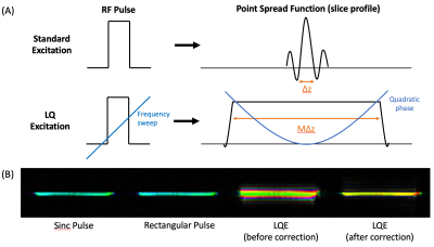 |
Volumetric T2-Weighted Spin-Echo MRI with Improved SNR Using Localized Quadratic Encoding
Dahan Kim1, Dinghui Wang1, Tzucheng Chao1, and James G Pipe1
1Department of Radiology, Mayo Clinic, Rochester, MN, United States
T2-weighted imaging typically employs either TSE or multi-pass 2D SE acquisitions, but using a thick slice and/or a slice gap is often necessary to overcome SNR inefficiencies of these methods, to achieve high-resolution scans in reasonable scan time. We studied efficient T2-weighted SE technique which employed localized quadratic encoding to realize SNR-efficient slice encoding scheme that produced contiguous volumetric coverage. Combined with long-readout spiral acquisitions, the proposed method, a hybrid of 2D and 3D imaging, demonstrated the expected SNR benefit compared to standard 2D scans and produced T2-weighted images with SNR equivalent as 2D-TSE scans but with larger, contiguous coverage.
|
|
| 15:30 | 0508 | Prepolarized MRI of Hard Tissues and Solid-State Matter
Jose Borreguero Morata1, Jose Manuel González Hernández1, Eduardo Pallás Lodeiro2, Juan Pablo Rigla Pérez1, Jose Miguel Algarín Guisado2, Rubén Bosch Esteve1, Fernando Galve Conde2, Daniel Grau Ruíz1, Rubén Pellicer Guridi2, Alfonso Ríos Alonso1, Jose María Benlloch Baviera2, and Joseba Alonso2
1Tesoro Imaging S.L., Valencia, Spain, 2MRI-lab, Institute for Molecular Imaging and Instrumentation (i3M), Spanish National Research Council (CSIC) and Universitat Politècnica de València (UPV), Valencia, Spain
Prepolarized Magnetic Resonance Imaging is a long established technique conceived to counteract the loss in signal strength inherent to low field MRI systems. When it comes to hard biological tissues and solid state matter, PMRI is severely restricted by their ultra short characteristic relaxation times. Here we demonstrate that hard tissue prepolarization is possible with a 0.26 T scanner designed for dental MRI. These results can be applied to clinical dental imaging, making low field PMRI scanners a possible replacement for hazardous X ray systems.
|
 Back to Meeting Home
 Back to the Program-at-a-Glance
The International Society for Magnetic Resonance in Medicine is accredited by the Accreditation Council for Continuing Medical Education to provide continuing medical education for physicians.
 Back to Meeting Home
 Back to the Program-at-a-Glance
 View the Presentation