Oral Session
Rapid, Dynamic & Motion Robust Acquisition
Joint Annual Meeting ISMRM-ESMRMB & ISMRT 31st Annual Meeting • 07-12 May 2022 • London, UK

Oral Session
Rapid, Dynamic & Motion Robust Acquisition
| 17:00 | 0755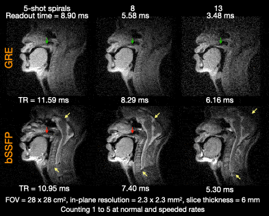 |
Real-time MRI of speech production at 0.55 Tesla Video Not Available
Yongwan Lim1 and Krishna S. Nayak1
1University of Southern California, Los Agneles, CA, United States
We demonstrate 2D mid-sagittal real-time MRI of speech production at 0.55T by employing a custom designed upper airway coil, spiral-based bSSFP and GRE sequences, and constrained image reconstruction. We found that bSSFP offered superior SNR and tissue contrast compared to GRE, and the quality of images at 0.55T was superior to that commonly reported at 1.5T and 3T field strengths. We attribute this improvement to the significantly reduced off-resonance from air-tissue boundaries at 0.55T, making it possible to leverage bSSFP without suffering from banding artifacts.
|
|
| 17:12 | 0756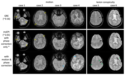 |
A rapid motion-corrected T2*-weighted multi-shot EPI technique for the Emergency Department setting
Zhiqiang Li1, Melvyn B Ooi2, and John P Karis1
1Neuroradiology, Barrow Neurological Institute, Phoenix, AZ, United States, 2Philips Healthcare, Gainesville, FL, United States
T2*w-MRI is routinely used for the diagnosis of hemorrhage and stroke in the Emergency Department, where time is critical and motion is often a concern. However, conventional T2*w-GRE have long scan times, and often suffer from motion artifacts. Parallel imaging and compressed sensing reduce the scan time, but are still prone to motion. ssEPI is fast and motion-insensitive, but suffers from severe geometric distortions. msEPI alleviates the distortion artifacts, but at the cost of increased motion sensitivity. This work presents an msEPI with a navigator echo for rapid motion-corrected T2*w-MRI. Volunteer and patient studies have demonstrated its robustness.
|
|
| 17:24 | 0757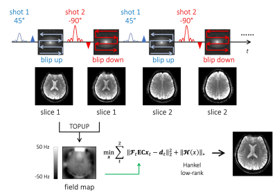 |
Variable Flip, Blip-Up and -Down Undersampling (VUDU) Enables Motion-Robust, Distortion-Free Multi-Shot EPI
Jaejin Cho1,2, Tae Hyung Kim1,2, Borjan Gagoski2,3, Zijing Zhang4, Avery Berman1,2, Congyu Liao5, and Berkin Bilgic1,2,6
1Athinoula A. Martinos Center for Biomedical Imaging, Charlestown, MA, United States, 2Department of Radiology, Harvard Medical School, Boston, MA, United States, 3Fetal-Neonatal Neuroimaging & Developmental Science Center, Boston Children’s Hospital, Boston, MA, United States, 4State Key Laboratory of Modern Optical Instrumentation, College of Optical Science and Engineering, Zhejiang University, Hangzhou, China, 5Radiological Sciences Laboratory, Stanford University, Palo Alto, CA, United States, 6Harvard/MIT Health Sciences and Technology, Cambridge, MA, United States
We demonstrate the motion-robustness of multi-shot diffusion imaging using VUDU (Variable flip, blip-Up, and -Down Undersampling) in the brain. VUDU employs FLEET-ordering to acquire all EPI shots of a given slice successively before proceeding to the next slice and maximizes the signal using variable flip angle (vfa) excitation. It also utilizes BUDA (Blip-Up and -Down Acquisition) to eliminate B0 distortion and employs the Hankel low-rank regularization to address shot-to-shot phase variations. VUDU was able to provide high-fidelity diffusion-weighted images from in vivo data acquired during intentional head motion.
|
|
| 17:36 | 0758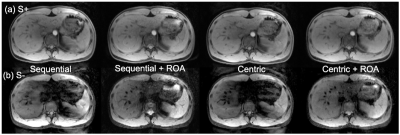 |
Motion insensitive T2-weighted dual-echo steady-state (DESS) imaging of the liver using alternating readout gradients
Daiki Tamada1 and Scott B. Reeder1,2,3,4,5
1Radiology, University of Wisconsin-Madison, Madison, WI, United States, 2Biomedical Engineering, University of Wisconsin-Madison, Madison, WI, United States, 3Medical Physics, University of Wisconsin-Madison, Madison, WI, United States, 4Emergency Medicine, University of Wisconsin-Madison, Madison, WI, United States, 5Medicine, University of Wisconsin-Madison, Madison, WI, United States
In this work, we present a novel method to reduce motion sensitivity for T2-weighted dual-echo steady-state (DESS) imaging, through the use of a readout alternation (ROA) approach. ROA can be achieved by inverting polarity of gradient pulses in the readout axis, alternating every TR. Bloch equation simulations that included small physiological motion in the liver, and volunteer liver imaging was performed using the proposed method. Simulation and volunteer studies demonstrated that ROA effectively suppresses signal loss and motion-related artifacts.
|
|
| 17:48 | 0759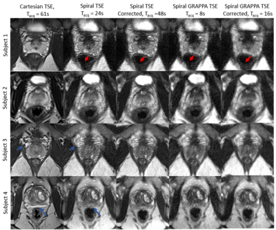 |
A Fast T2-weighted Approach for Prostate Imaging using non-Cartesian GRAPPA and Spiral TSE readout
James Ahad1, Yun Jiang2, Vikas Gulani2, and Nicole Seiberlich2
1Biomedical Engineering, Case Western Reserve University, CLEVELAND, OH, United States, 2Radiology, University of Michigan, Ann Arbor, MI, United States
In-gantry prostate biopsy relies on repeated time-consuming T2-weighted TSE imaging for needle guidance. Reducing the acquisition time for this scan has been challenging due to the spatial resolution and field-of-view requirements of these images, in addition to susceptibility and off resonance artifacts sources common to pelvic imaging. This work proposes a non-Cartesian GRAPPA approach with a short spiral in-out TSE readout to reduce acquisition time for needle guidance images. Comparisons of in vivo Cartesian and spiral TSE images with 300mm FOV, 1.17x1.17mm2 resolution, and 3mm slice thickness acquired in 61s and 8s respectively, are shown.
|
|
| 18:00 | 0760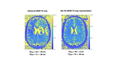 |
Respiration artifact-free 3D DESS T2 mapping of the human brain
Emile Clements Kadalie1, Aurélien J Trotier1, Nadège Corbin1, Sylvain Miraux1, and Emeline J Ribot1
1Centre de Résonance Magnétique des Systèmes Biologiques, UMR5536, CNRS, Bordeaux, France
The Dual Echo Steady State (DESS) sequence has the potential to be used for 3D human brain imaging and 3D T2 mapping. It is however known to be sensitive to respiration. The DESS sequence was therefore modified to include a self-gating approach in order to investigate the influence of breathing on brain images and then extract artifact-free images. To apply this method for high resolution T2 mapping at 3T, an acceleration method compatible with Compressed Sensing was implemented to maintain a clinically acceptable acquisition time.
|
|
| 18:12 | 0761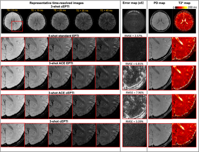 |
Circular Echo-Planar Time-resolved Imaging (cEPTI) for Rapid Time-Resolved and Quantitative Imaging
Nan Wang1, Congyu Liao1, Siddharth Srinivasan1, Xiaozhi Cao1, Justin Haldar2, and Kawin Setsompop1
1Stanford University, Stanford, CA, United States, 2University of Southern California, Los Angeles, CA, United States
EPTI is a highly accelerated time-resolved imaging method for rapid quantitative imaging. To improve the spatiotemporal encoding efficiency of EPTI, we developed a “circular” EPTI sampling trajectory (cEPTI), designed to efficiently traverse a tight circular k-space coverage with full ramp-sampling. A systematic approach was also developed to characterize the noise and effective resolution of the EPTI acquisition and used to guide the optimization of cEPTI’s k-t sampling trajectory. The optimized cEPTI was demonstrated to be capable of producing a 50ms time-resolved series of distortion-free sharp brain images with varying T2* weightings at 1 mm in-plane resolution from a 195ms scan.
|
 Back to Meeting Home
 Back to the Program-at-a-Glance
The International Society for Magnetic Resonance in Medicine is accredited by the Accreditation Council for Continuing Medical Education to provide continuing medical education for physicians.
 Back to Meeting Home
 Back to the Program-at-a-Glance
 View the Presentation