Oral Session
Signal Modelling for Quantitative MRI
Joint Annual Meeting ISMRM-ESMRMB & ISMRT 31st Annual Meeting • 07-12 May 2022 • London, UK

Oral Session
Signal Modelling for Quantitative MRI
| 09:15 | 0621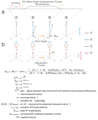 |
Bias-free T1 Mapping via Simultaneously Estimating Bloch-Siegert and Magnetization Transfer Effects
Albert Jang1, Paul K Han1, Chao Ma1, Georges El Fakhri1, and Fang Liu1
1Radiology, Massachusetts General Hospital, Harvard Medical School, Boston, MA, United States
T1 maps obtained using fast steady-state sequences are usually sensitive to B1+ inhomogeneity and MT effects. We propose a novel method to achieve simultaneously B1+- and MT-corrected T1 quantification by introducing off-resonance RF pulses that create two effects: 1) direct saturation of macromolecules and 2) B1+ dependent Bloch-Siegert phase shift. This method, termed BTS (Bloch-Siegert and magnetization transfer simultaneously), is presented and validated in both simulations and experiments, correcting for T1 bias induced from B1+ inhomogeneity and MT effects.
|
|
| 09:27 | 0622 |
Robust STARE (Steady-state T2 And Rf Estimation) for high-resolution quantitative T2 mapping at 7T MRI
Rita Schmidt1,2
1Brain Sciences, Weizmann Institute of Science, Rehovot, Israel, 2The Azrieli National Institute for Human Brain Imaging and Research, Weizmann Institute of Science, Rehovot, Israel
T2 mapping can provide a valuable tool for monitoring and characterization of the tissue changes in the brain. Its implementation in 7T MRI will improve SNR and resolution, but also needs to overcome an increase in SAR and B1 inhomogeneity. We developed a Steady-state T2 And Rf Estimation (STARE) method capable to deliver T2 and RF-field maps in 7T brain imaging. This work summarizes progress towards robust high-resolution whole brain T2 mapping. The method was examined in phantoms and human imaging. Whole brain T2 maps with 1mm resolution were acquired within 3:27 minutes, sufficiently fast to be used in clinics.
|
|
| 09:39 | 0623 |
Rapid quantitative magnetization transfer imaging: utilizing the hybrid state and the generalized Bloch model Video Not Available
Jakob Assländer1,2, Cem Gultekin3, Xiaoxia Zhang1,2, Quentin Duchemin4, Kangning Liu5, Robert WE Charlson6, Timothy Shepherd1, Carlos Fernandez-Granda3,5, and Sebastian Flassbeck1,2
1Center for Biomedical Imaging, New York University School of Medicine, New York, NY, United States, 2Center for Advanced Imaging Innovation and Research, New York University School of Medicine, New York, NY, United States, 3Courant Institute of Mathematical Sciences, New York University, New York, NY, United States, 4Laboratoire d’analyse et de mathématiques appliquées, Université Gustave Eiffel, Paris, France, 5Center for Data Science, New York University, New York, NY, United States, 6Multiple Sclerosis Comprehensive Care Center, Department of Neurology, New York University School of Medicine, New York, NY, United States
We combine two recently proposed biophysical models, the hybrid state and the generalized-Bloch model, to describe magnetization transfer. Combined into a single model, they facilitate quantitative magnetization transfer imaging of the whole brain with 1mm isotropic resolution in 12 minutes. We optimized an MR-Fingerprinting-like pulse sequence, and validated the approach in two multiple sclerosis patients and two controls.
|
|
09:51 |
0624 |
Efficient Time-dependent Diffusion MRI Using Multiple Stimulated Echoes
Guangyu Dan1,2, Kaibao Sun1, Qingfei Luo1, and Xiaohong Joe Zhou1,2,3
1Center for Magnetic Resonance Research, University of Illinois at Chicago, Chicago, IL, United States, 2Department of Biomedical Engineering, University of Illinois at Chicago, Chicago, IL, United States, 3Departments of Radiology and Neurosurgery, University of Illinois at Chicago, Chicago, IL, United States
Studies on temporal diffusion characteristics can reveal a wealth of tissue microstructural information for disease evaluation, but typically require a long scan time because multiple diffusion times are need with each requiring a separate acquisition acquire. We herein report a time-efficient pulse sequence that acquires multiple diffusion-weighted images with different diffusion times in a single shot by utilizing multiple stimulated echoes with variable flip angles. This technique has been implemented on a 3T human scanner and successfully demonstrated in a quantitative diffusion phantom, the human brain gray and white matters, and the prostate peripheral zone and central grand.
|
|
| 10:03 |
0625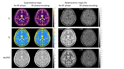 |
Increasing the T2 sensitivity of MR-STAT sequences by small quadratic RF phase increments
Hongyan Liu1, Tom Bruijnen1, Maaike van Haandel1, Oscar van den Heide1, Miha Fuderer1, Cornelis A.T. van den Berg1, and Alessandro A.T. Sbrizzi1
1Computational Imaging Group for MR diagnostics & therapy, Center for Image Sciences, UMC Utrecht, Utrecht, Netherlands
We propose a new method to increase the T2 encoding ability of MR-STAT sequences, according to a recently developed strategy for T2 mapping. Recent work has shown that RF phase modulated GRE sequences with small quadratic phase increments can effectively encode T2 information into the phase of the signal. In this abstract, we show that by incorporating a simple and similar RF modulation strategy in 2D MR-STAT sequences, T2 sensitivity of transient-state gradient-spoiled sequences is improved, and therefore T2 maps as well as the proton density maps (PD) can be reconstructed with higher accuracy.
|
|
| 10:15 | 0626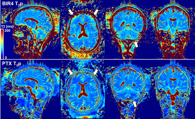 |
Parallel Transmit T2 Preparation for 3D T2 Mapping at Ultra-High Field Video Permission Withheld
Gabriele Bonanno1,2,3, Patrick Liebig4, Kurt Majewsky5, Tobias Kober6,7,8, and Tom Hilbert6,7,8
1Advanced Clinical Imaging Technology, Siemens Healthcare AG, Bern, Switzerland, 2Magnetic Resonance Methodology, Institute of Diagnostic and Interventional Neuroradiology, University of Bern, Bern, Switzerland, 3Translational Imaging Center, sitem-insel, Bern, Switzerland, 4Siemens Healthcare GmbH, Erlangen, Germany, 5Siemens AG, Munich, Germany, 6Advanced Clinical Imaging Technology, Siemens Healthcare AG, Lausanne, Switzerland, 7Department of Radiology, University Hospital (CHUV) and University of Lausanne (UNIL), Lausanne, Switzerland, 8LTS5, École Polytechnique Fédérale de Lausanne, Lausanne, Switzerland
T2 relaxometry at ultra-high field has the potential to become an important quantitative MRI biomarker thanks to its sensitivity to pathology. However, acquiring high-resolution isotropic T2 maps is challenging due to signal-to-noise and specific absorption rate constraints. We present a T2-mapping method for ultra-high-field MRI based on a parallel transmit T2-preparation (T2p) module integrated in a compressed sensing accelerated segmented 3D FLASH sequence. The proposed parallel transmit T2p module is investigated in phantom and in vivo experiments and compared to an adiabatic T2p module. Preliminary tests show feasibility for sub-millimeter T2 relaxometry at 7 T.
|
|
| 10:27 | 0627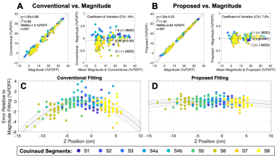 |
Addressing Concomitant Gradient Phase Errors in Chemical Shift-Encoded MRI with a Pass-Specific Phase Fitting Method
Nathan Tibbitts Roberts, BS1,2, Diego Hernando, PhD1,2,3,4, Nikolaos Panagiotopoulos, MD1, and Scott B Reeder, MD, PhD1,3,4,5,6
1Radiology, University of Wisconsin - Madison, Madison, WI, United States, 2Electrical and Computer Engineering, University of Wisconsin - Madison, Madison, WI, United States, 3Medical Physics, University of Wisconsin - Madison, Madison, WI, United States, 4Biomedical Engineering, University of Wisconsin - Madison, Madison, WI, United States, 5Medicine, University of Wisconsin - Madison, Madison, WI, United States, 6Emergency Medicine, University of Wisconsin - Madison, Madison, WI, United States
This work presents a complex-based fitting method to address concomitant gradient (CG) phase errors for time-interleaved (i.e., multi-pass) chemical shift-encoded (CSE) MRI estimation of proton density fat fraction (PDFF) and R2* through a joint estimation of pass-specific phase terms. The proposed method does not require prior knowledge of gradient waveforms. The proposed fitting method removed significant PDFF and R2* estimation errors in both phantom and in vivo evaluations away from isocenter. An evaluation in 29 clinical liver datasets demonstrated reduced PDFF bias and variability (8.4% improvement in the coefficient of variation), even when the imaging volume was centered at isocenter.
|
 Back to Meeting Home
 Back to the Program-at-a-Glance
The International Society for Magnetic Resonance in Medicine is accredited by the Accreditation Council for Continuing Medical Education to provide continuing medical education for physicians.
 Back to Meeting Home
 Back to the Program-at-a-Glance
 View the Presentation