Oral Session
Diffusion MRI in Gray Matter: Exchange & Restriction
Joint Annual Meeting ISMRM-ESMRMB & ISMRT 31st Annual Meeting • 07-12 May 2022 • London, UK

Oral Session
Diffusion MRI in Gray Matter: Exchange & Restriction
| 09:15 | 0250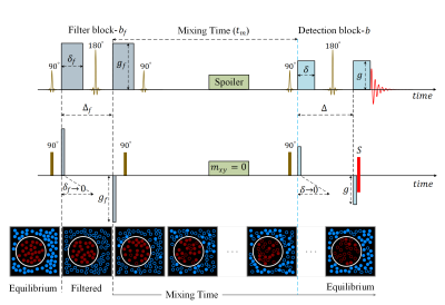 |
What Does FEXI Measure?
Mohammad Khateri1, Marco Reisert 2, Alejandra Sierra1, Jussi Tohka1, and Valerij G. Kiselev2
1A.I.Virtanen Institute for Molecular Sciences, University of Eastern Finland, Kuopio, Finland, 2Department of Radiology, University Medical Center Freiburg, Freiburg, Germany
Filter-EXchange Imaging (FEXI) evaluates the permeability of cell membranes, which is already used in clinical research. It relies on suppressing the extracellular signal using strong diffusion weighting (mobility filter causing a reduction in the overall diffusivity) and monitoring the subsequent diffusivity recovery. Using simulations, we demonstrate the presence of a spurious exchange: In compartments with complex geometry, there are locations where spins remain unaffected by the mobility filter. Moving to other locations afterward contributes to the diffusivity recovery, thus mimicking exchange. The discovered phenomenon opens large room for investigation towards characterizing the compartment geometry and crystallizing the genuine membrane permeation.
|
|
| 09:27 | 0251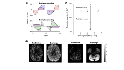 |
Mapping water exchange in the human brain using diffusion MRI with free waveform encoding
Arthur Chakwizira1, Filip Szczepankiewicz2, Carl-Fredrik Westin3, Geraline Vis4, Linda Knutsson1,5,6, Pia Sundgren7,8,9,10, and Markus Nilsson2
1Medical Radiation Physics, Lund, Lund University, Lund, Sweden, 2Clinical Sciences Lund, Lund University, Lund, Sweden, 3Department of Radiology, Brigham and Women's hospital, Harvard Medical School, Boston, MA, United States, 4Department of Diagnostic Radiology, Clinical Sciences Lund, Lund University, Lund, Sweden, 5Russell H. Morgan Department of Radiology and Radiological Science, Johns Hopkins University School of Medicine, Baltimore, MD, United States, 6F. M. Kirby Research Center for Functional Brain Imaging, Kennedy Krieger Institute, Baltimore, MD, United States, 7Department of Diagnostic Radiology, Lund University, Lund, Sweden, 8Lund University Bioimaging Center, Lund University, Lund, Sweden, 9Department for Medical Imaging and Physiology, Skåne University Hospital, Lund, Sweden, 10Department of Radiology, University of Michigan, Ann Arbor, MI, United States
Temporal velocity correlations of diffusing particles carry information about the structure of the diffusion environment. Studying high-order velocity autocorrelation functions can aid in understanding how signal is influenced by restricted diffusion and exchange, and in turn how to design experiments that are sensitive/independent to these phenomena. In this work, we employ numerical simulations on a variety of substrates to demonstrate this notion. We find that the fourth order velocity autocorrelation bears a distinctive signature of exchanging systems. In addition, we highlight that the effect of exchange on the second order velocity autocorrelation is negligible when exchange is barrier-limited.
|
|
| 09:39 | 0252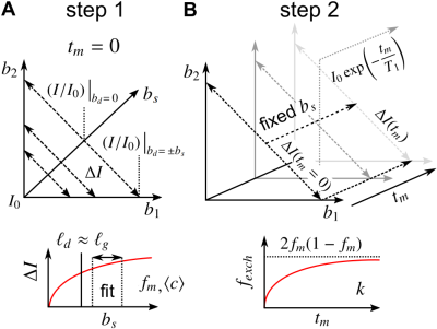 |
Isolating restriction and exchange in gray matter using double and single diffusion encodings with equal diffusion weighting
Teddy Xuke Cai1,2, Nathan Hu Williamson2,3, Rea Ravin2,4, and Peter Joel Basser2
1Department of Clinical Neurosciences, Wellcome Centre for Integrative Neuroimaging, FMRIB, University of Oxford, Oxford, UK, United Kingdom, 2Section on Quantitative Imaging and Tissue Sciences, Eunice Kennedy Shriver National Institute of Child Health and Human Development, Bethesda, MD, United States, 3National Institute of General Medical Sciences, Bethesda, MD, United States, 4Celoptics, Inc., Rockville, MD, United States
The diffusion MR signal in complex tissue such as gray matter exhibits non-Gaussian signal attenuation due to exchange and restrictions. Existing signal models typically ignore one or both effects by assuming Gaussian diffusion or negligible exchange. We propose a more rigorous signal model that incorporates both effects. Subsequently, an acquisition scheme utilizing equal double diffusion encodings ($$$b_1=b_2$$$) at various mixing times, and single diffusion encodings with the same total weighting $$$b=b_1+b_2$$$, is designed to independently characterize the effects of restriction and exchange. The method is tested on live and fixed gray matter specimen using a low-field, high-gradient MR system.
|
|
| 09:51 | 0253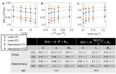 |
Ex vivo gray matter is complex: exchange & disorder exponents
Ileana O Jelescu1,2 and Quentin Uhl1
1Radiology, Lausanne University Hospital (CHUV), Lausanne, Switzerland, 2CIBM Center for Biomedical Imaging, Ecole Polytechnique Federale de Lausanne, Lausanne, Switzerland
Fixation alters tissue properties significantly and in vivo vs ex vivo models should be adapted accordingly. Unlike in vivo studies of rodent gray matter (GM) where diffusion time-dependence D(t) was absent for t > 10 ms, allowing an interpretation of time-dependent kurtosis K(t) as resulting from inter-compartment exchange, here we show that ex vivo rodent GM displays marked D(t), with non-Gaussianity arising most probably from extracellular water. K(t) could thus result from combined effect of disorder and exchange. High-b data where extracellular water is preferentially suppressed may still enable the unconfounded estimation of exchange.
|
|
| 10:03 | 0254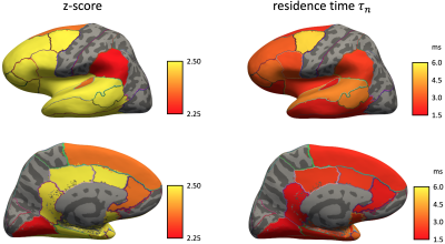 |
Revealing diffusion time-dependence and exchange effect in the in vivo human brain gray matter by using high gradient diffusion MRI
Hong Hsi Lee1,2, Jonas L Olesen3,4, Qiyuan Tian1,2, Gabriel Ramos Llorden1,2, Sune N Jespersen3,4, and Susie Y Huang1,2
1Athinoula A. Martinos Center for Biomedical Imaging, Massachusetts General Hospital, Charlestown, MA, United States, 2Department of Radiology, Harvard Medical School, Boston, MA, United States, 3Center of Functionally Integrative Neuroscience (CFIN) and MINDLab, Department of Clinical Medicine, Aarhus University, Aarhus, Denmark, 4Department of Physics and Astronomy, Aarhus University, Aarhus, Denmark
The characteristic signal time-dependence in the brain gray matter indicates a significant exchange effect, modeled by an extension of the Kärger exchange model. Here we perform in vivo diffusion measurements with varying time scales (21-40 ms) up to high b-values (≤22 ms/µm2) in a healthy subject on an HCP scanner, and observe the signal time-dependence in frontal lobe, temporal lobe, and limbic system. The estimated residence time is 2-7 ms in the brain gray matter, suggesting that diffusion in neurite and extra-cellular space is coarse-grained at clinical diffusion time scale.
|
|
| 10:15 | 0255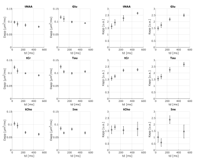 |
Investigating exchange, structural disorder and restriction in Gray Matter via water and metabolites diffusivity and kurtosis time-dependence
Eloïse MOUGEL1, Julien Valette1, and Marco Palombo2,3,4
1Université Paris-Saclay, Commissariat à l’Energie Atomique et aux Energies Alternatives (CEA), Centre National de la Recherche Scientifique(CNRS), Molecular Imaging Research Center (MIRCen), Laboratoire des Maladies Neurodégénératives, Fontenay aux Roses, France, 2Centre for Medical Image Computing (CMIC), Department of Computer Science, University College London, London, United Kingdom, 3Cardiff University Brain Research Imaging Centre (CUBRIC), School of Psychology, Cardiff University, Cardiff, United Kingdom, 4School of Computer Science and Informatics, Cardiff University, Cardiff, United Kingdom
This work reports first-time measurements of the time-dependent diffusivity and kurtosis of both water and metabolites in vivo in the mouse gray matter (GM). Our aim is to exploit the complementary information provided by the diffusion of water and intracellular metabolites to investigate and potentially disentangle the role of exchange, structural disorder and restriction in GM. Our results show evidence that water diffusion-time dependence in GM is mostly driven by 1D short-range disorder, potentially alongside exchange. Conversely, metabolites’ diffusion-time dependence is exclusively driven by cellular restrictions, paving a new way to quantify noninvasively microstructural restrictions in GM.
|
|
| 10:27 | 0256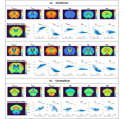 |
Correlation of Soma and Neurite Density Imaging (SANDI) metrics with Allen Mouse Brain Atlas
Andrada Ianus1, Joana Carvalho1, Francisca F. Fernandes1, Renata Cruz1, Cristina Chavarrias1, Marco Palombo2,3,4, and Noam Shemesh1
1Champalimaud Research, Champalimaud Centre for the Unknown, Lisbon, Portugal, 2Centre for Medical Image Computing (CMIC), Dept of Computer Science, University College London, London, United Kingdom, 3Cardiff University Brain Research Imaging Centre (CUBRIC), School of Psychology, Cardiff University, Cardiff, United Kingdom, 4School of Computer Science and Informatics, Cardiff University, Cardiff, United Kingdom
Cross modal validation of micro-architectural features derived from diffusion MRI with contrast from other imaging modalities is critical for developing reliable imaging biomarkers. Here, we map apparent soma and neurite densities via the SANDI methodology in the in-vivo mouse brain at 9.4T and compared the derived metrics with the contrast from the Allen Mouse Brain Atlas which reflects cell body density in the brain. Our findings suggest that soma signal fraction correlates well with atlas intensities in different parts of the brain.
|
|
| 10:39 | 0257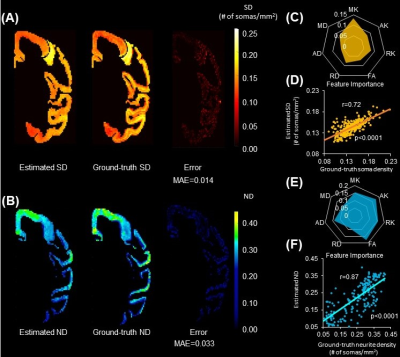 |
Diffusion-MRI based Estimation of Cortical Architecture using Machine-learning (DECAM)
Tianjia Zhu1,2, Minhui Ouyang1, and Hao Huang1,3
1Department of Radiology, Children's Hospital of Philadelphia, Philadelphia, PA, United States, 2Department of Bioengineering, University of Pennsylvania, Philadelphia, PA, United States, 3Department of Radiology, University of Pennsylvania, Philadelphia, PA, United States
Advanced diffusion MRI (dMRI) has enabled noninvasive microstructural assessment that can be only conventionally measured with histology1-9. However, analytical dMRI models are limited by their restrictive model assumptions, lack of validation, and biased microstructural measures. We have developed Diffusion-MRI based Estimation of Cortical Architecture using Machine-learning (DECAM), a data-driven dMRI-based method accurately estimating cortical soma and neurite densities (SD and ND) in the cortex10 leveraging a variety of complementary dMRI contrasts. By providing high-fidelity estimated soma and neurite density maps validated with histology, DECAM paves the way for data-driven noninvasive virtual histology for potential applications such as Alzheimer’s diseases.
|
 Back to Meeting Home
 Back to the Program-at-a-Glance
The International Society for Magnetic Resonance in Medicine is accredited by the Accreditation Council for Continuing Medical Education to provide continuing medical education for physicians.
 Back to Meeting Home
 Back to the Program-at-a-Glance
 View the Presentation