Oral Session
Beyond Conventional Diffusion Encoding
Joint Annual Meeting ISMRM-ESMRMB & ISMRT 31st Annual Meeting • 07-12 May 2022 • London, UK

Oral Session
Beyond Conventional Diffusion Encoding
| 14:30 | 0509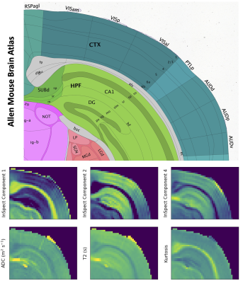 |
Separating Tissue Components in Ex-vivo Mouse Brain with Joint Diffusion-Relaxation MRI and InSpect
Paddy J. Slator1, Noam Shemesh2, and Andrada Ianus2
1Centre for Medical Image Computing, Department of Computer Science, University College London, London, United Kingdom, 2Champalimaud Research, Champalimaud Centre for the Unknown, Lisbon, Portugal
We identify and map intra-voxel tissue components in the mouse brain by applying InSpect, a data-driven approach for quantitative MRI analysis, to high resolution ex-vivo T2-diffusion MRI. Our approach reveals distinct tissue microenvironments with minimal model assumptions, revealing features that cannot be seen in diffusion kurtosis maps.
|
|
| 14:42 | 0510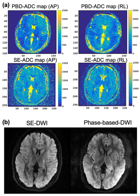 |
Phase-based 3D diffusion mapping using RF phase-modulated gradient echo imaging
Daiki Tamada1 and Scott B. Reeder1,2,3,4,5
1Radiology, University of Wisconsin-Madison, Madison, WI, United States, 2Biomedical Engineering, University of Wisconsin-Madison, Madison, WI, United States, 3Medical Physics, University of Wisconsin-Madison, Madison, WI, United States, 4Emergency Medicine, University of Wisconsin-Madison, Madison, WI, United States, 5Medicine, University of Wisconsin-Madison, Madison, WI, United States A novel 3D diffusion mapping method using RF phase-modulated gradient echo imaging was developed by encoding diffusion weighting into signal phase. We developed closed form signal equations based on configuration theory. A four-pass acquisition and lookup table-based reconstruction is performed to estimate diffusion and T2 simultaneously. Simulation results revealed excellent agreement of signal magnitude and phase between Bloch equation and the developed equations. Phantom and in vivo studies demonstrated the feasibility of the proposed approach. These results demonstrate that the proposed method can enable qualitative and quantitative diffusion imaging with smaller gradient amplitude compared to the conventional diffusion-weighted imaging. |
|
| 14:54 | 0511 |
DEXSY can measure water exchange linked to cellular homeostasis and active states in central nervous system tissue
Nathan H. Williamson1,2, Rea Ravin1, Teddy X. Cai1,3, Melanie Falgairolle4, Michael J. O'Donovan4, and Peter J. Basser1
1National Institute of Child Health and Human Development, Potomac, MD, United States, 2National Institute of General Medical Sciences, Potomac, MD, United States, 3Department of Clinical Neurosciences, Wellcome Centre for Integrative Neuroimaging, FMRIB, University of Oxford, Oxford, United Kingdom, 4National Institute of Neurological Disorders and Stroke, Potomac, MD, United States
We propose a new functional MR method based on steady-state transmembrane water exchange rate measurements with diffusion exchange spectroscopy (DEXSY). Rapid DEXSY methods are implemented on a low-field MR system with a strong static gradient to test for an active component of water exchange in ex vivo neonatal mouse spinal cords. Temperature-dependent Arrhenius activation energies for water exchange are significantly greater in viable “live” samples than in the same samples after fixation, suggesting a connection to ATP-driven enzymatic processes. Moreover, exchange rates in live samples significantly decrease after blocking Na+/K+-ATPase pump activity, revealing an active component of water exchange.
|
|
| 15:06 | 0512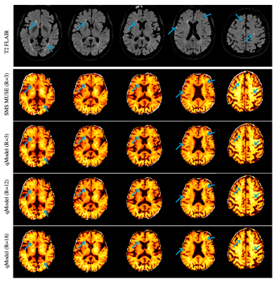 |
qModeL: A Model-based Deep Learning Framework for the Recovery of Diffusion-based Microstructural Parameters from Healthy and Lesion Data
Merry Mani1, Baolian Yang2, Girish Bathla1, Vincent Magnotta1, and Mathews Jacob1
1University of Iowa, Iowa City, IA, United States, 2GE Healthcare, Waukesha, WI, United States
A flexible deep learning based framework is presented for the recovery of microstructural parameter maps from advanced diffusion models. The method is shown to work well across field strengths, and healthy and diseased tissue without needing separate training of the DL network. The DL framework is embedded in a model-based reconstruction, which enables the framework to handle variations in data acquisition settings such as various acceleration factors and noise levels, without having to change the network. k-q accelerations in the range of 12-18 fold is demonstrated for single and multi-shell diffusion data.
|
|
| 15:18 | 0513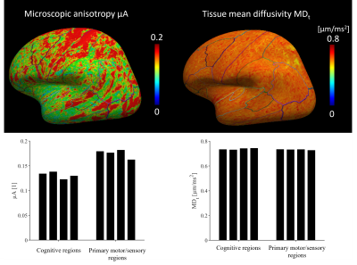 |
Gray matter microstructure imaging by b-tensor encoding at high b-values and high spatial resolution
Geraline Vis1, Nicola Spotorno2, Björn Lampinen1, Filip Szczepankiewicz1, and Markus Nilsson1
1Department of Diagnostic Radiology, Clinical Sciences Lund, Lund University, Lund, Sweden, 2Clinical Memory Research Unit, Department of Clinical Sciences, Lund University, Lund, Sweden
Studying gray matter microstructure with dMRI is challenging because of partial volume effects and high orientation dispersion. In this work, we propose a novel method that combines spherical b-tensor encoding, strong diffusion weighting and super-resolution reconstruction to perform high-resolution microstructure imaging of gray matter. We map the tissue mean diffusivity and the microscopic anisotropy which, by their design, are unaffected by partial volume effects and intra-voxel orientation dispersion. We demonstrate the approach in human brain in vivo at a resolution of 1.4 mm isotropic.
|
|
15:30 |
0514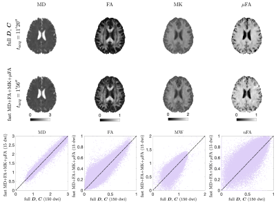 |
Fast acquisitions for mean kurtosis and microscopic fractional anisotropy
Santiago Coelho1, Filip Szczepankiewicz2, Els Fieremans1, and Dmitry S. Novikov1
1Center for Advanced Imaging Innovation and Research (CAI2R), Department of Radiology, New York University, School of Medicine, New York, NY, United States, 2Department of Diagnostic Radiology, Clinical Sciences, Lund University, Lund, Sweden
We propose two acquisition protocols that minimize the number of directions needed for estimating MD, FA, MK, and $$$\mu$$$FA. Using symmetric trace-free decompositions of the diffusion, kurtosis, and covariance tensors, we design acquisitions that cancel unnecessary tensor elements, thereby reducing the number of directions needed to generate the maps. Our protocols consist of $$$3b_1+6b_2=9$$$ DWIs for MD+MK in a 1-minute scan, and $$$6b_1^\text{LTE}+6b_2^\text{LTE}+3b_{1.5}^\text{STE}=15$$$ DWIs for MD+FA+MK+$$$\mu$$$FA in a 2-minute scan. We show the feasibility of these approaches on a healthy volunteer.
|
|
| 15:42 | 0515 |
Correlation Tensor MRI reveals dynamic changes in diffusion kurtosis sources along stroke progression
Rita Alves1, Rafael Neto Henriques1, Sune Nørhøj Jespersen2,3, and Noam Shemesh1
1Champalimaud Research, Champalimaud Centre for the Unknown, Lisbon, Portugal, 2Center of Functionally Integrative Neuroscience (CFIN) and MINDLab, Department of Clinical Medicine, Aarhus University, Aarhus, Denmark, 3Department of Physics and Astronomy, Aarhus University, Aarhus, Portugal
Correlation Tensor Imaging is emerging as a novel methodology for resolving diffusional kurtosis sources. Here, we decouple the anisotropic (Kaniso), isotropic (Kiso), and microscopic kurtosis (μK) sources and map them along the progression of experimental ischemia. Our results indicate that μK and Kaniso, associated with cross sectional variance (tentatively associated with neurite beading) and Kiso, likely reflecting edema formation, are pivotal markers for pathology. Our findings are promising for more specific characterizations of stroke lesions and a better understanding of tissue injury.
|
|
| 15:54 | 0516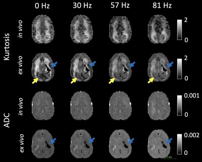 |
Oscillating Gradient (OGSE) and Microscopic Anisotropy Diffusion in the in vivo and ex vivo Marmoset Brain
Tales Santini1, Naila Rahman1, Alyson Shim1, Wataru Inoue1, Stefan Everling1, and Corey Baron1
1Western University, London, ON, Canada
We investigated OGSE and μFA diffusion techniques for the first time in the common marmoset (Callithrix jacchus) brain, which revealed comparable values to those found in humans. The same animal was scanned in vivo and after fixation of the brain, allowing comparison of in vivo and ex vivo OGSE and μFA parameters for the first time in marmosets. There were differences in kurtosis, μFA, and μA that may be caused by slight neurite beading and tissue shrinkage during perfusion/fixation, and temperature differences.
|
 Back to Meeting Home
 Back to the Program-at-a-Glance
The International Society for Magnetic Resonance in Medicine is accredited by the Accreditation Council for Continuing Medical Education to provide continuing medical education for physicians.
 Back to Meeting Home
 Back to the Program-at-a-Glance
 View the Presentation