Oral Session
MR Elastography
Joint Annual Meeting ISMRM-ESMRMB & ISMRT 31st Annual Meeting • 07-12 May 2022 • London, UK

14:45 |
0117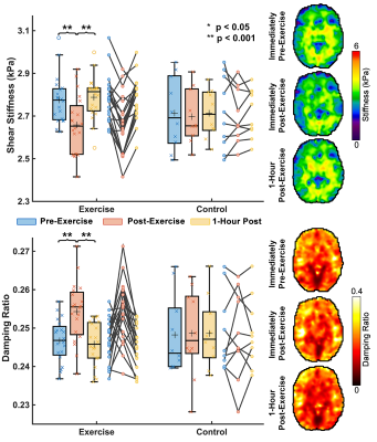 |
Acute Effects of High-Intensity Exercise on Brain Mechanical Properties and Cognitive Function
Grace McIlvain1, Emily M Magoon2, Rebecca Clements2, Alexis Merritt2, Lucy V Hiscox3, Hillary Schwarb4, and Curtis L Johnson2
1Biomedical Engineering, University of Delaware, Newark, DE, United States, 2University of Delaware, Newark, DE, United States, 3University of Bath, Bath, United Kingdom, 4University of Illinois at Urbana-Champaign, Champaign, IL, United States
Here we use magnetic resonance elastography (MRE) to quantify brain mechanical properties of 30 individuals aged 19-30 immediately before a high intensity interval training (HIIT) exercise, immediately after exercise, and 1 hour after exercise. We additionally tested subjects’ cognitive performance at these time points through a memory task and two executive function tasks. We found that whole brain stiffness decreased in nearly all subjects immediately after exercise by an average of 4.2% (p<0.0001) and that whole brain damping ratio increased by an average of 3.1% (p=0.001). Performance on all three cognitive tasks improved immediately after exercise (p<0.05).
|
|
| 14:57 | 0118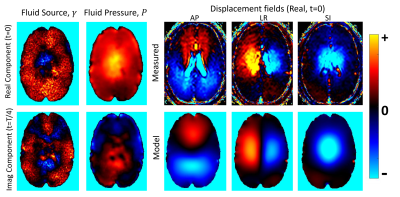 |
Imaging the pulsatile fluid source distribution and pressure gradients in the brain using a poroelastic model and intrinsic pulsatile motions.
Matthew McGarry1, John Weaver1,2, and Keith Paulsen1,2
1Dartmouth College, Hanover, NH, United States, 2Dartmouth-Hitchcock Medical Center, Lebanon, NH, United States
The pulsatile tissue motions from the cardiac cycle can be imaged using retrospectively gated phase contrast MRI. Modeling brain tissue as a poroelastic material allows a distribution of pulsatile fluid sources to be computed which reproduce the measured motions by estimating the RHS vector of a finite element representation of the poroelastic system. This Fluid source imaging (FSI) represents a completely new type of image parameterization that could offer unique diagnostic signatures. The associated pressure field gradients are also the driving force for interstitial fluid movement, where disruptions are thought to cause buildup of waste products which leads to dementia.
|
|
| 15:09 |
0119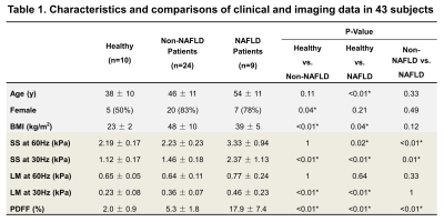 |
Influence of hepatic tissue water content on MRE-based biomarkers in clinical and preclinical studies
Jiahui Li1, Zheng Zhu1, Jingbiao Chen1, Alina M Allen2, Sudhakar K. Venkatesh1, Taofic Mounajjed3, Kevin J. Glaser1, Armando Manduca1, Vijay H. Shah2, Richard L. Ehman1, and Meng Yin1
1Radiology, Mayo Clinic, Rochester, MN, United States, 2Gastroenterology and Hepatology, Mayo Clinic, Rochester, MN, United States, 3Anatomic Pathology, Mayo Clinic, Rochester, MN, United States The water in hepatic tissue has intracellular, extracellular matrix bound, and extracellular interstitial free fluid components, which are affected by inflammation and fibrosis. This study assessed longitudinal changes in MRE-based biomarkers and their relationship with hepatic water content in clinical and preclinical models. In clinical studies, MRE-assessed loss modulus (LM) was elevated in obese patients and in NAFLD patients with severe inflammation. In preclinical models, changes in LM correlated with water content and disease progression in non-NAFLD mice. LM significantly increased with the development of NASH. MRE-assessed viscoelasticity strongly reflects extracellular free fluid associated with inflammation. |
|
| 15:21 | 0120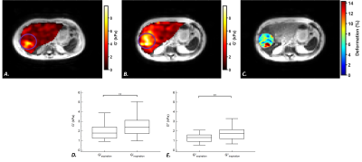 |
Microvascular invasion in patients with hepatocellular carcinoma: assessment with compression MR elastography
Gwenaël Pagé1, Philippe Garteiser1, Valérie Paradis2, Ralph Sinkus3, Valérie Vilgrain1,4, and Bernard E. Van Beers1,4
1Laboratory of imaging biomarkers, INSERM UMR 1149, Paris, France, 2Center for research on inflammation, INSERM UMR 1149, Paris, France, 3Laboratory for vascular translational science, INSERM UMR 1148, Paris, France, 4Department of Radiology, University Hospital Paris Nord, Clichy, France
The aim of this study was to investigate the value of compression MR elastography to assess microvascular invasion in hepatocellular carcinomas. MR elastographic and T2-weighted images were acquired at end expiration and end inspiration in 52 patients with hepatocellular carcinomas. The storage and loss moduli at inspiration did not differ between tumors with/without microvascular invasion. However, the compression stiffening rate, derived from the increase of tumor storage modulus between expiration and inspiration, had good diagnostic performance (area under the receiver operating characteristic curve 0.85) in detecting microvascular invasion.
|
|
| 15:33 | 0121 |
Self-Navigated Radial Free-Breathing Magnetic Resonance Elastography of the Liver with Rapid Motion Encoding in Children at 3T
Sevgi Gokce Kafali1,2, Bradley D. Bolster Jr.3, Shu-Fu Shih1,2, Grace J. Kim1, Joanna Yeh4, Robert S. Venick4, Shahnaz Ghahremani1, Kara L. Calkins4, and Holden H. Wu1,2
1Radiological Sciences, University of California, Los Angeles, Los Angeles, CA, United States, 2Bioengineering, University of California, Los Angeles, Los Angeles, CA, United States, 3MR Collaborations, Siemens Medical Solutions, Inc., Salt Lake City, UT, United States, 4Pediatrics, University of California, Los Angeles, Los Angeles, CA, United States
Hepatic stiffness measured by magnetic resonance elastography (MRE) is a biomarker for hepatic fibrosis. Conventional Cartesian gradient-echo MRE requires breath-holding (BH), which can be challenging for children. While radial free-breathing (FB) MRE overcomes the challenge of BH, the scan time is relatively longer (163 seconds/slice). This study’s objective was to reduce radial FB-MRE scan time by a factor of two with rapid motion encoding (81 seconds/slice) and to improve robustness by performing self-navigated motion compensation. Compared to Cartesian BH-MRE in children at 3T, the proposed rapid radial FB-MRE technique quantified hepatic stiffness with close agreement and similar repeatability.
|
|
15:45 |
0122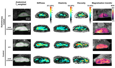 |
Early tumour response to radiotherapy assessed by magnetic resonance elastography
Clementine Lesbats1, Upasana Roy1, Emma L. Reeves1, Jessica K. R. Boult1, Yann Jamin2, Craig Cummings1, James Sullivan3, Ralph Sinkus4, Erik Wennerberg1, and Simon P. Robinson2
1Division of Radiotherapy & Imaging, The Institute of Cancer Research, Sutton, United Kingdom, 2The Institute of Cancer Research, Sutton, United Kingdom, 3The Royal Marsden NHS Foundation Trust, Sutton, United Kingdom, 4School of Biomedical Engineering and Imaging Sciences, King's College London, London, United Kingdom Radiotherapy is widely used to treat cancer. In this study, we used MR elastography to evaluate early tumour response to radiation therapy. A significant increase in stiffness |G*| and elasticity Gd were determined 3 days after orthotopic 4T1 breast tumours were irradiated with a single 8Gy dose, compared to untreated control tumour-bearing mice. The increase in tumour stiffness was associated with an increased macromolecule pool measured by magnetisation transfer, and increased collagen deposition observed histologically. |
|
| 15:57 | 0123 |
Feasibility of compressed sensing accelerated Ristretto magnetic resonance elastography in the pancreas
Anne-Sophie van Schelt1, Nienke P.M. Wassenaar1, Jurgen H. Runge1, Jules L. Nelissen1, Marian Troelstra1,2, Christian Guenthner3, Ralph Sinkus2,4, Jaap Stoker1, Aart J. Nederveen1, and Eric M. Schrauben1
1Radiology and Nuclear Medicine, Amsterdam UMC location AMC, Amsterdam, Netherlands, 2Department of Imaging Sciences and Biomedical Engineering, King’s College London, London, United Kingdom, 3Department of Biomed, ETH Zurich, Zurich, Switzerland, 4Inserm U1148, LVTS, Univeristy Paris Diderot, Paris, France
Magnetic resonance elastography (MRE) allows for non-invasive determination of pancreatic visco-elastic properties in four consecutive breath-holds. The aim of this study was to develop and test a single breath-hold MRE acquisition accelerated using prospective undersampling and compressed sensing (CS) reconstruction. Testing was done on a retrospective undersampled phantom dataset and in-vivo. These showed that CS accelerated (R< 8.7), single breath-hold MRE is feasible without hampering visco-elastic reconstruction in tissues with low shear stiffness (|G*|<1.6kPa). Further research is necessary to guarantee accuracy of the measured shear stiffness, notably for high stiffness tissues such as tumors, and at higher acceleration factors.
|
 Back to Meeting Home
 Back to the Program-at-a-Glance
The International Society for Magnetic Resonance in Medicine is accredited by the Accreditation Council for Continuing Medical Education to provide continuing medical education for physicians.
 Back to Meeting Home
 Back to the Program-at-a-Glance
 View the Presentation